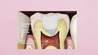
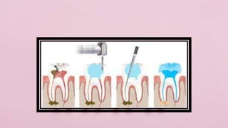
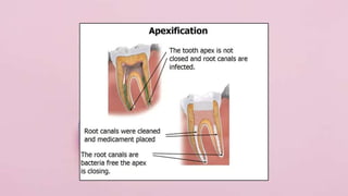
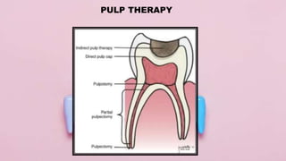
Apexogenesis & apexification in pediatric dentistryDr. Harsh Shah
SDDCH Parbhani
Presented by : Vipul GIratkar
Dept. of Pediatric dentitstry
Guided by . Dr. Rehan Khan
DIscussion regarding apexification and apexogenesis
The presentation features the pulp reparative and regenerative procedures which can be carried out in immature teeth. It involves development of mature tooth from an immature one by root formation and root fixation as a preparatory phase for root canal treatment.
it will provide u a detail description about direct pulp capping treatment,its indication ,contraindication,methods and materials used,techniqes,advantage and disadvantage and its limitation on primary teeth
Dental management of children with special health care needsaravindhanarumugam1
hope this will throw a light in understanding special children and dental management of the same particularly for pediatric dentistry PGs .children with genetic diseases and emotionally handicapped ( child abuse and neglect ) are not discussed here as they are separate topics.
dr. aravindhan
INTRODUCTION
DEFINITION
TYPES OF TRAUMA FROM OCCLUSION
GLICKMAN CONCEPT
WAERHAUG CONCEPT
STAGES OF TISSUE RESPONSE TO INJURY
CLINICAL AND RADIOGRAPHIC FEATURES OF TFO
CLINICAL DIAGNOSIS OF TFO
TFO AND IMPLANTS
TREATMENT OF TFO
CONCLUSION
REFRENCES
ARTIFICIAL INTELLIGENCE IN HEALTHCARE.pdfAnujkumaranit
Artificial intelligence (AI) refers to the simulation of human intelligence processes by machines, especially computer systems. It encompasses tasks such as learning, reasoning, problem-solving, perception, and language understanding. AI technologies are revolutionizing various fields, from healthcare to finance, by enabling machines to perform tasks that typically require human intelligence.
NVBDCP.pptx Nation vector borne disease control programSapna Thakur
NVBDCP was launched in 2003-2004 . Vector-Borne Disease: Disease that results from an infection transmitted to humans and other animals by blood-feeding arthropods, such as mosquitoes, ticks, and fleas. Examples of vector-borne diseases include Dengue fever, West Nile Virus, Lyme disease, and malaria.
Title: Sense of Taste
Presenter: Dr. Faiza, Assistant Professor of Physiology
Qualifications:
MBBS (Best Graduate, AIMC Lahore)
FCPS Physiology
ICMT, CHPE, DHPE (STMU)
MPH (GC University, Faisalabad)
MBA (Virtual University of Pakistan)
Learning Objectives:
Describe the structure and function of taste buds.
Describe the relationship between the taste threshold and taste index of common substances.
Explain the chemical basis and signal transduction of taste perception for each type of primary taste sensation.
Recognize different abnormalities of taste perception and their causes.
Key Topics:
Significance of Taste Sensation:
Differentiation between pleasant and harmful food
Influence on behavior
Selection of food based on metabolic needs
Receptors of Taste:
Taste buds on the tongue
Influence of sense of smell, texture of food, and pain stimulation (e.g., by pepper)
Primary and Secondary Taste Sensations:
Primary taste sensations: Sweet, Sour, Salty, Bitter, Umami
Chemical basis and signal transduction mechanisms for each taste
Taste Threshold and Index:
Taste threshold values for Sweet (sucrose), Salty (NaCl), Sour (HCl), and Bitter (Quinine)
Taste index relationship: Inversely proportional to taste threshold
Taste Blindness:
Inability to taste certain substances, particularly thiourea compounds
Example: Phenylthiocarbamide
Structure and Function of Taste Buds:
Composition: Epithelial cells, Sustentacular/Supporting cells, Taste cells, Basal cells
Features: Taste pores, Taste hairs/microvilli, and Taste nerve fibers
Location of Taste Buds:
Found in papillae of the tongue (Fungiform, Circumvallate, Foliate)
Also present on the palate, tonsillar pillars, epiglottis, and proximal esophagus
Mechanism of Taste Stimulation:
Interaction of taste substances with receptors on microvilli
Signal transduction pathways for Umami, Sweet, Bitter, Sour, and Salty tastes
Taste Sensitivity and Adaptation:
Decrease in sensitivity with age
Rapid adaptation of taste sensation
Role of Saliva in Taste:
Dissolution of tastants to reach receptors
Washing away the stimulus
Taste Preferences and Aversions:
Mechanisms behind taste preference and aversion
Influence of receptors and neural pathways
Impact of Sensory Nerve Damage:
Degeneration of taste buds if the sensory nerve fiber is cut
Abnormalities of Taste Detection:
Conditions: Ageusia, Hypogeusia, Dysgeusia (parageusia)
Causes: Nerve damage, neurological disorders, infections, poor oral hygiene, adverse drug effects, deficiencies, aging, tobacco use, altered neurotransmitter levels
Neurotransmitters and Taste Threshold:
Effects of serotonin (5-HT) and norepinephrine (NE) on taste sensitivity
Supertasters:
25% of the population with heightened sensitivity to taste, especially bitterness
Increased number of fungiform papillae
TEST BANK for Operations Management, 14th Edition by William J. Stevenson, Ve...kevinkariuki227
TEST BANK for Operations Management, 14th Edition by William J. Stevenson, Verified Chapters 1 - 19, Complete Newest Version.pdf
- Video recording of this lecture in English language: https://youtu.be/lK81BzxMqdo
- Video recording of this lecture in Arabic language: https://youtu.be/Ve4P0COk9OI
- Link to download the book free: https://nephrotube.blogspot.com/p/nephrotube-nephrology-books.html
- Link to NephroTube website: www.NephroTube.com
- Link to NephroTube social media accounts: https://nephrotube.blogspot.com/p/join-nephrotube-on-social-media.html
Ozempic: Preoperative Management of Patients on GLP-1 Receptor Agonists Saeid Safari
Preoperative Management of Patients on GLP-1 Receptor Agonists like Ozempic and Semiglutide
ASA GUIDELINE
NYSORA Guideline
2 Case Reports of Gastric Ultrasound
The prostate is an exocrine gland of the male mammalian reproductive system
It is a walnut-sized gland that forms part of the male reproductive system and is located in front of the rectum and just below the urinary bladder
Function is to store and secrete a clear, slightly alkaline fluid that constitutes 10-30% of the volume of the seminal fluid that along with the spermatozoa, constitutes semen
A healthy human prostate measures (4cm-vertical, by 3cm-horizontal, 2cm ant-post ).
It surrounds the urethra just below the urinary bladder. It has anterior, median, posterior and two lateral lobes
It’s work is regulated by androgens which are responsible for male sex characteristics
Generalised disease of the prostate due to hormonal derangement which leads to non malignant enlargement of the gland (increase in the number of epithelial cells and stromal tissue)to cause compression of the urethra leading to symptoms (LUTS
Title: Sense of Smell
Presenter: Dr. Faiza, Assistant Professor of Physiology
Qualifications:
MBBS (Best Graduate, AIMC Lahore)
FCPS Physiology
ICMT, CHPE, DHPE (STMU)
MPH (GC University, Faisalabad)
MBA (Virtual University of Pakistan)
Learning Objectives:
Describe the primary categories of smells and the concept of odor blindness.
Explain the structure and location of the olfactory membrane and mucosa, including the types and roles of cells involved in olfaction.
Describe the pathway and mechanisms of olfactory signal transmission from the olfactory receptors to the brain.
Illustrate the biochemical cascade triggered by odorant binding to olfactory receptors, including the role of G-proteins and second messengers in generating an action potential.
Identify different types of olfactory disorders such as anosmia, hyposmia, hyperosmia, and dysosmia, including their potential causes.
Key Topics:
Olfactory Genes:
3% of the human genome accounts for olfactory genes.
400 genes for odorant receptors.
Olfactory Membrane:
Located in the superior part of the nasal cavity.
Medially: Folds downward along the superior septum.
Laterally: Folds over the superior turbinate and upper surface of the middle turbinate.
Total surface area: 5-10 square centimeters.
Olfactory Mucosa:
Olfactory Cells: Bipolar nerve cells derived from the CNS (100 million), with 4-25 olfactory cilia per cell.
Sustentacular Cells: Produce mucus and maintain ionic and molecular environment.
Basal Cells: Replace worn-out olfactory cells with an average lifespan of 1-2 months.
Bowman’s Gland: Secretes mucus.
Stimulation of Olfactory Cells:
Odorant dissolves in mucus and attaches to receptors on olfactory cilia.
Involves a cascade effect through G-proteins and second messengers, leading to depolarization and action potential generation in the olfactory nerve.
Quality of a Good Odorant:
Small (3-20 Carbon atoms), volatile, water-soluble, and lipid-soluble.
Facilitated by odorant-binding proteins in mucus.
Membrane Potential and Action Potential:
Resting membrane potential: -55mV.
Action potential frequency in the olfactory nerve increases with odorant strength.
Adaptation Towards the Sense of Smell:
Rapid adaptation within the first second, with further slow adaptation.
Psychological adaptation greater than receptor adaptation, involving feedback inhibition from the central nervous system.
Primary Sensations of Smell:
Camphoraceous, Musky, Floral, Pepperminty, Ethereal, Pungent, Putrid.
Odor Detection Threshold:
Examples: Hydrogen sulfide (0.0005 ppm), Methyl-mercaptan (0.002 ppm).
Some toxic substances are odorless at lethal concentrations.
Characteristics of Smell:
Odor blindness for single substances due to lack of appropriate receptor protein.
Behavioral and emotional influences of smell.
Transmission of Olfactory Signals:
From olfactory cells to glomeruli in the olfactory bulb, involving lateral inhibition.
Primitive, less old, and new olfactory systems with different path
Acute scrotum is a general term referring to an emergency condition affecting the contents or the wall of the scrotum.
There are a number of conditions that present acutely, predominantly with pain and/or swelling
A careful and detailed history and examination, and in some cases, investigations allow differentiation between these diagnoses. A prompt diagnosis is essential as the patient may require urgent surgical intervention
Testicular torsion refers to twisting of the spermatic cord, causing ischaemia of the testicle.
Testicular torsion results from inadequate fixation of the testis to the tunica vaginalis producing ischemia from reduced arterial inflow and venous outflow obstruction.
The prevalence of testicular torsion in adult patients hospitalized with acute scrotal pain is approximately 25 to 50 percent
Ethanol (CH3CH2OH), or beverage alcohol, is a two-carbon alcohol
that is rapidly distributed in the body and brain. Ethanol alters many
neurochemical systems and has rewarding and addictive properties. It
is the oldest recreational drug and likely contributes to more morbidity,
mortality, and public health costs than all illicit drugs combined. The
5th edition of the Diagnostic and Statistical Manual of Mental Disorders
(DSM-5) integrates alcohol abuse and alcohol dependence into a single
disorder called alcohol use disorder (AUD), with mild, moderate,
and severe subclassifications (American Psychiatric Association, 2013).
In the DSM-5, all types of substance abuse and dependence have been
combined into a single substance use disorder (SUD) on a continuum
from mild to severe. A diagnosis of AUD requires that at least two of
the 11 DSM-5 behaviors be present within a 12-month period (mild
AUD: 2–3 criteria; moderate AUD: 4–5 criteria; severe AUD: 6–11 criteria).
The four main behavioral effects of AUD are impaired control over
drinking, negative social consequences, risky use, and altered physiological
effects (tolerance, withdrawal). This chapter presents an overview
of the prevalence and harmful consequences of AUD in the U.S.,
the systemic nature of the disease, neurocircuitry and stages of AUD,
comorbidities, fetal alcohol spectrum disorders, genetic risk factors, and
pharmacotherapies for AUD.
Flu Vaccine Alert in Bangalore Karnatakaaddon Scans
As flu season approaches, health officials in Bangalore, Karnataka, are urging residents to get their flu vaccinations. The seasonal flu, while common, can lead to severe health complications, particularly for vulnerable populations such as young children, the elderly, and those with underlying health conditions.
Dr. Vidisha Kumari, a leading epidemiologist in Bangalore, emphasizes the importance of getting vaccinated. "The flu vaccine is our best defense against the influenza virus. It not only protects individuals but also helps prevent the spread of the virus in our communities," he says.
This year, the flu season is expected to coincide with a potential increase in other respiratory illnesses. The Karnataka Health Department has launched an awareness campaign highlighting the significance of flu vaccinations. They have set up multiple vaccination centers across Bangalore, making it convenient for residents to receive their shots.
To encourage widespread vaccination, the government is also collaborating with local schools, workplaces, and community centers to facilitate vaccination drives. Special attention is being given to ensuring that the vaccine is accessible to all, including marginalized communities who may have limited access to healthcare.
Residents are reminded that the flu vaccine is safe and effective. Common side effects are mild and may include soreness at the injection site, mild fever, or muscle aches. These side effects are generally short-lived and far less severe than the flu itself.
Healthcare providers are also stressing the importance of continuing COVID-19 precautions. Wearing masks, practicing good hand hygiene, and maintaining social distancing are still crucial, especially in crowded places.
Protect yourself and your loved ones by getting vaccinated. Together, we can help keep Bangalore healthy and safe this flu season. For more information on vaccination centers and schedules, residents can visit the Karnataka Health Department’s official website or follow their social media pages.
Stay informed, stay safe, and get your flu shot today!
How STIs Influence the Development of Pelvic Inflammatory Disease.pptx
Pulp therapy
1. P U L P T H E R A P Y
IN
PRIMARY DENTITION
GLO ARBY ARGUELLES,
DMD
2. P U L P T H E R A P Y
The primary goal of pulp therapy is to maintain the integrity and health of the
teeth and their supporting tissues while maintaining the vitality of the pulp of a
tooth affected by caries, traumatic injury, or other causes.
Especially in young permanent teeth with immature roots, the pulp is integral to
continue apexogenesis.
Long term retention of a permanent tooth requires a root with a favorable
crown/root ratio and dentinal walls that are thick enough to withstand normal
function.
Therefore, pulp preservation is a primary goal for treatment of the young
permanent dentition
3. P U L P T H E R A P Y
The indications, objectives, and type of pulpal therapy are based on the health status of
the pulp tissue which is classified as: normal pulp (symptom free and normally
responsive to vitality testing), reversible pulpitis (pulp is capable of healing),
symptomatic or asymptomatic irreversible pulpitis (vital inflamed pulp is incapable of
healing), or necrotic pulp. The clinical diagnosis derived from:
1. a comprehensive medical history.
2. a review of past and present dental history and treatment, including current symptoms
and chief complaint.
3. a subjective evaluation of the area associated with the current symptoms/chief
complaint by questioning the patient/parent on the location, intensity, duration,
stimulus, relief, and spontaneity.
4. an objective extraoral examination as well as examination of the intraoral soft and
hard tissues.
5. if obtainable, radiograph(s) to diagnose periapical or periradicular changes.
6. clinical tests such as palpation, percussion, and mobility; however, electric pulp and
4. P U L P T H E R A P Y
Teeth exhibiting provoked pain of short duration relieved with over-the-counter
analgesics, by brushing, or upon the removal of the stimulus and without signs or
symptoms of irreversible pulpitis have a clinical diagnosis of reversible pulpitis and are
candidates for vital pulp therapy.
Teeth diagnosed with a normal pulp requiring pulp therapy or with reversible pulpitis
should be treated with vital pulp therapy.
Teeth exhibiting signs or symptoms such as a history of spontaneous unprovoked pain, a
sinus tract, soft tissue inflammation not resulting from gingivitis or periodontitis,
excessive mobility not associated with trauma or exfoliation, furcation/ apical
radiolucency, or radiographic evidence of internal/ external resorption have a clinical
diagnosis of irreversible pulpitis or necrosis and are candidates for nonvital pulp
treatment.
7. WHAT IS PULP
CAPPING?
Placing a suitable dressing either directly on the exposed pulp or on a thin
residual layer of slightly soft dentin, to maintain a vital pulp.
8. Indirect Pulp Capping
• It is a procedure performed in a tooth with a deep caries lesion approximating the pulp but
without evidence of radicular pathology.
• “Indirect pulp treatment is a procedure that leaves the deepest caries adjacent to the pulp
undisturbed in an effort to avoid a pulp exposure.
• This caries-affected dentin is covered with a biocompatible material to produce a biological
seal.”
• A radiopaque liner such as a dentin bonding agent, resin modified glass ionomer, calcium
hydroxide, or MTA (or any other biocompatible material) is placed over the remaining carious
dentin to stimulate healing and repair.
• The liner that is placed over the dentin (calcium hydroxide, glass ionomer, or bonding agents)
does not affect the IPT success.
• The tooth then is restored with a material that seals the tooth from microleakage.
9. Indirect Pulp Capping
CONTRAINDICATIONS:
Sharp, penetrating pain that persists after removal of stimulus.
Prolonged, spontaneous pain, particularly at night.
OE:
Excessive tooth mobility
Non responsive to pulp testing techniques
Discolored
RE:
Large carious lesion, with apparent pulp exposure
Widened PDL space, interrupted an broken lamina dura
Radiolucency at the root apices or furcation areas
10.
11. Direct Pulp Capping
• When a pinpoint exposure (one millimeter or less) of the pulp is encountered during cavity preparation or
following a traumatic injury, a biocompatible radiopaque base such as MTA or calcium hydroxide may be placed in
contact with the exposed pulp tissue.
• The tooth is restored with a material that seals the tooth from microleakage.
• Indications:
This procedure is indicated in a primary tooth with a normal pulp following a small pulpal exposure of one
millimeter or less when conditions for a favorable response are optimal.
• Objectives:
The tooth’s vitality should be maintained. No post-treatment signs or symptoms such as sensitivity, pain, or
swelling should be evident. Pulp healing and reparative dentin formation should result. There should be no
radiographic signs of pathologic external or progressive internal root resorption or furcation/apical
radiolucency. There should be no harm to the succedaneous tooth.
12. Direct Pulp Capping
CONTRAINDICATIONS:
Carious exposure of primary tooth.
Large, carious exposure in symptomatic teeth.
Severe pain at night.
Spontaneous pain.
OE:
Excessive tooth mobility
Non responsive to pulp testing techniques
Discolored
Excessive hemorrhage
Purulent or serous exudate
RE:
Large carious lesion, with apparent pulp exposure
Widened PDL space, interrupted an broken lamina dura
Radiographic evidence of furcal or periradicular degenration
13.
14. Pulpotomy
• A pulpotomy is performed in a primary tooth when caries removal results in a pulp exposure in a tooth with a normal
pulp or reversible pulpitis or after a traumatic pulp exposure and there is no radiographic sign of infection or pathologic
resorption.
• The coronal pulp is amputated, pulpal hemorrhage controlled, and the remaining vital radicular pulp tissue surface is
treated with a long-term clinically-successful medicament.
• Only MTA and formocresol are recommended as the medicament of choice for teeth expected to be retained for 24
months or more.
• The AAPD’s Use of vital Pulp Therapies in Primary Teeth with Deep Caries Lesions recommended against the use of
calcium hydroxide for pulpotomy.
• After the coronal pulp chamber is filled with a suitable base, the tooth is restored with a restoration that seals the tooth
from microleakage.
• If there is sufficient supporting enamel remaining, amalgam or composite resin can provide a functional alternative
when the primary tooth has a life span of two years or less. However, for multisurface lesions, a stainless steel crown is
the restoration of choice.
15. Pulpotomy
• Indications:
The pulpotomy procedure is indicated when caries removal results in pulp exposure in a primary tooth with a
normal pulp or reversible pulpitis or after a traumatic pulp exposure,and when there are no radiographic signs of
infection or pathologic resorption. When the coronal tissue is amputated, the remaining radicular tissue must be
judged to be vital without suppuration, purulence, necrosis, or excessive hemorrhage that cannot be controlled by a
cotton pellet after several minutes.
• Objectives:
The radicular pulp should remain asymptomatic without adverse clinical signs or symptoms such as sensitivity,
pain, or swelling. There should be no postoperative radiographic evidence of pathologic external root resorption.
Internal root resorption may be self-limiting and stable. The clinician should monitor the internal resorption, removing
the affected tooth if perforation causes loss of supportive bone and/or clinical signs of infection and inflammation.
There should be no harm to the succedaneous tooth.
16. Pulpotomy
CONTRAINDICATIONS:
A history of spontaneous toothache.
Non restorable tooth where coronal seal is inadequate.
OE:
Pulp that does not bleed (necrotic)
Inability to control radicular hemorrhage
A pulp with purulent or serous drainage
RE:
Evidence of pathologic root resorption.
Evidence of periapical pathosis
17.
18. Pulpectomy
• Pulpectomy is a root canal procedure for pulp tissue that is irreversibly inflamed or necrotic due to
caries or trauma. The root canals are debrided and shaped with hand or rotary files and then irrigated.
• A recent systematic review showed no difference in success when irrigating with chlorhexidine or one
to five percent sodium hypochlorite or sterile water/saline. Because it is a potent tissue irritant, sodium
hypochlorite must not be extruded beyond the apex. After the canals are dried, a resorbable material
such as nonreinforced zinc/oxide eugenol, iodoform-based paste or a combination paste of iodoform
and calcium hydroxide is used to fill the canals.
• A recent systematic review reports that ZOE performed better long term than iodoform-based pastes.
• The tooth then is restored with a restoration that seals the tooth from microleakage.
• Clinicians should evaluate non-vital pulp treatments for success and adverse events clinically and
radiographically at least every 12 months.
19. Pulpectomy
• Indications:
A pulpectomy is indicated in a primary tooth with irreversible pulpitis or necrosis or a tooth
treatment planned for pulpotomy in which the radicular pulp exhibits clinical signs of irreversible pulpitis
or pulp necrosis (e.g., suppuration, purulence) The roots should exhibit minimal or no resorption. When
there is no root resorption present, pulpectomy is recommended.
• Objectives:
Following treatment, the radiographic infectious process should resolve in six months as evidenced
by bone deposition in the pretreatment radiolucent areas, and pretreatment clinical signs and symptoms
should resolve within a few weeks. There should be radiographic evidence of successful filling without
gross overextension or underfilling. The treatment should permit resorption of the primary tooth root and
filling material to permit normal eruption of the succedaneous tooth. There should be no pathologic root
resorption or furcation/apical radiolucency.
20. Pulpectomy
CONTRAINDICATIONS:
Non restorable tooth
RE:
Internal resorption of roots.
Excessive pathologic root resorption involving more than one-third of the root
Excessive pathologic loss of bone support and periodontal attachment
21.
22. Apexogenesis
• Apexogenesis is a histological term used to describe the continued physiologic
development and formation of the root’s apex.
• Formation of the apex in vital young permanent teeth can be accomplished by
implementing the appropriate vital pulp therapy described in this section (i.e., indirect pulp
treatment, direct pulp capping, partial pulpotomy for carious exposures and traumatic
exposures).
23.
24. Apexification
• Apexification is a method of inducing root end closure of an incompletely formed non-vital permanent tooth by
removing the coronal and non-vital radicular tissue just short of the root end and placing a biocompatible agent
such as calcium hydroxide in the canals for two weeks to one month to disinfect the canal space.
• Root end closure is accomplished with an apical barrier such as MTA. In instances when complete closure cannot
be accomplished by MTA, an absorbable collagen wound dressing can be placed at the root end to allow MTA to
be packed within the confines of the canal space. Gutta percha is used to fill the remaining canal space. If the canal
walls are thin, the canal space can be filled with MTA or composite resin instead of gutta percha to strengthen the
tooth against fracture.
25. Apexification
• Indications:
• This procedure is indicated for non-vital permanent teeth with incompletely formed roots.
• Objectives:
• This procedure should induce root end closure (apexification) at the apices of immature roots or result in an
apical barrier as confirmed by clinical and radiographic evaluation. Adverse post-treatment clinical signs or
symptoms of sensitivity, pain, or swelling should not be evident. There should be no radiographic evidence of
external root resorption, lateral root pathosis, root fracture, or breakdown of periradicular supporting tissues
during or following therapy. The tooth should continue to erupt, and the alveolus should continue to grow in
conjunction with the adjacent teeth.