Download to read offline


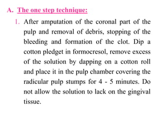
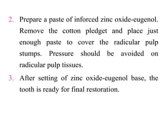
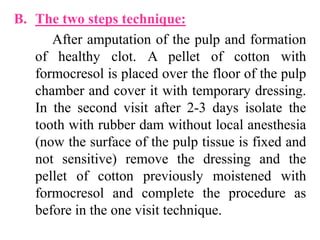
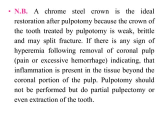


The document covers various pulp therapy techniques in pediatric dentistry, including direct pulp capping, pulpotomy, partial pulpectomy, and complete pulpectomy, detailing their definitions, indications, and techniques. The effectiveness and materials used for each procedure are discussed, highlighting the role of calcium hydroxide, zinc oxide-eugenol, and formocresol in pulp treatment. The document emphasizes the importance of careful technique and management of complications to ensure successful outcomes in treating dental pulp in children.


![CASE_PRESENTATION_ON_subdural_hematoma(SDH)[1 FINAL PPT]-1.pptx](https://cdn.slidesharecdn.com/ss_thumbnails/casepresentationonsubduralhematomasdh1finalppt-1-260129172522-d405d375-thumbnail.jpg?width=640&height=640&fit=bounds)


