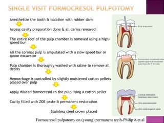
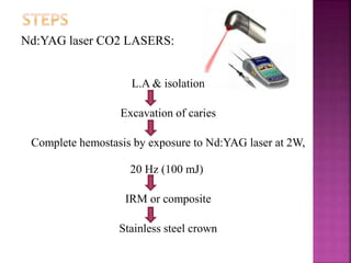
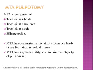
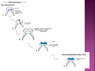
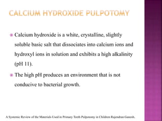
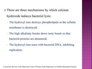
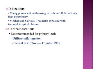
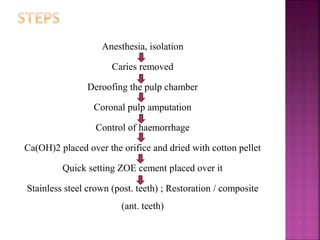
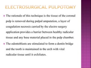
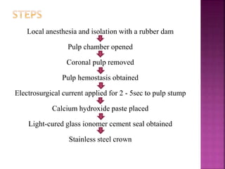
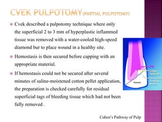
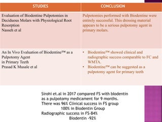
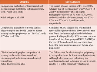
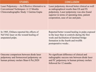
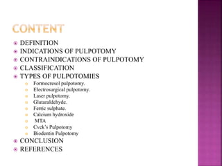
This document discusses pulpotomy, including definitions, indications, contraindications, classifications, and types. Pulpotomy involves removing the coronal portion of the dental pulp and placing a medicament to promote healing and preserve vitality. Types discussed include formocresol pulpotomy, electrosurgical pulpotomy, laser pulpotomy, glutaraldehyde, ferric sulphate, calcium hydroxide, MTA, and Cvek's pulpotomy. Success is defined as asymptomatic pulp without pathology. Pulpotomy is indicated for carious exposure in a vital tooth with healthy supporting tissues and no signs of infection.


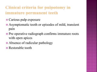
![ The formocresol pulpotomy technique was first
advocated by SWEET [1930]
He used a multiple sitting technique, which has
been subsequently modified to either a single or
two stage technique.
BUCKLEY’S FORMALDEHYDE FORMULA :-
19% Formaldehyde
35% cresol
15% Glycerin &
31%Water
To prepare 1.5% concentration of this formula,
first mix 3 parts of glycerin with 1 part of distilled
water , then add 4 parts of this preparation to 1 part
buckley's formocresol,and throughly mix again.](https://image.slidesharecdn.com/14pulpotomy-230425122150-91cc6596/85/PULPOTOMY-pptx-16-320.jpg)