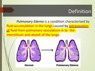
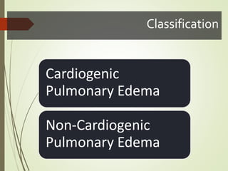
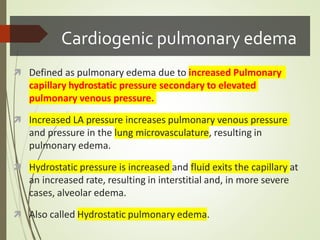
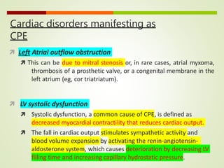
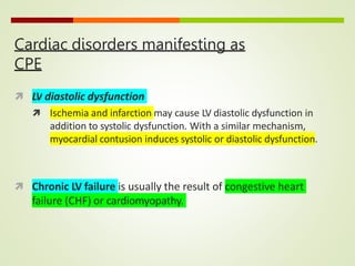
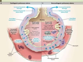
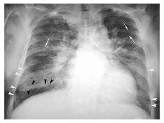
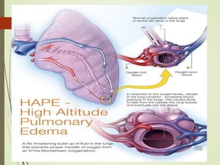
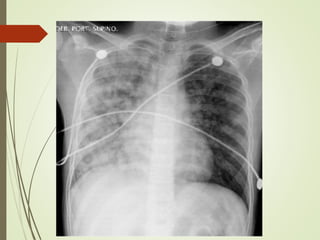
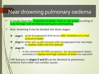
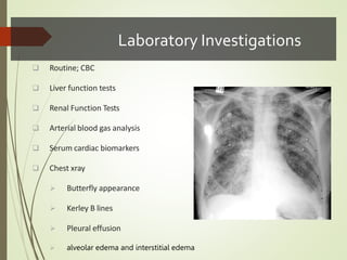
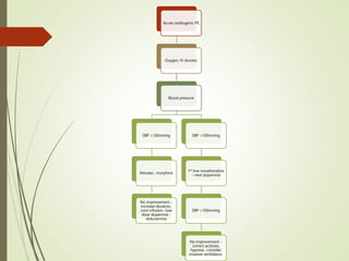

Acute Pulmonary Edema
Presenter: Gokullshautri
Overview
 Definition
 Epidemiology
 Etiopathogenesis
 Classification & Staging
 Clinical Picture
 Complications
 Diagnosis & Management
 Prognosis
Definition
Pulmonary Edema is a condition characterized by
fluid accumulation in the lungs caused by extravasation
of fluid from pulmonary vasculature in to the
interstitium and alveoli of the lungs.
Etiopathogenesis
 Pulmonary edema can be caused by the following major
pathophysiologic mechanisms:
 Imbalance of Starling forces
• increased pulmonary capillary pressure,
• decreased plasma oncotic pressure,
• increased negative interstitial pressure
 Damage to the alveolar-capillary barrier
 Lymphatic obstruction
 Idiopathic (unknown) mechanism
Classification
Cardiogenic
Pulmonary Edema
Non-Cardiogenic
Pulmonary Edema
Cardiogenic pulmonary edema
 Defined as pulmonary edema due to increased Pulmonary
capillary hydrostatic pressure secondary to elevated
pulmonary venous pressure.
 Increased LA pressure increases pulmonary venous pressure
and pressure in the lung microvasculature, resulting in
pulmonary edema.
 Hydrostatic pressure is increased and fluid exits the capillary at
an increased rate, resulting in interstitial and, in more severe
cases, alveolar edema.
 Also called Hydrostatic pulmonary edema.
Cardiac disorders manifesting as
CPE
 Left Atrial outflow obstruction
 This can be due to mitral stenosis or, in rare cases, atrial myxoma,
thrombosis of a prosthetic valve, or a congenital membrane in the
left atrium (eg, cor triatriatum).
 LV systolic dysfunction
 Systolic dysfunction, a common cause of CPE, is defined as
decreased myocardial contractility that reduces cardiac output.
 The fall in cardiac output stimulates sympathetic activity and
blood volume expansion by activating the renin-angiotensin-
aldosterone system, which causes deterioration by decreasing LV
filling time and increasing capillary hydrostatic pressure.
Cardiac disorders manifesting as
CPE
 LV diastolic dysfunction
 Ischemia and infarction may cause LV diastolic dysfunction in
addition to systolic dysfunction. With a similar mechanism,
myocardial contusion induces systolic or diastolic dysfunction.
 Chronic LV failure is usually the result of congestive heart
failure (CHF) or cardiomyopathy.
 Causes of acute exacerbations of CPE
 Acute myocardial infarction (MI) or ischemia
 Patient noncompliance with dietary restrictions (eg, dietary salt
restrictions)
 Patient noncompliance with medications (eg, diuretics)
 Severe anemia with underlying cardiac ilness
 Sepsis
 Thyrotoxicosis
 Myocarditis
 Myocardial toxins (eg, alcohol, cocaine, chemotherapeutic agents
such as doxorubicin [Adriamycin], trastuzumab [Herceptin])
 Chronic valvular disease, aortic stenosis, aortic regurgitation, and
mitral regurgitation
Cardiogenic PEStaging
The progression of fluid accumulation in CPE can be identified as 3
distinct physiologic stages.
 Stage 1
 elevated LA pressure causes distention and opening of
small pulmonary vessels.
 At this stage, increase fluid transfer into lung interstitium, at the
same time there is increase in lymphatic absorption.
 Stage 2
 Fluid and colloid shift into the lung interstitium from the pulmonary
capillaries, but an initial increase in lymphatic outflow efficiently
removes the fluid.
 The continuing filtration of liquid and solutes may overpower the
drainage capacity of the lymphatics. In this case, the fluid initially
collects in the relatively compliant interstitial compartment,
 The accumulation of liquid in the interstitium may compromise the
small airways, leading to mild hypoxemia.
 Hypoxemia at this stage is rarely of sufficient magnitude to stimulate
tachypnea.
 Stage 3
 As fluid filtration continues to increase and the filling of loose
interstitial space occurs, fluid accumulates in the relatively
noncompliant interstitial space.
 The interstitial space can contain up to 500mL of fluid. With
further accumulations, the fluid crosses the alveolar epithelium in
to the alveoli, leading to alveolar flooding.
 At this stage, abnormalities in gas exchange are noticeable, vital
capacity and other respiratory volumes are substantially reduced,
and hypoxemia becomes more severe.
Clinical features ofCPE
 Early signs of pulmonary edema include exertional dyspnea
and orthopnea.
 Chest radiographs show peribronchial thickening, prominent
vascular markings in the upper lung zones, and Kerley B lines.
 As the pulmonary edema worsens, alveoli fill with fluid; the
chest radiograph shows patchy alveolar filling, typically in a
perihilar distribution, which then progresses to diffuse alveolar
infiltrates.
 Increasing airway edema is associated with rhonchi and
wheezes.
Non cardiogenic pulmonary edema
  caused by changes in permeability
of the pulmonary capillary
membrane as a result of either a
direct or an indirect pathologic insult.
 Physiologically, noncardiogenic pulmonary edema is
characterized by intrapulmonary shunt with hypoxemia and
decreased pulmonary compliance leading to lower functional
residual capacity.
 Clinically, the picture ranges from mild dyspnea to respiratory
failure.
 Auscultation of the lungs may be relatively normal despite chest
radiographs that show diffuse alveolar infiltrates
HAPE - Pathogenesis
 Altered permeability of the alveolar-capillary barrier secondary
to intense pulmonary vasoconstriction and high capillary
pressure.
 This in turn induces endothelial leakage, which results in
interstitial and alveolar oedema without diffuse alveolar
damage.
 Reported clinical manifestations include:
 dyspnea at rest
 cough with frothy pink sputum production
 neurological disturbances associated with
oedema.
concomitant brain
Neurogenic Pulmonary Edema
 (NPE) is a clinical syndrome characterized by the acute onset of pulmonary
edema following a significant insult to the CNS.
 The etiology is thought to be a surge of catecholamines that results in
cardiopulmonary dysfunction.
 CNS events associated with NPE :
 spinal cord injury,
 subarachnoid hemorrhage (SAH),
 traumatic brain injury (TBI),
 intracranial hemorrhage,
 status epilepticus,
 meningitis, and
 subdural hemorrhage
 Although NPE was identified over 100 years ago, it is still underappreciated
in the clinical arena.
Near drowning pulmonary oedema
 It results from the inhalation of either fresh or sea water resulting in
lung damage and ventilation-perfusion mismatching.
 Near drowning It can be divided into three stages:
 stage I: acute laryngospasm that occurs after inhalation of a small
amount of water
 stage II: victim still usually presents with laryngospasm but may begin
to swallow water into the stomach
 stage III:
 in the remaining 85-90% of patients, the laryngospasm relaxes
secondary to hypoxia and large amounts of water are aspirated
 CXR features in stages II and III can be identical to pulmonary
oedema from other non-cardiac causes
Approach a Patient with Pulm.Edema
HISTORY EXAMINATION
SOB Air hunger, agitated, confused
Profuse sweating Blood pressure :
• Hypertension
• Hypotension (severe LV dysfunction
or cardiogenic shock)
Dyspnea Pulse rate : tachycardia
Orthopnea / PND RR : tachypnea
Cough Spo2 : desaturation
Hoarseness of voice Lungs :
Fine crepts
Bubbling heard at distance
Working wit accessory muscle
Chest pain Jugular vein distended
Anxiety / feeling drowning Skin : cool extremities (low O2, poor
perfusion)
Hypoxemia
Cardiogenic
 Is due to ventilation
perfusion miss match
 respond to administration of
oxygen
Non-cardiogenic
 Is due to intrapulmonary
shunting
 persists despite oxygen
supplimentation
CardiogenicVs. Non-cardiogenic Pul.Edema
 Finding suggesting cardiogenic edema
 S3 gallop
 elevated JVP
 Peripheral edema
 Findings suggesting non-cardiogenic edema
 Pulmonary findings may be relatively normal in the early
stages
 Clinical picture ranges from mild dyspnea to respiratory
failure despite CXR showing diffuse alveolar infiltrates.
Laboratory Investigations
 Routine; CBC
 Liver function tests
 Renal Function Tests
 Arterial blood gas analysis
 Serum cardiac biomarkers
 Chest xray
 Butterfly appearance
 Kerley B lines
 Pleural effusion
 alveolar edema and interstitial edema
Distinguishing features inX ray …..
 Cardiogenic cause
  Cardiomegaly
  Kerley B lines and loss
of distinct vascular
margins
  Cephalization:
engorgement of
vasculature to the apices
  Perihilar
alveolar infiltrate
  Pleural effusion
 Non cardiogenic cause
  Heart size is normal
  Uniform
alveolar infiltrate
  pleural
effusion is
uncommon
  lack of cephalization
Treatment approach
A. Principle of management
 Rapid recognition of the condition
 Stabilization of hemodynamic
 Improvement in clinical symptoms and
signs
 Identification and treatment of the
underlying cause
B. Therapy APO
1. Oxygen – 5-6L/min by mask with aim spo2>95%
2. Frusemide – IV frusemide 40-120mg, IVI 5-40
mg/hour
3. Morphine sulphate – IV 3.0-5.0 mg bolus up to 10mg
4. Nitrates – IVI GTN 5ug/min increasing 3-5 in by
5ug/min up to 100-200ug/min, can admin sublingually
or IV route
C. Treatment of adequate BP
 Frusemide – IV infusion 5-40mg/hour
 Inotropes – dopamine : low dose <2ug/kg/min to improve renal flow and promo
diuresis
- Dobutamine 2-5 ug/kg/min
 Vasodilators
D. Treatment if SBP <100mmhg at initial presentation or during treatment
 Noradrenaline infusion
 Next line : dopamine
 Avoid vasodilators (nitrates) & morphine
 Correct hypovolemia
Treatment approach
Acute cardiogenic PE
Oxygen, IV diuretic
Blood pressure
SBP >100mmhg
Nitrates , morphine
No improvement –
increase diurectic,
cont infusion –low
dose dopamine -
dobutamine
SBP <100mmhg
1st line noradrenaline
– next dopamine
SBP <100mmhg
No improvement –
correct acidosis,
hypoxia , consider
invasive ventilation,

Thank You
References:
 Harrison’s principles of Internal Medicine 19th Ed.
 ACCP Pulmonary Medicine 25th Ed.
 Fishman’s Pulmonary Diseases & Disorders 4th Ed.
 Braunwald’s Heart Disease 10th Ed.
 OnlineSource – PubmedCentral

pulmonary edema gokull.pptx

  • 1.
  • 2.
    Overview  Definition  Epidemiology Etiopathogenesis  Classification & Staging  Clinical Picture  Complications  Diagnosis & Management  Prognosis
  • 3.
    Definition Pulmonary Edema isa condition characterized by fluid accumulation in the lungs caused by extravasation of fluid from pulmonary vasculature in to the interstitium and alveoli of the lungs.
  • 4.
    Etiopathogenesis  Pulmonary edemacan be caused by the following major pathophysiologic mechanisms:  Imbalance of Starling forces • increased pulmonary capillary pressure, • decreased plasma oncotic pressure, • increased negative interstitial pressure  Damage to the alveolar-capillary barrier  Lymphatic obstruction  Idiopathic (unknown) mechanism
  • 5.
  • 6.
    Cardiogenic pulmonary edema Defined as pulmonary edema due to increased Pulmonary capillary hydrostatic pressure secondary to elevated pulmonary venous pressure.  Increased LA pressure increases pulmonary venous pressure and pressure in the lung microvasculature, resulting in pulmonary edema.  Hydrostatic pressure is increased and fluid exits the capillary at an increased rate, resulting in interstitial and, in more severe cases, alveolar edema.  Also called Hydrostatic pulmonary edema.
  • 7.
    Cardiac disorders manifestingas CPE  Left Atrial outflow obstruction  This can be due to mitral stenosis or, in rare cases, atrial myxoma, thrombosis of a prosthetic valve, or a congenital membrane in the left atrium (eg, cor triatriatum).  LV systolic dysfunction  Systolic dysfunction, a common cause of CPE, is defined as decreased myocardial contractility that reduces cardiac output.  The fall in cardiac output stimulates sympathetic activity and blood volume expansion by activating the renin-angiotensin- aldosterone system, which causes deterioration by decreasing LV filling time and increasing capillary hydrostatic pressure.
  • 8.
    Cardiac disorders manifestingas CPE  LV diastolic dysfunction  Ischemia and infarction may cause LV diastolic dysfunction in addition to systolic dysfunction. With a similar mechanism, myocardial contusion induces systolic or diastolic dysfunction.  Chronic LV failure is usually the result of congestive heart failure (CHF) or cardiomyopathy.
  • 9.
     Causes ofacute exacerbations of CPE  Acute myocardial infarction (MI) or ischemia  Patient noncompliance with dietary restrictions (eg, dietary salt restrictions)  Patient noncompliance with medications (eg, diuretics)  Severe anemia with underlying cardiac ilness  Sepsis  Thyrotoxicosis  Myocarditis  Myocardial toxins (eg, alcohol, cocaine, chemotherapeutic agents such as doxorubicin [Adriamycin], trastuzumab [Herceptin])  Chronic valvular disease, aortic stenosis, aortic regurgitation, and mitral regurgitation
  • 11.
    Cardiogenic PEStaging The progressionof fluid accumulation in CPE can be identified as 3 distinct physiologic stages.  Stage 1  elevated LA pressure causes distention and opening of small pulmonary vessels.  At this stage, increase fluid transfer into lung interstitium, at the same time there is increase in lymphatic absorption.
  • 12.
     Stage 2 Fluid and colloid shift into the lung interstitium from the pulmonary capillaries, but an initial increase in lymphatic outflow efficiently removes the fluid.  The continuing filtration of liquid and solutes may overpower the drainage capacity of the lymphatics. In this case, the fluid initially collects in the relatively compliant interstitial compartment,  The accumulation of liquid in the interstitium may compromise the small airways, leading to mild hypoxemia.  Hypoxemia at this stage is rarely of sufficient magnitude to stimulate tachypnea.
  • 13.
     Stage 3 As fluid filtration continues to increase and the filling of loose interstitial space occurs, fluid accumulates in the relatively noncompliant interstitial space.  The interstitial space can contain up to 500mL of fluid. With further accumulations, the fluid crosses the alveolar epithelium in to the alveoli, leading to alveolar flooding.  At this stage, abnormalities in gas exchange are noticeable, vital capacity and other respiratory volumes are substantially reduced, and hypoxemia becomes more severe.
  • 14.
    Clinical features ofCPE Early signs of pulmonary edema include exertional dyspnea and orthopnea.  Chest radiographs show peribronchial thickening, prominent vascular markings in the upper lung zones, and Kerley B lines.  As the pulmonary edema worsens, alveoli fill with fluid; the chest radiograph shows patchy alveolar filling, typically in a perihilar distribution, which then progresses to diffuse alveolar infiltrates.  Increasing airway edema is associated with rhonchi and wheezes.
  • 16.
    Non cardiogenic pulmonaryedema   caused by changes in permeability of the pulmonary capillary membrane as a result of either a direct or an indirect pathologic insult.
  • 19.
     Physiologically, noncardiogenicpulmonary edema is characterized by intrapulmonary shunt with hypoxemia and decreased pulmonary compliance leading to lower functional residual capacity.  Clinically, the picture ranges from mild dyspnea to respiratory failure.  Auscultation of the lungs may be relatively normal despite chest radiographs that show diffuse alveolar infiltrates
  • 21.
    HAPE - Pathogenesis Altered permeability of the alveolar-capillary barrier secondary to intense pulmonary vasoconstriction and high capillary pressure.  This in turn induces endothelial leakage, which results in interstitial and alveolar oedema without diffuse alveolar damage.  Reported clinical manifestations include:  dyspnea at rest  cough with frothy pink sputum production  neurological disturbances associated with oedema. concomitant brain
  • 23.
    Neurogenic Pulmonary Edema (NPE) is a clinical syndrome characterized by the acute onset of pulmonary edema following a significant insult to the CNS.  The etiology is thought to be a surge of catecholamines that results in cardiopulmonary dysfunction.  CNS events associated with NPE :  spinal cord injury,  subarachnoid hemorrhage (SAH),  traumatic brain injury (TBI),  intracranial hemorrhage,  status epilepticus,  meningitis, and  subdural hemorrhage  Although NPE was identified over 100 years ago, it is still underappreciated in the clinical arena.
  • 24.
    Near drowning pulmonaryoedema  It results from the inhalation of either fresh or sea water resulting in lung damage and ventilation-perfusion mismatching.  Near drowning It can be divided into three stages:  stage I: acute laryngospasm that occurs after inhalation of a small amount of water  stage II: victim still usually presents with laryngospasm but may begin to swallow water into the stomach  stage III:  in the remaining 85-90% of patients, the laryngospasm relaxes secondary to hypoxia and large amounts of water are aspirated  CXR features in stages II and III can be identical to pulmonary oedema from other non-cardiac causes
  • 25.
    Approach a Patientwith Pulm.Edema HISTORY EXAMINATION SOB Air hunger, agitated, confused Profuse sweating Blood pressure : • Hypertension • Hypotension (severe LV dysfunction or cardiogenic shock) Dyspnea Pulse rate : tachycardia Orthopnea / PND RR : tachypnea Cough Spo2 : desaturation Hoarseness of voice Lungs : Fine crepts Bubbling heard at distance Working wit accessory muscle Chest pain Jugular vein distended Anxiety / feeling drowning Skin : cool extremities (low O2, poor perfusion)
  • 26.
    Hypoxemia Cardiogenic  Is dueto ventilation perfusion miss match  respond to administration of oxygen Non-cardiogenic  Is due to intrapulmonary shunting  persists despite oxygen supplimentation
  • 27.
    CardiogenicVs. Non-cardiogenic Pul.Edema Finding suggesting cardiogenic edema  S3 gallop  elevated JVP  Peripheral edema  Findings suggesting non-cardiogenic edema  Pulmonary findings may be relatively normal in the early stages  Clinical picture ranges from mild dyspnea to respiratory failure despite CXR showing diffuse alveolar infiltrates.
  • 28.
    Laboratory Investigations  Routine;CBC  Liver function tests  Renal Function Tests  Arterial blood gas analysis  Serum cardiac biomarkers  Chest xray  Butterfly appearance  Kerley B lines  Pleural effusion  alveolar edema and interstitial edema
  • 29.
    Distinguishing features inXray …..  Cardiogenic cause   Cardiomegaly   Kerley B lines and loss of distinct vascular margins   Cephalization: engorgement of vasculature to the apices   Perihilar alveolar infiltrate   Pleural effusion  Non cardiogenic cause   Heart size is normal   Uniform alveolar infiltrate   pleural effusion is uncommon   lack of cephalization
  • 30.
    Treatment approach A. Principleof management  Rapid recognition of the condition  Stabilization of hemodynamic  Improvement in clinical symptoms and signs  Identification and treatment of the underlying cause B. Therapy APO 1. Oxygen – 5-6L/min by mask with aim spo2>95% 2. Frusemide – IV frusemide 40-120mg, IVI 5-40 mg/hour 3. Morphine sulphate – IV 3.0-5.0 mg bolus up to 10mg 4. Nitrates – IVI GTN 5ug/min increasing 3-5 in by 5ug/min up to 100-200ug/min, can admin sublingually or IV route
  • 31.
    C. Treatment ofadequate BP  Frusemide – IV infusion 5-40mg/hour  Inotropes – dopamine : low dose <2ug/kg/min to improve renal flow and promo diuresis - Dobutamine 2-5 ug/kg/min  Vasodilators D. Treatment if SBP <100mmhg at initial presentation or during treatment  Noradrenaline infusion  Next line : dopamine  Avoid vasodilators (nitrates) & morphine  Correct hypovolemia Treatment approach
  • 32.
    Acute cardiogenic PE Oxygen,IV diuretic Blood pressure SBP >100mmhg Nitrates , morphine No improvement – increase diurectic, cont infusion –low dose dopamine - dobutamine SBP <100mmhg 1st line noradrenaline – next dopamine SBP <100mmhg No improvement – correct acidosis, hypoxia , consider invasive ventilation,
  • 33.
     Thank You References:  Harrison’sprinciples of Internal Medicine 19th Ed.  ACCP Pulmonary Medicine 25th Ed.  Fishman’s Pulmonary Diseases & Disorders 4th Ed.  Braunwald’s Heart Disease 10th Ed.  OnlineSource – PubmedCentral