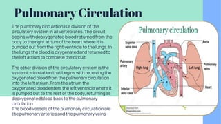
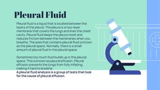
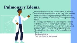
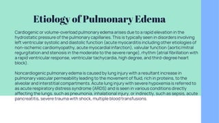
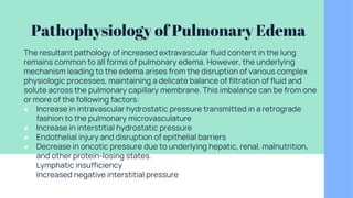
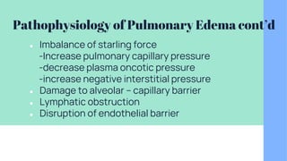
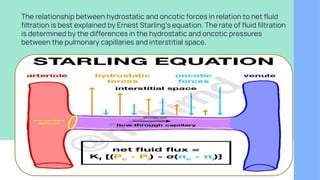
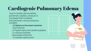
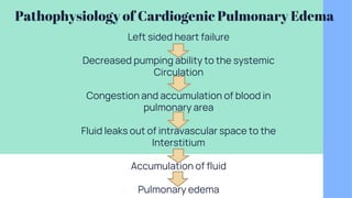
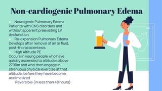
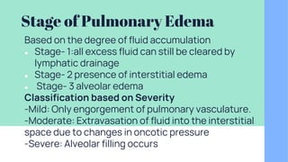
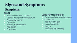
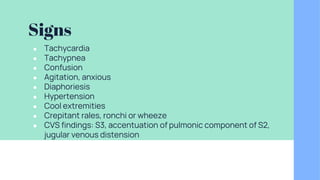
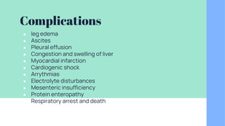
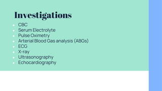
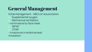
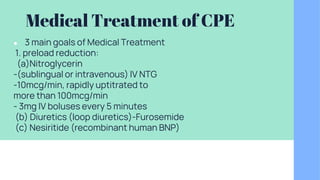
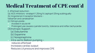
PULMOMONARY
CIRCULATION , PLEURAL
FLUID AND PULMONARY
EDEMA
Pulmonary
Circulation
Pulmonary Circulation
The pulmonary circulation is a division of the
circulatory system in all vertebrates. The circuit
begins with deoxygenated blood returned from the
body to the right atrium of the heart where it is
pumped out from the right ventricle to the lungs. In
the lungs the blood is oxygenated and returned to
the left atrium to complete the circuit.
The other division of the circulatory system is the
systemic circulation that begins with receiving the
oxygenated blood from the pulmonary circulation
into the left atrium. From the atrium the
oxygenated blood enters the left ventricle where it
is pumped out to the rest of the body, returning as
deoxygenated blood back to the pulmonary
circulation.
The blood vessels of the pulmonary circulation are
the pulmonary arteries and the pulmonary veins
Pleural Fluid
Pleural fluid is a liquid that is located between the
layers of the pleura. The pleura is a two-layer
membrane that covers the lungs and lines the chest
cavity. Pleural fluid keeps the pleura moist and
reduces friction between the membranes when you
breathe. The area that contains pleural fluid is known
as the pleural space. Normally, there is a small
amount of pleural fluid in the pleural space.
Sometimes too much fluid builds up in the pleural
space. This is known as pleural effusion. Pleural
effusion prevents the lungs from fully inflating,
making it hard to breathe.
A pleural fluid analysis is a group of tests that look
for the cause of pleural effusion.
Pulmonary Edema
Pulmonary edema is the accumulation of fluids in
the interstitium and alveoli of the lung. This process
leads to diminished gas exchange at the alveolar
level, progressing to potentially causing respiratory
failure.
Its etiology is either due to a cardiogenic process
with the inability to remove sufficient blood away
from the pulmonary circulation or non-cardiogenic
precipitated by injury to the lung parenchyma.
There are two main types of Pulmonary Edema.
These are
-Cardiogenic and
-Non-cardiogenic pulmonary
Etiology of Pulmonary Edema
Cardiogenic or volume-overload pulmonary edema arises due to a rapid elevation in the
hydrostatic pressure of the pulmonary capillaries. This is typically seen in disorders involving
left ventricular systolic and diastolic function (acute myocarditis including other etiologies of
non-ischemic cardiomyopathy, acute myocardial infarction), valvular function (aortic/mitral
regurgitation and stenosis in the moderate to the severe range), rhythm (atrial fibrillation with
a rapid ventricular response, ventricular tachycardia, high degree, and third-degree heart
block).
Noncardiogenic pulmonary edema is caused by lung injury with a resultant increase in
pulmonary vascular permeability leading to the movement of fluid, rich in proteins, to the
alveolar and interstitial compartments. Acute lung injury with severe hypoxemia is referred to
as acute respiratory distress syndrome (ARDS) and is seen in various conditions directly
affecting the lungs, such as pneumonia, inhalational injury, or indirectly, such as sepsis, acute
pancreatitis, severe trauma with shock, multiple blood transfusions.
Pathophysiology of Pulmonary Edema
The resultant pathology of increased extravascular fluid content in the lung
remains common to all forms of pulmonary edema. However, the underlying
mechanism leading to the edema arises from the disruption of various complex
physiologic processes, maintaining a delicate balance of filtration of fluid and
solute across the pulmonary capillary membrane. This imbalance can be from one
or more of the following factors:
● Increase in intravascular hydrostatic pressure transmitted in a retrograde
fashion to the pulmonary microvasculature
● Increase in interstitial hydrostatic pressure
● Endothelial injury and disruption of epithelial barriers
● Decrease in oncotic pressure due to underlying hepatic, renal, malnutrition,
and other protein-losing states.
● Lymphatic insufficiency
● Increased negative interstitial pressure
Pathophysiology of Pulmonary Edema cont’d
● Imbalance of starling force
-Increase pulmonary capillary pressure
-decrease plasma oncotic pressure
-increase negative interstitial pressure
● Damage to alveolar – capillary barrier
● Lymphatic obstruction
● Disruption of endothelial barrier
The relationship between hydrostatic and oncotic forces in relation to net fluid
filtration is best explained by Ernest Starling’s equation. The rate of fluid filtration
is determined by the differences in the hydrostatic and oncotic pressures
between the pulmonary capillaries and interstitial space.
Cardiogenic Pulmonary Edema
Due to cardiac abnormalities,
pulmonary capillary pressure is
increased that increases
the pulmonary venous pressure.
CAUSES :
● LV failure is the most common
● Dysrhythmia
● LV hypertrophy and cardiomyopathy
● LV volume overload
● Myocardial infarction
● LV outflow obstruction
Pathophysiology of Cardiogenic Pulmonary Edema
Left sided heart failure
Decreased pumping ability to the systemic
Circulation
Congestion and accumulation of blood in
pulmonary area
Fluid leaks out of intravascular space to the
Interstitium
Accumulation of fluid
Pulmonary edema
Non-cardiogenic Pulmonary Edema
● Neurogenic Pulmonary Edema
Patients with CNS disorders and
without apparent preexisting LV
dysfunction
● Re-expansion Pulmonary Edema
Develops after removal of air or fluid,
post-thoracocentesis.
● High Altitude PE
Occurs in young people who have
quickly ascended to altitudes above
2700m and who then engage in
strenuous physical exercise at that
altitude, before they have become
acclimatized
● Reversible (in less than 48 hours)
Stage of Pulmonary Edema
Based on the degree of fluid accumulation
● Stage- 1:all excess fluid can still be cleared by
lymphatic drainage
● Stage- 2 presence of interstitial edema
● Stage- 3 alveolar edema
Classification based on Severity
-Mild: Only engorgement of pulmonary vasculature.
-Moderate: Extravasation of fluid into the interstitial
space due to changes in oncotic pressure
-Severe: Alveolar filling occurs
Signs and Symptoms
ACUTE
● Severe shortness of breath
● Cough- with pink frothy sputum
● Profuse sweating
● Cyanosis
● Anxiety, restlessness
● Palpitation
● Chest pain
LONG TERM (CHRONIC)
● Paroxysmal nocturnal dyspnea
● Orthopnea
● Rapid weight gain
● Loss of appetite
● Fatigue
● Ankle and leg swelling
Symptoms
Signs
● Tachycardia
● Tachypnea
● Confusion
● Agitation, anxious
● Diaphoriesis
● Hypertension
● Cool extremities
● Crepitant rales, ronchi or wheeze
● CVS findings: S3, accentuation of pulmonic component of S2,
jugular venous distension
Complications
● leg edema
● Ascites
● Pleural effusion
● Congestion and swelling of liver
● Myocardial infarction
● Cardiogenic shock
● Arrythmias
● Electrolyte disturbances
● Mesenteric insufficiency
● Protein enteropathy
● Respiratory arrest and death
Investigations
● CBC
● Serum Electrolyte
● Pulse Oximetry
● Arterial Blood Gas analysis (ABGs)
● ECG
● X-ray
● Ultrasonography
● Echocardiography
General Management
Initial management - ABCs of resuscitation
● Supplemental oxygen
● Mechanical ventilation
- noninvasive by face mask
● BiPAP
● CPAP
- invasive as in endotracheal
intubation
Medical Treatment of CPE
● 3 main goals of Medical Treatment
1. preload reduction:
(a)Nitroglycerin
-(sublingual or intravenous) IV NTG
-10mcg/min, rapidly uptitrated to
more than 100mcg/min
- 3mg IV boluses every 5 minutes
(b) Diuretics (loop diuretics)-Furosemide
(c) Nesiritide (recombinant human BNP)
Medical Treatment of CPE cont’d
2. Afterload reduction:
(a) ACE inhibitors –enalapril 1.25mg IV,captopril 25mg sublingually
(b) Angiotensin II receptor blockers –
Valsartan and candesartan
(c) Nitroprusside-
● Avoided in acute MI
● Prolonged use causes cyanide toxicity ,tolerance and reflex tachycardia
(3)Inotropic Support :
(a) Dobutamine
(b) Dopamine
(c) Norepinephrine
● Intra-aortic Balloon pumping –
● reduces afterload
● Increases cardiac output
● Reduces LA pressure and improves CPE
Medical treatment of Neurogenic PE
Resolves within 48-72 hours in majority of patients
Medical care:
● oxygen supplementation
● Diuretics
● Inotropic support
● Surgical Care : directed at the neurological insult
(e.g., intracerebral hemorrhage, subdural
hematoma, etc.)
Medical Treatment of High Altitude PE
● Descent and supplemental O2
● Tab nifedipine 10mg sublingual or 20mg sustained
release 6hrly.
● Hydralazine
● Inhaled nitrous oxide
● Acetazolamide
References
● https://www.sciencedirect.com/topics/neuroscience/pulmonary-edema
● https://www.slideshare.net/upload?download_ id=70671502
● https://www.ncbi.nlm.nih.gov/books/NBK557611/
● https://www.vin.com/apputil/content/defaultadv1.aspx?pId=11196&catId=
30763&id=3854264
● Guyton and Hall Textbook of Medical Physiology
Thank You!

Pulmonary Edema.pptx

  • 1.
  • 2.
  • 3.
    Pulmonary Circulation The pulmonarycirculation is a division of the circulatory system in all vertebrates. The circuit begins with deoxygenated blood returned from the body to the right atrium of the heart where it is pumped out from the right ventricle to the lungs. In the lungs the blood is oxygenated and returned to the left atrium to complete the circuit. The other division of the circulatory system is the systemic circulation that begins with receiving the oxygenated blood from the pulmonary circulation into the left atrium. From the atrium the oxygenated blood enters the left ventricle where it is pumped out to the rest of the body, returning as deoxygenated blood back to the pulmonary circulation. The blood vessels of the pulmonary circulation are the pulmonary arteries and the pulmonary veins
  • 4.
    Pleural Fluid Pleural fluidis a liquid that is located between the layers of the pleura. The pleura is a two-layer membrane that covers the lungs and lines the chest cavity. Pleural fluid keeps the pleura moist and reduces friction between the membranes when you breathe. The area that contains pleural fluid is known as the pleural space. Normally, there is a small amount of pleural fluid in the pleural space. Sometimes too much fluid builds up in the pleural space. This is known as pleural effusion. Pleural effusion prevents the lungs from fully inflating, making it hard to breathe. A pleural fluid analysis is a group of tests that look for the cause of pleural effusion.
  • 5.
    Pulmonary Edema Pulmonary edemais the accumulation of fluids in the interstitium and alveoli of the lung. This process leads to diminished gas exchange at the alveolar level, progressing to potentially causing respiratory failure. Its etiology is either due to a cardiogenic process with the inability to remove sufficient blood away from the pulmonary circulation or non-cardiogenic precipitated by injury to the lung parenchyma. There are two main types of Pulmonary Edema. These are -Cardiogenic and -Non-cardiogenic pulmonary
  • 6.
    Etiology of PulmonaryEdema Cardiogenic or volume-overload pulmonary edema arises due to a rapid elevation in the hydrostatic pressure of the pulmonary capillaries. This is typically seen in disorders involving left ventricular systolic and diastolic function (acute myocarditis including other etiologies of non-ischemic cardiomyopathy, acute myocardial infarction), valvular function (aortic/mitral regurgitation and stenosis in the moderate to the severe range), rhythm (atrial fibrillation with a rapid ventricular response, ventricular tachycardia, high degree, and third-degree heart block). Noncardiogenic pulmonary edema is caused by lung injury with a resultant increase in pulmonary vascular permeability leading to the movement of fluid, rich in proteins, to the alveolar and interstitial compartments. Acute lung injury with severe hypoxemia is referred to as acute respiratory distress syndrome (ARDS) and is seen in various conditions directly affecting the lungs, such as pneumonia, inhalational injury, or indirectly, such as sepsis, acute pancreatitis, severe trauma with shock, multiple blood transfusions.
  • 7.
    Pathophysiology of PulmonaryEdema The resultant pathology of increased extravascular fluid content in the lung remains common to all forms of pulmonary edema. However, the underlying mechanism leading to the edema arises from the disruption of various complex physiologic processes, maintaining a delicate balance of filtration of fluid and solute across the pulmonary capillary membrane. This imbalance can be from one or more of the following factors: ● Increase in intravascular hydrostatic pressure transmitted in a retrograde fashion to the pulmonary microvasculature ● Increase in interstitial hydrostatic pressure ● Endothelial injury and disruption of epithelial barriers ● Decrease in oncotic pressure due to underlying hepatic, renal, malnutrition, and other protein-losing states. ● Lymphatic insufficiency ● Increased negative interstitial pressure
  • 8.
    Pathophysiology of PulmonaryEdema cont’d ● Imbalance of starling force -Increase pulmonary capillary pressure -decrease plasma oncotic pressure -increase negative interstitial pressure ● Damage to alveolar – capillary barrier ● Lymphatic obstruction ● Disruption of endothelial barrier
  • 9.
    The relationship betweenhydrostatic and oncotic forces in relation to net fluid filtration is best explained by Ernest Starling’s equation. The rate of fluid filtration is determined by the differences in the hydrostatic and oncotic pressures between the pulmonary capillaries and interstitial space.
  • 10.
    Cardiogenic Pulmonary Edema Dueto cardiac abnormalities, pulmonary capillary pressure is increased that increases the pulmonary venous pressure. CAUSES : ● LV failure is the most common ● Dysrhythmia ● LV hypertrophy and cardiomyopathy ● LV volume overload ● Myocardial infarction ● LV outflow obstruction
  • 11.
    Pathophysiology of CardiogenicPulmonary Edema Left sided heart failure Decreased pumping ability to the systemic Circulation Congestion and accumulation of blood in pulmonary area Fluid leaks out of intravascular space to the Interstitium Accumulation of fluid Pulmonary edema
  • 12.
    Non-cardiogenic Pulmonary Edema ●Neurogenic Pulmonary Edema Patients with CNS disorders and without apparent preexisting LV dysfunction ● Re-expansion Pulmonary Edema Develops after removal of air or fluid, post-thoracocentesis. ● High Altitude PE Occurs in young people who have quickly ascended to altitudes above 2700m and who then engage in strenuous physical exercise at that altitude, before they have become acclimatized ● Reversible (in less than 48 hours)
  • 13.
    Stage of PulmonaryEdema Based on the degree of fluid accumulation ● Stage- 1:all excess fluid can still be cleared by lymphatic drainage ● Stage- 2 presence of interstitial edema ● Stage- 3 alveolar edema Classification based on Severity -Mild: Only engorgement of pulmonary vasculature. -Moderate: Extravasation of fluid into the interstitial space due to changes in oncotic pressure -Severe: Alveolar filling occurs
  • 14.
    Signs and Symptoms ACUTE ●Severe shortness of breath ● Cough- with pink frothy sputum ● Profuse sweating ● Cyanosis ● Anxiety, restlessness ● Palpitation ● Chest pain LONG TERM (CHRONIC) ● Paroxysmal nocturnal dyspnea ● Orthopnea ● Rapid weight gain ● Loss of appetite ● Fatigue ● Ankle and leg swelling Symptoms
  • 15.
    Signs ● Tachycardia ● Tachypnea ●Confusion ● Agitation, anxious ● Diaphoriesis ● Hypertension ● Cool extremities ● Crepitant rales, ronchi or wheeze ● CVS findings: S3, accentuation of pulmonic component of S2, jugular venous distension
  • 16.
    Complications ● leg edema ●Ascites ● Pleural effusion ● Congestion and swelling of liver ● Myocardial infarction ● Cardiogenic shock ● Arrythmias ● Electrolyte disturbances ● Mesenteric insufficiency ● Protein enteropathy ● Respiratory arrest and death
  • 17.
    Investigations ● CBC ● SerumElectrolyte ● Pulse Oximetry ● Arterial Blood Gas analysis (ABGs) ● ECG ● X-ray ● Ultrasonography ● Echocardiography
  • 18.
    General Management Initial management- ABCs of resuscitation ● Supplemental oxygen ● Mechanical ventilation - noninvasive by face mask ● BiPAP ● CPAP - invasive as in endotracheal intubation
  • 19.
    Medical Treatment ofCPE ● 3 main goals of Medical Treatment 1. preload reduction: (a)Nitroglycerin -(sublingual or intravenous) IV NTG -10mcg/min, rapidly uptitrated to more than 100mcg/min - 3mg IV boluses every 5 minutes (b) Diuretics (loop diuretics)-Furosemide (c) Nesiritide (recombinant human BNP)
  • 20.
    Medical Treatment ofCPE cont’d 2. Afterload reduction: (a) ACE inhibitors –enalapril 1.25mg IV,captopril 25mg sublingually (b) Angiotensin II receptor blockers – Valsartan and candesartan (c) Nitroprusside- ● Avoided in acute MI ● Prolonged use causes cyanide toxicity ,tolerance and reflex tachycardia (3)Inotropic Support : (a) Dobutamine (b) Dopamine (c) Norepinephrine ● Intra-aortic Balloon pumping – ● reduces afterload ● Increases cardiac output ● Reduces LA pressure and improves CPE
  • 21.
    Medical treatment ofNeurogenic PE Resolves within 48-72 hours in majority of patients Medical care: ● oxygen supplementation ● Diuretics ● Inotropic support ● Surgical Care : directed at the neurological insult (e.g., intracerebral hemorrhage, subdural hematoma, etc.)
  • 22.
    Medical Treatment ofHigh Altitude PE ● Descent and supplemental O2 ● Tab nifedipine 10mg sublingual or 20mg sustained release 6hrly. ● Hydralazine ● Inhaled nitrous oxide ● Acetazolamide
  • 23.
    References ● https://www.sciencedirect.com/topics/neuroscience/pulmonary-edema ● https://www.slideshare.net/upload?download_id=70671502 ● https://www.ncbi.nlm.nih.gov/books/NBK557611/ ● https://www.vin.com/apputil/content/defaultadv1.aspx?pId=11196&catId= 30763&id=3854264 ● Guyton and Hall Textbook of Medical Physiology
  • 24.