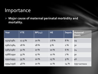
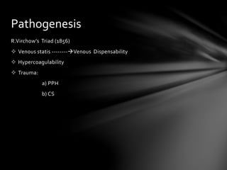
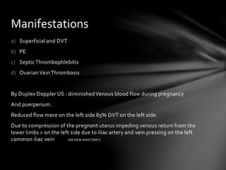
2. Major cause of maternal perinatal morbidity and
mortality.
Importance
Year VTE BP(High) HE Sepsis Maternal
Death
1979/1981 12.9 % 20 % 7.8 % 8 % 29
1982/1984 18 % 18 % 9 % 2 % 30
1985/1987 32 % 27 % 10 % 6 % 24
1988/1990 33 % 27 % 22 % 7 % 30
1991/1993 35 % 20 % 15 % 9 % 46
1994/1996 48 % 20 % 12 % 14 % 159 Total Patients
3. R.Virchow’s Triad (1856)
Venous statis --------Venous Dispensability
Hypercoagulability
Trauma:
a) PPH
b) CS
Pathogenesis
4. a) Superficial and DVT
b) PE
c) SepticThrombophlebitis
d) Ovarian VeinThrombosis
By Duplex Doppler US : diminishedVenous blood flow during pregnancy
And puerperium.
Reduced flow more on the left side 85% DVT on the left side.
Due to compression of the pregnant uterus impeding venous return from the
lower limbs > on the left side due to iliac artery and vein pressing on the left
common iliac vein (REVIEW ANATOMY)
Manifestations
5. 8.5 / 10,000
30 % antenatal 70 % postnatal
Untreated 25 % are liable to develop PE
15 % mortality ratio. While treated only 5 % will develop PE and mortality is < 1%
Hence prompt diagnosis is mandatory.
Incidence
6. Review coagulation cascade
Briefly the pathways are almost as follows
TF following vascular disruption complexes with F VII converting X to Xa ate
the same time the same complex of TF/VIIa will work indirectly to convert IX
to IXa which will activate factor X to Xa.
Xa complexes with Va to convert prothrombin (FII) to Thrombin (IIa)
Thrombin will activate platelets plus cross linking with fibrinogen to
generate fibrin monomers which will polymerize and cross linking by XIIIa to
form a stable clot. However clotting always is faced with anticlotting at one
and the same time with processes counteracting with each other eg. TFPI
will stop the complex TF /VIIa/Xa process of clotting but factor XIa works as
an alternative to stabilize the clot.
Also fibrinolysis disintegrates the fibrin clot by binding activated plasmin.
Plasmin itself will be inactivated by antiplasmin in the fibrin clot.
Haemostasis
7. 1. CS + Endomyometrisis (reduce protein C and S)
2. Trauma (placental separation)
3. Obesity
4. Nephrotic syndrome
5. Age (>35)
6. Bed rest and long travel
7. Orthopedic surgery
8. History of VTE
9. Smoking
Clinical risk factors
8. 1. Inherited
2. Acquired
Constitutes very high risk of VTE in pregnancy.
a) Most common is APCR (FVL mutation) with incidence of 5 % in the pregnant and 40 %
with VTE.
It is autosomal dominant.
FV polypeptide will undergo process of replacement of glutamine for arginine at position
506, this will prevent inactivation of factor V by APC and the increased factor VIII with
reduces protein S in pregnancy will exacerbate FVL mutation increasing risk of VTE from
0.07 in the non pregnant to 0.2 in the pregnant, however FVL heterozygote patient has got
5 fold increase of VTE while the homozygote has more than 100 % fold risk which is 17 %.
The APCR is best confirmed by PCR in the lab.
b) Prothrombin gene promoter mutation (G20210A) found in 2 to 3 % Europeans
And constitutes 17 % of VTE. In the heterozygous form carries a risk of 1 in 200 while in the
homozygous 15 % to 17 % however generally rare.
It is Autosomal dominant.
Thrombophilia
9. c) ATIII deficiency very serious deficiency though very rare.(incidence 1 in
3000)
Associated with > 50 % of VTE antepartum and post partum.
d) Protein C/S deficiency: less rare (1/200-1/1500) carries a 4% risk of VTE in
pregnancy.
e) Other associated thrombophilia conditions is hyperhomocysteimaemia +
increase of VII,VIII,IX which are of minimal importance.
10. Mainly caused by antiphospholipid antibodies (APA)
Constitutes 14 % of VTE in pregnancy.
Associated with pregnancy adverse outcomes: eg. Recurrent miscarriage
throbocytopenia ,SB,IUGR and severe preeclampsia,
Distinct classes of APA are closely linked with VTE a:LAC despite name is
associated with thrombosis. b:Anticardiolipin antibodies(ACA).
Are associated with medical disorders and SLE they promote directly
thrombosis by interference with a variety of an ionic phospholipids
eg.B2GP1,annexin,antithrombin,protein C and S.
Therefore newly diagnosed VTE or strong family history should thoroughly
investigated for FVL, prothrombin 20210A mutation, AT, protein C/S
deficiency,APA’s,B2GP1 antibodies.
Acquired thrombophilias
11. Classical presentation with swollen tender red limb should not necessarily
mean DVT nor a positive Homman’s sign(foot dorsiflexion)
It may diagnose only 50 % cases of DVT.
DIAGNOSIS:
A key investigation is the compression color doppler US which has a
sensitivity of 92 % and specifity of 98 % of popliteal and femoral vein
thrombosis .
A gold standard investigation is contrast venography which has a minimal
hazard when used with lead shielding.
MRI not widely used in pregnancy is emerging as an important investigation
for thigh and pelvic vein thrombosis.
Both investigations are used for equivocal doppler results.
DVT
12. Clinical diagnosed by :
1. Dyspnoea
2. Pleuritic pain
3. Haemoptysis
But these signs are non specific for PE.
The incidene of PE is 1:2500
ECG changes are variable and are insensitive for diagnosis except for a large
pulmonary artery occlusion.
The initial evaluation is however by V/Q scan (ventilation perfusion).
Only interpreted in the context of clinical probability- high or low
When in doubt use doppler US for evidence of DVT.
SVCT more specific but still for central pulmonary artery thrombosis
Negative CT with strong suspicion should prompt pulmonary angiography with
appropriate lead shielding.
Pulmonary Embolism (PE)
13. SPT very uncommon and controversial complication of PID or more after CS
than vaginal delivery.
Also associated with other gyn procedures.
Physical findings : quite non specific eg. Spiky fever not responding to
antibiotics.
MRI and CT may help in diagnosis.
Traditionally treat with anti coagulance and await clinical response within 7-
10 days.
PelvicThrombosis
14. Again non specific but typically presents with abdominal pain 2 – 3 days post
partum with no fever and resembles acute appendicitis .
Rarely occurs antepartum.
The incidence in 1/4000.
DIAGNOSIS:
With CT and MRI. Mostly found on the right side.
TREATMENT:
same as SPT.
OvarianVeinThrombosis
15. Heparin
Does not cross the placenta.
Does not enter the breast milk.
Acts by :
Increases AT activity.
Increases Xa inhibitory activity.
Enhances platelet aggregation.
Side effects:
Hemorrhage specially when combined with asprin.
Osteoporosis.
Thrombocytopenia.
Treatment of thromboembolism
16. o Haemorrhage is increased by asprin use,recent surgery,thrombocytopenia,and liver
disease.
o There are two types of thrombocytopenia caused by Heparin: HIT1 which is an
immediate and benign thrombocytopenia that does not need treatment.
The other is HIT2 which is a delayed and malignant thrombocytopenia which will require
immediate cessation of therapy.
It has less incidence with LMWH.
Follow up with regular platelet count for three weeks
o Osteoporosis occurs with dosage of 1500 BD for more than 6 months
Occurs less with LMWH.
The main goal of VTE therapy is
1.To maintain Heparin level at 0.2 - 0.4 or U/ml
2.Maintain an anti Xa activity between 0.6-1.2 U/ml
3.aPPT (of 1.2 – 2.5)
The dose varies among individuals because of Heparin binding protein power
17. 1. Unfractionated Heparin: dose is IV loading of 80/kg,followed by infusion
of 18/kg/hour continuous.
Adjust dose according to INR and aPTT
aPTT 6 hours after injection in early stages.
Continue for 5-6 days till clinical improvement then shift to subcutaneous
Heparin 10,000 BD.
Treatment will continue till end of pregnancy.
Consider use of Warfarin in puerperium.
LMWH: extracted from the unfractionated Heparin molecule.
There are three versions a) Dalteparin (100 anti Xa U/kg every 12 hours).
b) Enoxaparin : 1 mg/kg 12 hourly.
c) Tinzaparin : 40 U/kg.
Types of Heparin
18. Titrate to reach anti Xa level to reach 0.6 to 1.2 U/ml 4 hours after injection.
Spinal and epidural anesthesia are contra indicated within 18-24 hours of
LMWH administration.
Adjust dosage according to complications associated with DVT eg.
Thrombophelia
Restart treatment four hours after vaginal delivery and 8-12 hours after CS.
Prophylaxis: continue postpartum for 6-12 weeks with PE or complex
Ileofemoral DVT 4-6 months.
Oral warfarin therapy adjust dose to maintain INR at 2-2.5.
Use Warfarin /Heparin together for 4 days and then continue with Warfarin
alone to avoid paradoxical warfarin induced thromboembolism.
Give 1.5g Ca suppliment for patients in pregnancy.
Check bone densitometry for Heparin of more than 6 months.
19. Action : inhibit vitamin K action, which is a co-factor of FVII,IX,X and II.
Crosses placenta with 33% risk of embryophathy .
Causes abruptioplacentae.
Antidote: vitamin K or FF plasma.
WARFARIN
20. 1. Assumption of lateral ducupitus position(3rd trimester).
2. Elastic compression stockings.
3. Pneumatic compression.
Pharmapological prevention
1. Previous DVT with no thrombophelia : unwarranted.
2. DVT with non recurring cause eg. OC:unwarranted.
But both should receive post partum prophylaxis.
Prevention ofVTE