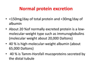
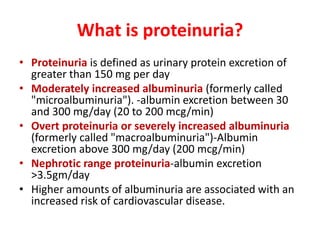
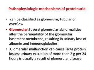
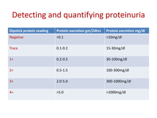
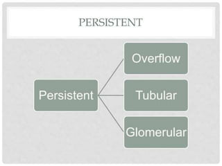
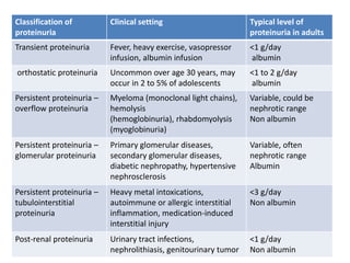
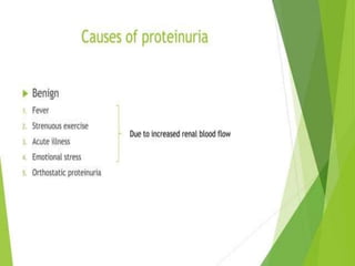
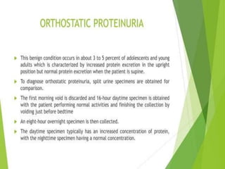
Proteinuria in Adults: A Diagnostic
Approach
Dr.G.Venkata ramana
HOD Family medicine
RDT hospital Bathalapalli
Normal protein excretion
• <150mg/day of total protein and <30mg/day of
albumin
• About 20 %of normally excreted protein is a low-
molecular-weight type such as immunoglobulins
(molecular weight about 20,000 Daltons)
• 40 % is high-molecular-weight albumin (about
65,000 Daltons)
• 40 % is Tamm-Horsfall mucoproteins secreted by
the distal tubule
What is proteinuria?
• Proteinuria is defined as urinary protein excretion of
greater than 150 mg per day
• Moderately increased albuminuria (formerly called
"microalbuminuria"). -albumin excretion between 30
and 300 mg/day (20 to 200 mcg/min)
• Overt proteinuria or severely increased albuminuria
(formerly called "macroalbuminuria")-Albumin
excretion above 300 mg/day (200 mcg/min)
• Nephrotic range proteinuria-albumin excretion
>3.5gm/day
• Higher amounts of albuminuria are associated with an
increased risk of cardiovascular disease.
Pathophysiologic mechanisms of proteinuria
• can be classified as glomerular, tubular or
overflow
• Glomerular:Several glomerular abnormalities
alter the permeability of the glomerular
basement membrane, resulting in urinary loss of
albumin and immunoglobulins.
• Glomerular malfunction can cause large protein
losses; urinary excretion of more than 2 g per 24
hours is usually a result of glomerular disease
• Tubular proteinuria occurs when
tubulointerstitial disease prevents the proximal
tubule from reabsorbing low-molecular-weight
proteins (part of the normal glomerular
ultrafiltrate).
• When a patient has tubular disease, usually less
than 2 g of protein is excreted in 24 hours.
• Tubular diseases include hypertensive
nephrosclerosis and tubulointerstitial
nephropathy caused by nonsteroidal anti-
inflammatory drugs.
• overflow proteinuria: low-molecular-weight
proteins overwhelm the ability of the proximal
tubules to reabsorb filtered proteins.
• Most often, this is a result of the
immunoglobulin overproduction that occurs in
multiple myeloma.
• The resultant light-chain immunoglobulin
fragments (Bence Jones proteins) produce a
monoclonal spike in the urine electrophoretic
pattern
URINE DIPSTICK TEST
Detecting and quantifying proteinuria
Dipstick protein reading Protein excretion gm/24hrs Protein excretion mg/dl
Negative <0.1 <10mg/dl
Trace 0.1-0.2 15-30mg/dl
1+ 0.2-0.5 30-100mg/dl
2+ 0.5-1.5 100-300mg/dl
3+ 2.0-5.0 300-1000mg/dl
4+ >5.0 >1000mg/dl
Classification of
proteinuria
Clinical setting Typical level of
proteinuria in adults
Transient proteinuria Fever, heavy exercise, vasopressor
infusion, albumin infusion
<1 g/day
albumin
orthostatic proteinuria Uncommon over age 30 years, may
occur in 2 to 5% of adolescents
<1 to 2 g/day
albumin
Persistent proteinuria –
overflow proteinuria
Myeloma (monoclonal light chains),
hemolysis
(hemoglobinuria), rhabdomyolysis
(myoglobinuria)
Variable, could be
nephrotic range
Non albumin
Persistent proteinuria –
glomerular proteinuria
Primary glomerular diseases,
secondary glomerular diseases,
diabetic nephropathy, hypertensive
nephrosclerosis
Variable, often
nephrotic range
Albumin
Persistent proteinuria –
tubulointerstitial
proteinuria
Heavy metal intoxications,
autoimmune or allergic interstitial
inflammation, medication-induced
interstitial injury
<3 g/day
Non albumin
Post-renal proteinuria Urinary tract infections,
nephrolithiasis, genitourinary tumor
<1 g/day
Non albumin
Causes of proteinuria
• False postives in dipstick testing
• Concentrated urine
• Alkaline urine (pH>7)
• Gross hematuria
• Mucus
• Semen
• White cells
DIAGNOSTIC EVALUATION OF PROTEINURIA
Microscopic finding Pathologic process
Fatty casts, free fat or oval fat bodies Nephrotic range proteinuria (> 3.5 g per
24 hours)
Leukocytes, leukocyte casts with bacteria Urinary tract infection
Leukocytes, leukocyte casts without
bacteria
Renal interstitial disease
Normal-shaped erythrocytes Suggestive of lower urinary tract lesion
Dysmorphic erythrocytes Suggestive of upper urinary tract lesion
Erythrocyte casts Glomerular disease
Waxy, granular or cellular casts Advanced chronic renal disease
Eosinophiluria Suggestive of drug-induced acute
interstitial nephritis
Hyaline casts No renal disease; present with
dehydration and with diuretic therapy
Diagnostic evaluation of proteinuria
proteinuria.pptx
proteinuria.pptx

proteinuria.pptx

  • 1.
    Proteinuria in Adults:A Diagnostic Approach Dr.G.Venkata ramana HOD Family medicine RDT hospital Bathalapalli
  • 3.
    Normal protein excretion •<150mg/day of total protein and <30mg/day of albumin • About 20 %of normally excreted protein is a low- molecular-weight type such as immunoglobulins (molecular weight about 20,000 Daltons) • 40 % is high-molecular-weight albumin (about 65,000 Daltons) • 40 % is Tamm-Horsfall mucoproteins secreted by the distal tubule
  • 4.
    What is proteinuria? •Proteinuria is defined as urinary protein excretion of greater than 150 mg per day • Moderately increased albuminuria (formerly called "microalbuminuria"). -albumin excretion between 30 and 300 mg/day (20 to 200 mcg/min) • Overt proteinuria or severely increased albuminuria (formerly called "macroalbuminuria")-Albumin excretion above 300 mg/day (200 mcg/min) • Nephrotic range proteinuria-albumin excretion >3.5gm/day • Higher amounts of albuminuria are associated with an increased risk of cardiovascular disease.
  • 6.
    Pathophysiologic mechanisms ofproteinuria • can be classified as glomerular, tubular or overflow • Glomerular:Several glomerular abnormalities alter the permeability of the glomerular basement membrane, resulting in urinary loss of albumin and immunoglobulins. • Glomerular malfunction can cause large protein losses; urinary excretion of more than 2 g per 24 hours is usually a result of glomerular disease
  • 7.
    • Tubular proteinuriaoccurs when tubulointerstitial disease prevents the proximal tubule from reabsorbing low-molecular-weight proteins (part of the normal glomerular ultrafiltrate). • When a patient has tubular disease, usually less than 2 g of protein is excreted in 24 hours. • Tubular diseases include hypertensive nephrosclerosis and tubulointerstitial nephropathy caused by nonsteroidal anti- inflammatory drugs.
  • 8.
    • overflow proteinuria:low-molecular-weight proteins overwhelm the ability of the proximal tubules to reabsorb filtered proteins. • Most often, this is a result of the immunoglobulin overproduction that occurs in multiple myeloma. • The resultant light-chain immunoglobulin fragments (Bence Jones proteins) produce a monoclonal spike in the urine electrophoretic pattern
  • 9.
  • 10.
    Detecting and quantifyingproteinuria Dipstick protein reading Protein excretion gm/24hrs Protein excretion mg/dl Negative <0.1 <10mg/dl Trace 0.1-0.2 15-30mg/dl 1+ 0.2-0.5 30-100mg/dl 2+ 0.5-1.5 100-300mg/dl 3+ 2.0-5.0 300-1000mg/dl 4+ >5.0 >1000mg/dl
  • 17.
    Classification of proteinuria Clinical settingTypical level of proteinuria in adults Transient proteinuria Fever, heavy exercise, vasopressor infusion, albumin infusion <1 g/day albumin orthostatic proteinuria Uncommon over age 30 years, may occur in 2 to 5% of adolescents <1 to 2 g/day albumin Persistent proteinuria – overflow proteinuria Myeloma (monoclonal light chains), hemolysis (hemoglobinuria), rhabdomyolysis (myoglobinuria) Variable, could be nephrotic range Non albumin Persistent proteinuria – glomerular proteinuria Primary glomerular diseases, secondary glomerular diseases, diabetic nephropathy, hypertensive nephrosclerosis Variable, often nephrotic range Albumin Persistent proteinuria – tubulointerstitial proteinuria Heavy metal intoxications, autoimmune or allergic interstitial inflammation, medication-induced interstitial injury <3 g/day Non albumin Post-renal proteinuria Urinary tract infections, nephrolithiasis, genitourinary tumor <1 g/day Non albumin
  • 18.
    Causes of proteinuria •False postives in dipstick testing • Concentrated urine • Alkaline urine (pH>7) • Gross hematuria • Mucus • Semen • White cells
  • 23.
    DIAGNOSTIC EVALUATION OFPROTEINURIA Microscopic finding Pathologic process Fatty casts, free fat or oval fat bodies Nephrotic range proteinuria (> 3.5 g per 24 hours) Leukocytes, leukocyte casts with bacteria Urinary tract infection Leukocytes, leukocyte casts without bacteria Renal interstitial disease Normal-shaped erythrocytes Suggestive of lower urinary tract lesion Dysmorphic erythrocytes Suggestive of upper urinary tract lesion Erythrocyte casts Glomerular disease Waxy, granular or cellular casts Advanced chronic renal disease Eosinophiluria Suggestive of drug-induced acute interstitial nephritis Hyaline casts No renal disease; present with dehydration and with diuretic therapy
  • 25.