- Nephrotic syndrome is defined as protein excretion greater than 3.5 g/24 hours, hypoalbuminemia less than 3.0 g/dL, and peripheral edema.
- Common causes include minimal change disease, focal segmental glomerulosclerosis, and membranous nephropathy. Secondary causes can be due to diseases like diabetes, lupus, amyloidosis.
- Metabolic consequences of nephrotic syndrome include hyperlipidemia, risk of infection due to urinary protein losses, hypocalcemia, hypercoagulability, and hypovolemia with severe hypoalbuminemia.

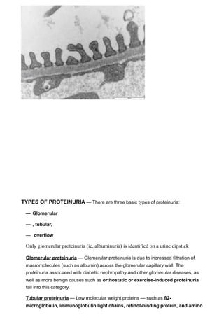

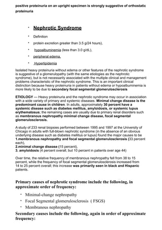
![Normal rate (albumin): up to 30mg/24h
Microalbuminuria : between 30 and 300 mg/day
Proteinuria : >300mg/24h
more than 3.5 grams per day = NEPHROTIC SYNDROME
- diabetics: disease that cause proteinuria- usually for first 10 yrs- doesnt
matter waht type- will have microalbuminuria- then will go to proteinuria,and
then nephrotic syndrome (more than 3.5 g /day)
- microalbuminuria: bad prognostic sign- we are about albumin- not total
proteinuria (unless pt has myeloma)
24-hour collection — The traditional method requires a 24-hour urine collection to
directly determine the daily total protein or albumin excretion. An extra benefit of this
approach, if creatinine is also measured, is that it provides the information necessary
to estimate the glomerular filtration rate (GFR) from the creatinine clearance.
Protein/creatinine ratio
An alternative method requires only a random urine specimen to estimate the degree
of proteinuria [9-12]. This test calculates the total protein-to-creatinine ratio (mg/mg).
This ratio correlates with daily protein excretion expressed in terms of g per 1.73m2 of
body surface area (figure 1). Thus, a ratio of 4.9 (as with respective urinary protein
and creatinine concentrations of 210 and 43 mg/dL) represents a daily protein
excretion of approximately 4.9 g per 1.73.
Thus, a ratio of 4.9 represents a daily protein excretion of approximately 4.9 g/
24h
- most imp test to detect early stage of renal disease: especially in diabetics
ACR Urine albumin to creatinine ratio — The urine albumin:creatinine ratio
(ACR), like the PC ratio, is measured using a random "spot" urine specimen.
The K/DOQI guidelines note that the relative merits of measuring and monitoring the
total protein-to-creatinine ratio versus the albumin-to-creatinine ratio to detect and
monitor kidney damage are unclear. However, given that albuminuria is a more](https://image.slidesharecdn.com/icmnephsynd234-120528142345-phpapp02/85/Icm-neph-synd-23-4-5-320.jpg)
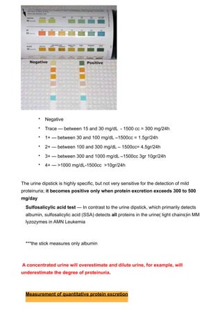
![• Diabetes mellitus
• Lupus erythematosus
• Amyloidosis and paraproteinemias (MM)
• Viral infections (eg, hepatitis B, hepatitis C, human
immunodeficiency virus [HIV] )
• Preeclampsia
Metabolic consequences of proteinuria
Metabolic consequences of the nephrotic syndrome include the following:
Infection
Urinary immunoglobulin losses
• Edema fluid acting as a culture medium
• Protein deficiency
• Decreased bactericidal activity of the leukocytes
• Immunosuppressive therapy
• Decreased perfusion of the spleen caused by hypovolemia
• Urinary loss of a complement factor (properdin factor B) that
opsonizes certain bacteria
•
• Hyperlipidemia and atherosclerosis
• It is related to the hypoproteinemia and low serum oncotic pressure
of nephrotic syndrome, which then leads to reactive hepatic protein
synthesis, including of lipoproteins.[7] In addition, reduced plasma
levels of lipoprotein lipase results in diminution of lipid catabolism
• Hypocalcemia and bone abnormalities
•
Hypocalcemia is common in the nephrotic syndrome, but rather than
being a true hypocalcemia, it is usually caused by a low serum
albumin level.
Nonetheless, low bone density and abnormal bone histology are reported
in association with nephrotic syndrome. This could be caused by urinary](https://image.slidesharecdn.com/icmnephsynd234-120528142345-phpapp02/85/Icm-neph-synd-23-4-8-320.jpg)
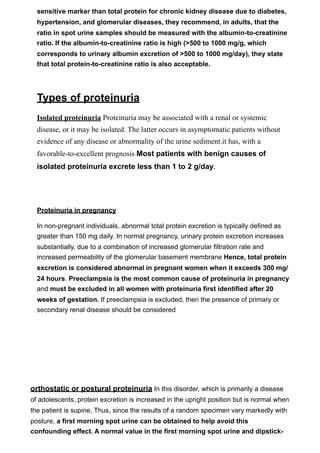
![losses of vitamin D–binding proteins, with consequent hypovitaminosis D
and, as a result, reduced intestinal calcium absorption.[9]
•
• Hypercoagulability
Venous thrombosis and pulmonary embolism are well-known
complications of the nephrotic syndrome. Hypercoagulability in these
cases appears to derive from urinary loss of anticoagulant proteins, such as
antithrombin III and plasminogen, along with the simultaneous increase
in clotting factors, especially factors I, VII, VIII, and X.
•
• Hypovolemia
•
• Hypovolemia occurs when hypoalbuminemia decreases the plasma
oncotic pressure, resulting in a loss of plasma water into the
interstitium and causing a decrease in circulating blood volume.
Hypovolemia is generally observed only when the patient's serum
albumin level is less than 1.5 g/dL](https://image.slidesharecdn.com/icmnephsynd234-120528142345-phpapp02/85/Icm-neph-synd-23-4-9-320.jpg)