This document discusses prosthetic valve thrombosis (PVT), including its definition, pathogenesis, incidence, diagnosis, and treatment. PVT occurs when a blood clot forms on an artificial heart valve, interfering with its function. It is more common with mechanical valves and in the mitral position. Diagnosis involves blood tests, imaging like echocardiography and fluoroscopy. For non-obstructive small clots on the left side, initial treatment is usually heparin. Larger or obstructive clots may require surgery or fibrinolysis.
A lecture on the echocardiographic evaluation of hypertrophic cardiomyopathy. Starts with an overview of the topic then a systematic approach to diagnosis and then a differential diagnosis followed by take-home messages and conclusion.
ECHOCARDIOGRAPHIC EVALUATION OF MITRAL VALVE DISEASEPraveen Nagula
MITRAL VALVE ANATOMY , M MODE FINDINGS IN MITRAL STENOSIS,EVALUATION OF THE SEVERITY OF LESION,CALCIFIC MS,CCMA,CONGENITAL LESIONS,GUIDELINES ALL IN DETAIL....
preop TEE assessment of atrial septal defect is very important for making decision for device closure, properly assessed adequate rims of ASD will reduce risk of device embolization to almost nil.
Percutaneous Balloon Mitral Valvuloplasty (PBMV) is a procedure to dilated the mitral valve in the setting of rheumatic mitral valve stenosis. A catheter is inserted into the femoral vein, advanced to the right atrium and across the interatrial septum. Then the mitral valve is crossed with a balloon and it is inflated to relieve the fusion of the mitral valve commissures effectively acting to increase the mitral valve area and reduce the degree of mitral stenosis. Mitral regurgitation is a potential complication and thus PBMV is contraindicated if moderate or severe regurgitation is present. The Wilkins score examines mitral valve morphology and is determined via echocardiography to assess the likelihood of using PBMV based on certain echocardiographic criteria.
Admixture lesions in congenital cyanotic heart diseaseRamachandra Barik
Admixture lesions in congenital cyanotic heart disease
Jaganmohan A Tharakan
Department of Cardiology, Sree Chitra Tirunal Institute for Medical Sciences and Technology, Trivandrum, India
Based on the principle that the distal coronary pressure measured during vasodilation is directly proportional to maximum vasodilated perfusion.
FFR is defined as the ratio of maximum blood flow in a stenotic artery to maximum blood flow in the same artery if there were no stenosis.
FFR is simply calculated as a ratio of mean pressure distal to a stenosis (Pd) to the mean pressure proximal stenosis, that is the mean pressure in the aorta (Pa), during maximal hyperaemia.
By the end of the module, you will be able to:
Define Arterio Venous Fistula and Arterio Venous Graft
Identify Complications and Management
Familiarise and use the Pre Needling Cannulation Tool
263778731218 Abortion Clinic /Pills In Harare ,sisternakatoto
263778731218 Abortion Clinic /Pills In Harare ,ABORTION WOMEN’S CLINIC +27730423979 IN women clinic we believe that every woman should be able to make choices in her pregnancy. Our job is to provide compassionate care, safety,affordable and confidential services. That’s why we have won the trust from all generations of women all over the world. we use non surgical method(Abortion pills) to terminate…Dr.LISA +27730423979women Clinic is committed to providing the highest quality of obstetrical and gynecological care to women of all ages. Our dedicated staff aim to treat each patient and her health concerns with compassion and respect.Our dedicated group ABORTION WOMEN’S CLINIC +27730423979 IN women clinic we believe that every woman should be able to make choices in her pregnancy. Our job is to provide compassionate care, safety,affordable and confidential services. That’s why we have won the trust from all generations of women all over the world. we use non surgical method(Abortion pills) to terminate…Dr.LISA +27730423979women Clinic is committed to providing the highest quality of obstetrical and gynecological care to women of all ages. Our dedicated staff aim to treat each patient and her health concerns with compassion and respect.Our dedicated group of receptionists, nurses, and physicians have worked together as a teamof receptionists, nurses, and physicians have worked together as a team wwww.lisywomensclinic.co.za/
Prix Galien International 2024 Forum ProgramLevi Shapiro
June 20, 2024, Prix Galien International and Jerusalem Ethics Forum in ROME. Detailed agenda including panels:
- ADVANCES IN CARDIOLOGY: A NEW PARADIGM IS COMING
- WOMEN’S HEALTH: FERTILITY PRESERVATION
- WHAT’S NEW IN THE TREATMENT OF INFECTIOUS,
ONCOLOGICAL AND INFLAMMATORY SKIN DISEASES?
- ARTIFICIAL INTELLIGENCE AND ETHICS
- GENE THERAPY
- BEYOND BORDERS: GLOBAL INITIATIVES FOR DEMOCRATIZING LIFE SCIENCE TECHNOLOGIES AND PROMOTING ACCESS TO HEALTHCARE
- ETHICAL CHALLENGES IN LIFE SCIENCES
- Prix Galien International Awards Ceremony
These simplified slides by Dr. Sidra Arshad present an overview of the non-respiratory functions of the respiratory tract.
Learning objectives:
1. Enlist the non-respiratory functions of the respiratory tract
2. Briefly explain how these functions are carried out
3. Discuss the significance of dead space
4. Differentiate between minute ventilation and alveolar ventilation
5. Describe the cough and sneeze reflexes
Study Resources:
1. Chapter 39, Guyton and Hall Textbook of Medical Physiology, 14th edition
2. Chapter 34, Ganong’s Review of Medical Physiology, 26th edition
3. Chapter 17, Human Physiology by Lauralee Sherwood, 9th edition
4. Non-respiratory functions of the lungs https://academic.oup.com/bjaed/article/13/3/98/278874
Report Back from SGO 2024: What’s the Latest in Cervical Cancer?bkling
Are you curious about what’s new in cervical cancer research or unsure what the findings mean? Join Dr. Emily Ko, a gynecologic oncologist at Penn Medicine, to learn about the latest updates from the Society of Gynecologic Oncology (SGO) 2024 Annual Meeting on Women’s Cancer. Dr. Ko will discuss what the research presented at the conference means for you and answer your questions about the new developments.
HOT NEW PRODUCT! BIG SALES FAST SHIPPING NOW FROM CHINA!! EU KU DB BK substit...GL Anaacs
Contact us if you are interested:
Email / Skype : kefaya1771@gmail.com
Threema: PXHY5PDH
New BATCH Ku !!! MUCH IN DEMAND FAST SALE EVERY BATCH HAPPY GOOD EFFECT BIG BATCH !
Contact me on Threema or skype to start big business!!
Hot-sale products:
NEW HOT EUTYLONE WHITE CRYSTAL!!
5cl-adba precursor (semi finished )
5cl-adba raw materials
ADBB precursor (semi finished )
ADBB raw materials
APVP powder
5fadb/4f-adb
Jwh018 / Jwh210
Eutylone crystal
Protonitazene (hydrochloride) CAS: 119276-01-6
Flubrotizolam CAS: 57801-95-3
Metonitazene CAS: 14680-51-4
Payment terms: Western Union,MoneyGram,Bitcoin or USDT.
Deliver Time: Usually 7-15days
Shipping method: FedEx, TNT, DHL,UPS etc.Our deliveries are 100% safe, fast, reliable and discreet.
Samples will be sent for your evaluation!If you are interested in, please contact me, let's talk details.
We specializes in exporting high quality Research chemical, medical intermediate, Pharmaceutical chemicals and so on. Products are exported to USA, Canada, France, Korea, Japan,Russia, Southeast Asia and other countries.
Lung Cancer: Artificial Intelligence, Synergetics, Complex System Analysis, S...Oleg Kshivets
RESULTS: Overall life span (LS) was 2252.1±1742.5 days and cumulative 5-year survival (5YS) reached 73.2%, 10 years – 64.8%, 20 years – 42.5%. 513 LCP lived more than 5 years (LS=3124.6±1525.6 days), 148 LCP – more than 10 years (LS=5054.4±1504.1 days).199 LCP died because of LC (LS=562.7±374.5 days). 5YS of LCP after bi/lobectomies was significantly superior in comparison with LCP after pneumonectomies (78.1% vs.63.7%, P=0.00001 by log-rank test). AT significantly improved 5YS (66.3% vs. 34.8%) (P=0.00000 by log-rank test) only for LCP with N1-2. Cox modeling displayed that 5YS of LCP significantly depended on: phase transition (PT) early-invasive LC in terms of synergetics, PT N0—N12, cell ratio factors (ratio between cancer cells- CC and blood cells subpopulations), G1-3, histology, glucose, AT, blood cell circuit, prothrombin index, heparin tolerance, recalcification time (P=0.000-0.038). Neural networks, genetic algorithm selection and bootstrap simulation revealed relationships between 5YS and PT early-invasive LC (rank=1), PT N0—N12 (rank=2), thrombocytes/CC (3), erythrocytes/CC (4), eosinophils/CC (5), healthy cells/CC (6), lymphocytes/CC (7), segmented neutrophils/CC (8), stick neutrophils/CC (9), monocytes/CC (10); leucocytes/CC (11). Correct prediction of 5YS was 100% by neural networks computing (area under ROC curve=1.0; error=0.0).
CONCLUSIONS: 5YS of LCP after radical procedures significantly depended on: 1) PT early-invasive cancer; 2) PT N0--N12; 3) cell ratio factors; 4) blood cell circuit; 5) biochemical factors; 6) hemostasis system; 7) AT; 8) LC characteristics; 9) LC cell dynamics; 10) surgery type: lobectomy/pneumonectomy; 11) anthropometric data. Optimal diagnosis and treatment strategies for LC are: 1) screening and early detection of LC; 2) availability of experienced thoracic surgeons because of complexity of radical procedures; 3) aggressive en block surgery and adequate lymph node dissection for completeness; 4) precise prediction; 5) adjuvant chemoimmunoradiotherapy for LCP with unfavorable prognosis.
Ethanol (CH3CH2OH), or beverage alcohol, is a two-carbon alcohol
that is rapidly distributed in the body and brain. Ethanol alters many
neurochemical systems and has rewarding and addictive properties. It
is the oldest recreational drug and likely contributes to more morbidity,
mortality, and public health costs than all illicit drugs combined. The
5th edition of the Diagnostic and Statistical Manual of Mental Disorders
(DSM-5) integrates alcohol abuse and alcohol dependence into a single
disorder called alcohol use disorder (AUD), with mild, moderate,
and severe subclassifications (American Psychiatric Association, 2013).
In the DSM-5, all types of substance abuse and dependence have been
combined into a single substance use disorder (SUD) on a continuum
from mild to severe. A diagnosis of AUD requires that at least two of
the 11 DSM-5 behaviors be present within a 12-month period (mild
AUD: 2–3 criteria; moderate AUD: 4–5 criteria; severe AUD: 6–11 criteria).
The four main behavioral effects of AUD are impaired control over
drinking, negative social consequences, risky use, and altered physiological
effects (tolerance, withdrawal). This chapter presents an overview
of the prevalence and harmful consequences of AUD in the U.S.,
the systemic nature of the disease, neurocircuitry and stages of AUD,
comorbidities, fetal alcohol spectrum disorders, genetic risk factors, and
pharmacotherapies for AUD.
2. • Introduction
• Definition
• Pathophysiology
• Incidence
• Epidemiology
• Diagnosis
• (i) Blood test
• (ii) Imaging
• Treatment
1. Medical Rx
2. Surgical Rx
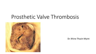
3. Prosthetic Valve Thrombosis (PVT)
• Prosthetic valve thrombosis (PVT) is a rare but serious complication of
valve replacement, most often encountered with mechanical prostheses.
• The significant morbidity and mortality associated with this condition
warrants rapid diagnostic evaluation.
• However, diagnosis can be challenging, mainly because of variable clinical
presentations and the degree of valvular obstruction.
4. Definition
• Prosthetic valve thrombosis (PVT) is defined by any thrombus,
• in the absence of infection,
• attached to or near an operated valve,
• occluding part of the blood flow or interfering with valvular function.
6. 1. Molecular interactions
• The first mechanism involves the molecular interaction
• between corpuscular blood components, plasma and artificial
surfaces.
• The initial adsorption of plasma proteins (fibrinogen) on the artificial
surface is generally followed by platelet adhesion.
7. 2. Influence of transprosthetic blood flow
• The second mechanism can occur in three ways.
• First, unphysiological flow that involves rapidly changing flow
directions (‘turbulent flow’) may result in a blood-borne increase in
shear stress and consequently in a structurally and metabolically
damaged endocardium, causing it to lose its resistance to thrombosis
8. • Second, blood stasis in recirculation areas downstream from the
prosthesis.
• Clot formation starts on the outflow side of the Prosthesis.
• Because the velocity of the transprosthetic flow near the valve housing is
low.
• Finally, chronic, subclinical haemolysis
• occur as a consequence of the accelerated destruction of thrombocytes
and erythrocytes with shortened intravascular lifespans.
9. 3. Local hypercoagulability
• All factors that promote clotting or platelet aggregation, permanently or temporarily,
increase the risk for local thrombosis.
• these factors are inadequate or unstable anticoagulation,
• loss of atrial contraction (atrial fibrillation),
• sporadic use of a variety of drugs,
• malignancies or systemic diseases,
• defects on the prosthetic surface,
• incomplete endothelialization of the sewing ring, and other hypercoagulable states.
10. Incidence
• Average 0·2% per patient-year after aortic and
• 1·8% per patient-year after mitral valve replacement,
• The risk for PVT appears to be twice as high in mitral as in aortic valve
replacement, and
• is more than three times higher for a tricuspid valve prosthesis
11. Epidemiology
• Mechanical valve thrombosis
• The incidence of obstructive PVT varies between 0.3–1.3% patient per years.
• Thromboembolic complications are more frequent and occur at a rate of 0.7–
6% patient years.
• Non‐obstructive PVT is a relatively frequent finding in the postoperative
period, with a reported incidence as high as 10% in recent (TEE) studies.
12. • Bioprosthetic valve thrombosis
• Rare occurrence when compared to mechanical prostheses.
• Bioprosthetic PVT is usually diagnosed in the early postoperative period,
when endothelialisation of the suture zone is not yet complete.
13. Diagnosis
• Clinical presentation
• Highly variable, depending on the presence or absence of obstruction.
• Severe obstructive PVT is typically associated with overt heart failure,
• whereas non‐obstructive PVT is often an incidental finding or can present as an
embolic episode.
• Partial obstruction (for example, obstruction of one leaflet) can manifest itself with
abnormal dyspnoea, or systemic embolism and rarely fever.
• In the presence of fever, diagnostic blood cultures should be performed to rule out
infectious endocarditis.
14. • The typical clinical finding in PVT
• cardiac auscultation may detect absence of prosthetic click or
• presence of new systolic or diastolic murmur.
15. Blood tests
• Routine periodic blood testing after mechanical valve replacement should include
• INR (prothrombin time) to monitor adequacy of anticoagulation
• (Inadequate anticoagulation may be associated with increased risk of prosthetic
valve thrombosis)
• Lactate dehydrogenase (to detect possible hemolysis)
• Plasma D‐dimer level has high specificity but low sensitivity for detecting
prosthetic heart valve thrombosis
16. Imaging studies
• Fluoroscopy
• As all mechanical valves available are radio‐opaque, fluoroscopy is an
important part of the diagnostic evaluation of suspected PVT.
• However, this technique will not be helpful in identifying
non‐obstructive PVT or differentiating pannus from thrombus.
• Hence, additional diagnostic procedures are often necessary.
17. Benefits of fluoroscopy
• Noninvasive
• May be used to evaluate opening and closing angle of prosthetic valve
leaflets
• Can detect motion of leaflets and base ring
• Can detect leaflet restriction
• Can assess integrity of mechanical prosthetic heart valve components
18. • Patient should be positioned so that leaflets are perpendicular to the
x‐ray tube.
• Not suitable for evaluation of bioprosthetic heart valves because of the
radiolucent properties of biological leaflets.
• Better than TTE and TEE for visualization of leaflet motion in aortic
position
• Comparable to TTE and TEE for visualization of leaflet motion in mitral
position
19.
20. • Transthoracic echocardiography ( TTE )
• By providing direct visualization of the prosthesis and a measure of
transvalvular gradients, TTE is an essential part of diagnostic assessment
• Using color Doppler, abnormal transprosthetic flow or central regurgitation,
indicating abnormal valve closure, can be observed.
• Pulmonary artery pressures and cardiac output should also be measured.
• Direct signs of PVT include abnormal movement of the prosthesis (immobile
hemi‐disc, incomplete or delayed opening) or visualization of a paraprosthetic
thrombus.
21. • For mitral prostheses, a mean gradient >8 mm Hg and an effective area
⩽1.3 cm2 is indicative of PVT .
• For aortic prostheses, criteria for PVT are a mean gradient >45 mm Hg and an
obstructive index <0.25.
• In the case of small size aortic prostheses (for example, mechanical valve
sizes 19 or 21), diagnosis can be difficult when considering that these
prostheses often have “normally” elevated gradients, due to local turbulent
flow across the main orifice and potential patient–prosthesis mismatch.
22. • Limitations of TTE include
• quality of the acoustic window,
• artefacts associated with the prosthesis, and non‐obstructive PVT (where
TTE will usually be normal).
• In conditions of low cardiac output, transvalvular gradients can be within the
normal range despite significant prosthetic valve obstruction, so‐called
“silent Doppler PVT”.
• Hence if clinical suspicion remains, the investigation should be completed
with a TEE study.
23.
24. • Transesophageal echocardiography ( TEE )
• Direct signs of PVT include immobility or reduced leaflet mobility, and the
presence of thrombus on either side of the prosthesis, with or without
obstruction.
• Thrombi have to be differentiated from a fibrous pannus, which is usually
annular in location.
• Indirect TOE signs of PVT are the disappearance of the normal physiological
prosthesis regurgitant flow, the presence of central prosthesis regurgitation,
and pronounced spontaneous echo contrasts in the left atrium
25.
26. • Benefits of TEE include
• Proximity of probe to anatomical structures improves visualization of
anatomic abnormalities and dysfunction,
• better visualization of leaflet thickening, leaflet prolapse, and flail cusps
• Evaluation of PHV leaflet motion and assessment of regurgitation,
• identification of prosthetic and bioprosthetic valve obstruction.
• Better visualization of valve thrombosis, especially in the mitral position
• May be combined with Doppler ultrasound
27. • Limitations of TEE include
• Semi‐invasive
• Prosthetic valves may produce acoustic shadowing
• May not differentiate active from healed vegetations in patients with
infective endocarditis, thickened valves or nodules from vegetations
and
• thrombosis from pannus or vegetation
• difficulty visualizing leaflet motion in aortic position
28.
29. Thrombus or pannus?
• Some distinguishing features between thrombus and pannus in terms of when they
occur after mechanical valve replacement surgery and the echocardiographic
appearance.
• Inadequate anticoagulation with warfarin should raise suspicion of thrombus.
• Pannus formation usually occurs many years after mechanical valve replacement,
compared with thrombus, which may occur at any time.
• Pannus is more commonly associated with mechanical valves in the aortic position
but can occur in association with any mechanical valves. When observed on mitral
prosthetic valves, they most often occur on the atrial side of the prosthesis.
• Typically presenting as a very dense immobile echo
30.
31.
32.
33. TREATMENT
• Once diagnosis of PVT is confirmed, several therapeutic modalities can be
considered:
• Surgery, fibrinolysis, heparin treatment, or optimisation of anticoagulant and
antiplatelet therapy.
• Treatment can be separated according to presence of obstruction and
prosthesis location.
• The type of prosthesis (mechanical or biological) does not have particular
therapeutic implications, as the choice between surgery and medical
treatment will have to take into account the same considerations.
34. • Left-sided mechanical valves
• Valve obstruction from a small thrombus (less than 1 cm in diameter or 0.8
cm2 in area),
• recent onset (fewer than 14 days) and
• associated with mild symptoms (NYHA, class I-II)
• can be treated initially with a trial of 24 to 48 hours of intravenous
unfractionated heparin
• with a goal activated partial thromboplastin time (APTT) of 1.5 to 2 times the
control value.
35. • The patient should be monitored clinically,
• TTE should be performed every two to four hours, and
• TEE should be performed daily to assess the valve status.
• Improvement in valve function and gradient should be followed by
warfarin and continued intravenous heparin
• until the INR is in the range of 3 to 4 for aortic prostheses and 3.5 to
4.5 for mitral prostheses.
36. • If there is no improvement in valve function and gradient after 24
hours,
• thrombolytic therapy with recombinant tissue plasminogen activator
(rTPA)
• (10-mg intravenous bolus followed by 90-mg intravenous infusion over
two hours or,
• alternatively, 20-mg intravenous bolus followed by 30-mg infusion over
three hours) should be considered.
37. • Should be followed by warfarin and intravenous unfractionated heparin
until the INR is (3 to 4 for aortic valve prostheses) and (3.5 to 4.5 for mitral
prostheses).
• Patients in whom thrombolytic therapy is partially successful (the valve
gradient is improved but not normalized) present a challenge.
• Options include both subcutaneous unfractionated heparin twice daily to
achieve an APTT of 1.5 to 2 times control and warfarin therapy with a target
INR of 2.5 to 3.5 for three months.
38. • Emergency surgery is recommended for patients with
1. severe symptoms (NYHA class III-IV) and left-sided valve thrombosis,
regardless of thrombus size and
2. the thrombus size is large (more than 1 cm in diameter or greater
than 0.8 cm2 in area), regardless of symptoms.
39.
40. • Right-sided mechanical valves
• A trial of IV heparin followed by thrombolytic therapy is the mainstay of
treatment.
• followed by warfarin and intravenous unfractionated heparin until INR is in
the range of 3.5 to 4.5.
• Partially successful thrombolytic therapy may be followed by subcutaneous
unfractionated heparin twice daily to achieve an APTT of 1.5 to 2 times
control.
• Surgery should be considered if heparin and thrombolytic therapy fail.
41. Medications
Thrombolytic therapy
Indications for thrombolytic therapy (AHA/ACC) 2014 recommendations
For left‐sided thromboses
• recent‐onset (< 14 days) ,(NYHA) functional class I‐II symptoms, and small thrombus (<
0.8 cm2) (AHA/ACC Class IIa, Level B)
• first‐line therapy for NYHA functional class III‐IV symptoms, and small clot burden if
surgery is high risk or not available (AHA/ACC Class IIb, Level B)
• NYHA functional class II‐IV symptoms, and large clot burden if emergency surgery is
high risk or not available (AHA/ACC Class IIb, Level C)
42. • Right‐sided thromboses
• Consider for thrombosed right‐sided prosthetic heart valve (AHA/ACC
Class IIa, Level B)
• Consider for thrombosed right‐sided prosthetic heart valves with
NYHA functional class III‐IV symptoms or large clot burden (AHA/ACC
Class IIa, Level C)
43. Choice of thrombolytic agent
• Options include streptokinase, urokinase, and recombinant tissue
plasminogen activator (rt‐PA)
• Considerations when choosing thrombolytic agent
• Cost
• Time to attain maximal pharmacologic effect (rt‐PA is faster)
• Half‐life of thrombolytic agent (rt‐PA has faster effect reversion), important if urgent
surgery might be required due to failure of thrombolysis
• Experience with use of particular agent
44. Dosage and delivery of thrombolytic agents
• rt‐PA
• initial dose 10 mg bolus
• maintenance dose 90 mg infusion over 2 hours
• streptokinase
• loading dose 500,000 units over 20 minutes
• maintenance dose 1,500,000 units over 10 hours
• urokinase less effective than rt‐PA or streptokinase
45. • In patients with hemodynamic instability, short protocol
recommended for patients
• rtPA 10 mg bolus IV plus 90 mg rtPA in 90 minutes with
unfractionated heparin
• Streptokinase 1,500,000 units in 60 minutes without UFH
• Longer infusion duration may be used in stable patients
46. Contraindications to thrombolytic therapy
• A history of intracranial bleeding
• Recent cranial trauma
• Gastrointestinal or genitourinary bleeding within 21 days
• Hemorrhagic retinopathy;
• The presence of large, mobile thrombi
• Severe hypertension
• Hypotension or cardiogenic shock
• Major surgery within the previous two weeks
47. Anticoagulant therapy
• Start anticoagulant therapy after successful thrombolytic therapy
• Start unfractionated heparin IV until vitamin K antagonist (VKA) achieves
INR of 3‐4 for aortic prosthetic valve and 3.5‐4.5 for mitral prosthetic valve
• Continuous infusion unfractionated heparin indicated at end of
thrombolytic therapy
• Usual starting dose of heparin 1,000 units/hour
48. • Monitor activated partial thromboplastin time (aPTT) every 6‐8 hours and
adjust heparin infusion rate accordingly for first 48 hours
• after discontinuing thrombolysis resume oral anticoagulation therapy and
combine with heparin until optimum INR achieved
• Heparin infusion with frequent measurement of aPTT begins when aPTT
more than 2 times control levels and can be converted to warfarin (INR
2.5‐3.5) plus aspirin (81‐100 mg/day)
49. Surgery and procedures
• For prosthetic valve thrombosis
• Emergency operation is recommended for patients
• with thrombosed left‐sided prosthetic valve and (NYHA) functional class III‐IV
symptoms (AHA/ACC Class I, Level B)
• Emergency operation is reasonable for patients
• with thrombosed left‐sided prosthetic valve and mobile or large thrombus (area ≥
0.8 cm2) (AHA/ACC Class IIa, Level C)
50. • Surgery may be considered first for patients with
• Left atrial thrombus
• Active bleeding
• Within first 4 days after valve replacement
• Surgical options include
• Replacement of prosthesis
• if prosthesis functioning normally, thrombectomy may be preferred
• Surgery can be performed 24 hours after discontinuation of thrombolysis or 2 hours after
thrombolytic activity reversed with protease inhibitors.