
Recommended
More Related Content
What's hot
What's hot (20)
Similar to Prosthetic Heart Valve Thrombosis Diagnosis and Management
Similar to Prosthetic Heart Valve Thrombosis Diagnosis and Management (20)
Recently uploaded
Recently uploaded (20)
Prosthetic Heart Valve Thrombosis Diagnosis and Management
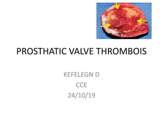
- 1. PROSTHATIC VALVE THROMBOIS KEFELEGN D CCE 24/10/19
- 2. OUTLINE • History • Types • Pathophysiology • Diagnoses • Management ,Literatures And Guidelines
- 3. History • The pioneering efforts of Dr. Charles Hufnagel, who made the first successful placement of a totally mechanical valvular prosthesis, started the era of artificial heart valves. • Hufnagel achieved this feat in 1952, by inserting a Plexiglas cage containing a ball occluder into the descending thoracic aorta. • The first implant of a mitral valve replacement in its anatomic position took place in 1960, when the Starr-Edwards prosthesis was put the clinical use
- 4. • Prosthetic valve thrombolysis: history – In 1971, Luluaga et al • were the first to use the thrombolytic therapy in prosthetic valve thrombosis. • Streptokinase was used for treating thrombosis of the tricuspid valve prosthesis. • Three years later, Baille et al reported the use of that thrombolytic agent in the aortic valve prosthesis.
- 8. Types of prosthetic valves and thrombogenicity Type of valve Model Thrombogenicity Mechanical Caged ball Starr-Edwards ++++ Single tilting disc Bjork-Shiley,Medtronic Hall +++ Bileaflet St Jude Medical,Sorin Bicarbon,Carbomedic s ++ Bioprosthetic Heterografts Carpentier- Edwards,Tissue Med (Aspire), Hancock II + to ++ Homografts +
- 9. Bileaflet valve Adv – • Low bulk - flat profile • Less thrombogenicy • Central laminar flow • two semicircular discs that pivot between open and closed positions • No need for supporting struts • Good hemodynamics even in small sizes • 2 lat ,1 central minor orifice , no chance of sudden catastro thrombosis Disadv- • Anticoagulation mandatory • risk of thrombosis St. Jude Medical mechanical heart valve Carbomedics Titanium housing Pyrolytic carbon
- 11. PROSTHETIC HEART VALVE THROMBOSIS: MECHANISMS
- 12. CLINICAL PRESENTATION • Patients with PV dysfunction with or without thrombosis may present with progressive dyspnea and signs of heart failure or systemic embolization. • Alternatively, PV thrombosis may be an incidental finding at the time of echocardiographic follow-up. • PV dysfunction should be suspected in patients with symptoms of acute or subacute onset associated with an increase in transprosthetic gradient compared with the last echocardiographic follow-up • Although arterial TE after surgical or transcatheter heart valve replacement should be considered prosthesis-related until proven otherwise. it may also arise from different cause. • Reduced or absent click, murmur.
- 14. DIAGNOSIS • LAB TEST • IMAGING
- 17. TTE • Regardless of the anatomic location of the prosthesis, the first-line imaging test for PV dysfunction is TTE. • Although it is helpful for evaluating prosthetic valve hemodynamics and valve motion, the test is limited for morphological characterization of the etiology of PV dysfunction. • Acoustic shadowing caused by the prosthesis may limit visualization of thrombus, vegetations, and pannus. • The diagnostic accuracy of TTE is influenced by other factors, such as the presence of pericardial effusion, emphysema, obesity, or prior sternotomy
- 21. TEE • TEE should be considered to better evaluate the pathological substrate of PV dysfunction. • In particular, TEE should always be performed if the transthoracic echocardiography is technically suboptimal, if the findings are not definitive, or if there is strong clinical suspicion of PV dysfunction. • TEE is superior to TEE for evaluating PV dysfunction, regardless of the valve type. • Although it is superior to TTE for identifying the mechanism of PV degeneration, even TEE cannot reliably discriminate between PV thrombosis and fibrotic pannus ingrowth
- 23. Cinefluoroscopy
- 29. MULTIDETECTOR COMPUTED TOMOGRAPHY • In patients with inconclusive TTE and TEE findings (which may be rather frequent), multidetector computed tomography could provide an accurate evaluation of the prosthetic valve structure and functional status • MDCT scanning may also help to differentiate between PV dysfunction and patient–prosthesis mismatch for 2 main reasons: • 1) it will detect thrombus, vegetations, or other masses; and • 2) it provides a more accurate assessment of the geometry of the left ventricular outflow tract and the effective orifice area for prostheses implanted in the aortic position
- 31. Management
- 42. • Outcome of treatment has been generally categorized into “complete success,” “partial success,” or “ineffective.” • The search eventually yielded 17 studies, comprising 756 patients , which were related to thrombolytic agents in OTPHV and 13 studies, comprising 662 patients, which were related to surgery in OTPHV
- 43. PANUS/THROMBUS • Ten studies presented data regarding findings of thrombus and/or pannus at the time of surgery for a total of 518 patients 41% had thrombus only, 38% had pannus only, and 21% had both thrombus and pannus.
- 44. How was INR ??
- 49. • The infusion dosage of streptokinase was a bolus of 250,000 U in 30 minutes, followed by 100,000 U/h. • Doppler echocardiography was used to monitor the time of infusion of the thrombolytic agent and to assess its efficacy
- 50. • The criteria for interrupting infusion were as follows: • 1.Hemodynamic improvement, assessed on echocardiography; • 2. Occurrence of major bleedings or hemorrhagic stroke. • 3. Infusion time of 72 hours.
- 51. • Complete hemodynamic improvement was observed in 81.8% of the patients, partial improvement in 10%,and treatment failure in 8.2%. • An embolic event occurred in 19.1% of the patients during treatment. • The success of thrombolysis was not influenced by the age of the patients, the time of symptom onset, the time of surgery, and the type or position of the valve prosthesis. • Atrial fibrillation was a predictor of embolic events.
- 53. • The thrombolytic agent should be interrupted at the 24 th hour of treatment, if no hemodynamic improvement (improvement in the gradient) occurs. • It should be interrupted after 72 hours, even if the improvement is partial, or should be interrupted earlier, if the hemodynamic improvement is complete
- 55. ACC 2017
- 61. CONCLUSION • Prosthetic valve thrombosis rate higher than the actual incidence • Approach could be to individualize the treatment depending on NYHA class, thrombus burden, and availability of surgery. • Newer regimen of very low-dose, slow infusion leads to equal efficacy with lower complication in majority of patients • According to recent guidelines thrombolysis considered in all classes of NYHA if individual patient characteristics support the recommendation of one treatment over the other.
Editor's Notes
- The various designs differ in the composition and purity of the pyrolytic carbon, the shape and opening angle of the leaflets, the design of the pivots, the size and shape of the housing, and the design of the sewing ring
- Eighteen studies provided information regarding anticoagulation status of patients at time of diagnosis of OTPHV (7–15,17,20–22,24–28). Of 1,005 patients, 61% had stated anticoagulation levels as adequate in the cited studies, and 39% had stated anticoagulation levels as inadequate in the cited studies
- Complete success was achieved in 81% of patients presenting in NYHA functional classes I/II and 74% of patients presenting in NYHA functional classes III/IV. Streptokinase was used in 12 of the 17 studies. The recurrence rate was 13%. The rate of cerebrovascular accident (CVA) or embolic phenomenon to other arterial sites was 14%. So