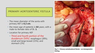
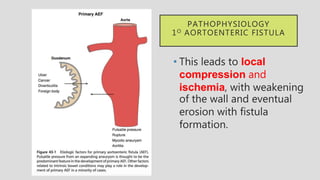
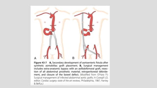
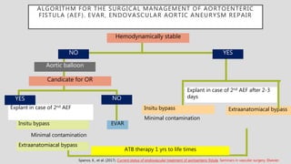
AORTOENTERIC FISTULA
(AEF)
F1 : Parach Sirisriro
18 June 2018
REFERENCE
Rutherford's Vascular Surgery and Endovascular Therapy, Chapter
49, 615-623.e
Textbook
Journal
1. Low RN, Wall SD, Jeffrey RB, et. al.: Aortoenteric fistula and perigraft
infection: evaluation with CT. Radiology 1990; 175: pp. 157-162.
2. Chung, J. (2018). "Management of Aortoenteric Fistula." Advances in
Surgery.
3. Spanos, K., et al. (2017). Current status of endovascular treatment of
aortoenteric fistula. Seminars in vascular surgery, Elsevier.
OUTLINE
• PRIMARY AORTOENTERIC FISTULA
Incidence, Etiology, and Pathogenesis
• SECONDARY AORTOENTERIC FISTULA
Incidence and Etiology
• PATHOGENESIS
• CLINICAL PRESENTATION
• DIAGNOSIS
• TREATMENT
INTRODUCTION
Aortoenteric fistula (AEF) is defined as a communication
between the aorta and gastrointestinal (GI) tract.
DEFINITION
• Primary AEF is a communication between the native aorta and
GI tract
• Secondary AEF is a communication between a reconstructed
aorta (for either aneurysmal or occlusive disease) and the GI tract.
PATHOPHYSIOLOGY
Chung, J. (2018). "Management of Aortoenteric Fistula." Advances in Surgery.
Primary and secondary AEF
occur via similar mechanisms
Infection seems to be the main
underlying cause of AEF.
PRIMARY AORTOENTERIC FISTULA
The incidence of primary AEF varies depending on the study base
 0.04% to 0.07% among patients who died of massive GI
hemorrhage : in large autopsy series
 0.69% to 2.36% among patients with abdominal aortic
aneurysms (AAAs)
In the majority of cases (83%), an aneurysmal aorta is associated
with
primary AEF , foreign bodies, tumors , radiotherapy , infection , And
GI tract disease
PRIMARY AORTOENTERIC FISTULA
• The mean diameter of the aorta with
primary AEF is 6.2 cm;
• the mean age of patients is 64 years, with a
male-to-female ratio of 3 : 1.
• Location for primary AEF
• Third and fourth portion of the
duodenum (54%). esophagus (28%),
small and large bowel (15%), and
stomach (2%)
• This leads to local
compression and
ischemia, with weakening
of the wall and eventual
erosion with fistula
formation.
PATHOPHYSIOLOGY
1O AORTOENTERIC FISTULA
SECONDARY AORTOENTERIC FISTULA
SECONDARY
AORTOENTERIC FISTULA
• Secondary AEF is more
common than primary
AEF and is related to
prior vascular surgery,
with an incidence of
0.36% to 1.6%
• Onset of symptoms is on
average 2 to 6 years after
graft placement.
SECONDARY
AORTOENTERIC FISTULA
• The most common location
described is the distal
duodenum and
proximal jejunum.
• Secondary AEF has been
described at multiple GI
sites, depending on the
location of the prosthetic
graft.
SECONDARY AORTOENTERIC FISTULA
GRAFT ENTERIC FISTULA
The graft enteric fistula’s
involvement of the suture line will
disrupt the arterial anastomosis
and cause dramatic hemorrhage.
• May be manifested first with
infectious symptoms —>direct
contact of the graft material
with the GI tract—>bacterial
translocation between the
interstices of the graft
GRAFT ENTERIC EROSION
PRESENTATION
SECONDARY AORTOENTERIC FISTULA
• Pathogenesis
• Infection : main cause By initial surgery or through
bacteremia
• Pulsatile pressure
Pressure from a noncompliant prosthesis
Suture line disruption
• Technical error
PATHOGENESIS
Technical Error
• The bowel can be injured during the initial dissection and
exposure
• Directly with sharp injury
• Heat transfer from cautery
• Indirectly by overretraction and tension.
AEF AFTER ENDOVASCULAR REPAIR
• Persistent endoleak with growth of the residual sac,
• Multiple coiling efforts to repair an endoleak,
• Erosion of the stent- graft through the aorta,
• Endotension
• Infection at the time of graft placement.
CLINICAL PRESENTATION
• The classic clinical triad for primary AEF described
by Sir Astley Cooper in 1829
• 64% to 94% for GI bleeding.
• 32% to 48% for abdominal pain.
• 17% to 25% for a pulsatile abdominal mass.
All three of these symptoms occurring concurrently in only 11%of cases
CLINICAL PRESENTATION
• Other symptoms reported include back pain, fever, and sepsis.
• Herald bleed
—> A minor bleed that is self-limited because of vasospasm and
thrombus formation.
The risk of fatal rebleeding is high
30% of patients rebleed within 6 hours
50% of patients rebleed within 24 hours.
DIAGNOSIS
Diagnosis
Hemodynamic
unstable
Hemodynamic
stable
Exploratory
laparotomy 1. Computed tomography (CT)
2. Esophagogastroduodenoscopy
3. Angiography
CT scan —> a preferred initial diagnostic test;
CT scans are less invasive than EGD or angiography,
are easy to obtain, and do not risk thrombus dislodgement.16
INVESTIGATION
• CT scan : The detection rate of CT for AEF is the highest of all
modalities (61%)
• EGD : The detection rate of AEF by EGD is 25%
• Angiography : The angiography detection rate for AEF is 26%
• Other tests, leukocyte and erythrocyte tagged nuclear scanning,
magnetic resonance imaging, ultrasound, and contrast enhanced GI
studies, are potentially useful but usually less accurate and more time
intensive.
CT scanEGD
Angiography
TREATMENT
TREATMENT
• Conservative nonoperative therapy for AEF is fatal
• Surgery is the only treatment option with the potential for
success.
TREATMENT
• The factors to effective surgical management
• Active hemorrhage
• The classification of the AEF (primary or secondary)
• The presence of sepsis,
• The anatomic distribution of the patient’s aneurysmal or
occlusive disease.
TREATMENT
1. Urgent Surgical Treatment of Hemorrhage.
2. Surgical Treatment in Stable Patients.
URGENT SURGICAL
TREATMENT OF
HEMORRHAGE
• A midline incision,
• Rapid proximal aortic
control. Infrarenal aorta
control is preferable
• Distal control may be
obtained with iliac clamping
or occlusion balloons.
• The bowel should be sharply
dissected off of the
aneurysm/prosthesis.
URGENT SURGICAL TREATMENT OF
HEMORRHAGE
• Evaluation of the bowel defect
• Small defect —> simple closure
• Large defect —> resection and anastomosis should be
performed after the aorta has been managed.
• The aorta may be resected and reconstruction options
—> evidence of infection??
URGENT SURGICAL
TREATMENT OF
HEMORRHAGE
• No evidence of infection :
in-situ repair may be
performed with prosthetic
graft, cadaveric artery, or
femoral vein.
• Care should be taken to
separate the graft from the GI
tract with the posterior
peritoneum or omentum.
• stump should be oversewn and covered
with omentum
• followed by retroperitoneal
débridement, drain placement
• extra- anatomic bypass.
URGENT SURGICAL TREATMENT OF
HEMORRHAGE
Evidence of local infection
SURGICAL TREATMENT IN STABLE
PATIENTS
• Primary Aortoenteric Fistula.
• Secondary Aortoenteric Fistula.
PRIMARY AORTOENTERIC
FISTULA.
• Depends on whether infection is present in the aortic bed
• Mild contamination —>in-situ replacement of the aorta
with local débridement and long-term antibiotic therapy
• Gross retroperitoneal contamination and sepsis—>
aneurysm resection, omentoplasty, retroperitoneal débridement,
and extraanatomic bypass should be performed.
PRIMARY AORTOENTERIC FISTULA.
• In the postoperative period. Broad-spectrum antibiotics should
be used until culture results are available.
• In the case of
• a negative culture—> antibiotic therapy can be
discontinued after 1 week
• positive cultures —> a 4 to 6 week course.
SECONDARY AORTOENTERIC
FISTULA.
• All infected synthetic material must be resected.
• The defect in the GI tract should be repaired with
conventional techniques appropriate for the location of the
fistula.
• A flap of omentum should be placed to separate the bowel
from the aortic stump closure and to fill the dead space
resulting from excision of the graft.
SECONDARY AORTOENTERIC
FISTULA.
• Operative mortality ranges from 13% to 86%, with an average
mortality of 30% to 40%.
• Amputation rates hover at 10%, and long-term survival
approximates 50% at 3 years.
SECONDARY AORTOENTERIC
FISTULA.
• The available operative choices
• Graft excision alone
• In-situ aortic graft replacement
• Graft excision and reconstruction with autogenous vein (i.e., the
neo-aortoiliac system procedure)
• Extraanatomic revascularization and graft excision
• Endovascular repair with lifelong antibiotics
GRAFT EXCISION ALONE
• If the graft has been placed for occlusive disease or has been chronically
occluded, it is possible to resect the graft without reconstruction.
• The concern in these clinical scenarios is that removal of the graft will lead
to profound ischemia and the risk of amputation.
• The patient needs to have sufficient collateralization —> CT or
angiography may be helpful.
IN-SITU AORTIC GRAFT
REPLACEMENT
• use of cryopreserved
allografts, antibiotic-soaked
synthetic graft, or silver-
coated Dacron.
• This approach remains
controversial; however, in
recent years it has become an
increasingly accepted strategy.
IN-SITU AORTIC GRAFT
REPLACEMENT
• Benefits:
• Improved longterm patency
• Decreased risk of stump blowout
• Decreased risk of thrombosis ascending to the renal arteries after
resection
• Maintenance of blood supply to the colon and pelvis.
IN-SITU AORTIC GRAFT
REPLACEMENT
• Studies have also begun to demonstrate the potential benefit of in-situ
reconstruction.
• A meta-analysis in 2006 demonstrated a lower operative mortality for in-
situ reconstruction with rifampin-bonded prosthesis compared with extra-
anatomic reconstruction in patients with aortic graft infections.
• A retrospective study of in-situ reconstruction for secondary AEFs
published in 2011 demonstrated no difference in operative mortality
between in-situ reconstruction and extra-anatomic reconstruction
-Batt M, et al: Early and late results of contemporary management of 37
secondary aortoenteric fistulae. Eur J Vasc Endovasc Surg 41:748, 2011.
-O’Connor S, et al: A systematic review and meta-analysis of
treatments of aortic graft infection. J Vasc Surg 44:38, 2006.
GRAFT EXCISION AND RECONSTRUCTION
WITH AUTOGENOUS VEIN
• Use the femoropopliteal vein to reconstruct an autogenous neo-
aortoiliac system.
• Benefit in this technique
• Infection-resistant autologous tissue
• avoids the pitfalls of the aortic stump.
• Relatively inexpensive
• It does not require lifelong anticoagulation
GRAFT EXCISION AND RECONSTRUCTION
WITH AUTOGENOUS VEIN
• The disadvantages of this procedure
• The length of time required to harvest the deep vein and that it
cannot be staged.
• The risk of venous insufficiency in the harvest limb leading to
compartment syndrome and fasciotomy.
OUTCOME
• Thirty-day operative mortality is <10%, with 5-year mortality rates of 30% to
50%.
• Thirty-day major amputation rates range from 2% to 7.4%, with 5-year limb-
salvage rates ranging between 89% and 96%.
• Early occlusion of the grafts are rare, with <4% undergoing thrombosis within
the first 30 days; 5-year primary patency ranges from 75% to 91%, with
secondary patency approaching 91% at 5 years.
Chung J, et al: Neoaortoiliac system (NAIS) procedure for the
treatment of the infected aortic graft. Semin Vasc Surg 24:220, 2011.
OUTCOME
• Recurrent infection is very rare, occurring in <2% of patients.
• Venous morbidity is similarly low,
• fasciotomy rates of 12%
• only 15% of patients experiencing chronic venous insufficiency at 5
years.
Chung J, et al: Neoaortoiliac system (NAIS) procedure for the
treatment of the infected aortic graft. Semin Vasc Surg 24:220, 2011.
EXTRA-ANATOMIC REVASCULARIZATION
AND GRAFT EXCISION
• Performed with externally supported
(ringed) PTFE and may be performed
after abdominal closure with a fresh set
of clean instruments to restore flow to
the extremities.
• Performed before the aortic procedure,if
the patient is stable and not actively
bleeding.
EXTRA-ANATOMIC REVASCULARIZATION
AND GRAFT EXCISION
• Aortic stump disruption, which may occur late in the patient’s course,occur in
up to 12% of patients.
• 5-year patency of axillo-bifemoral bypass reported to be 70%,122
but infection of
the graft can occur in 6% to 20% of cases.
EMERGENCE OF EVAR
• First reported by Burks in 2001
• 2 of 7 patients with primary aortoenteric fistula
• 82-year-old, male, hypertension, coronary artery
disease, 10cm AAA
• Treated with Aortouniiliac stent graft
• Immediate cessation of bleeding achieved
• Died 13 months due to myocardial infarction
• Several case reports with similar success
ENDOVASCULAR REPAIR
• Rapid control of bleeding
• Minimal physiologic insult to patient
• Avoidance of operating in hostile abdomen
• Straightforward and speed of procedure
• Eliminating complications associated with open surgical repair
• Lower perioperative complication incidence
• Shorter hospital stay and more likely to discharge home
EVAR IN AORTODUODENAL FISTULA
- CANDIDATE SELECTION
• Pre-operative CT scan: diagnosis and planning
• Significant co-morbidities / High-risk for conventional
operation
– Medical: cardiopulmonary, renal, etc.
– Surgical: hostile abdomen
• Expertise for emergency EVAR
• Stent graft in immediate availability
EVAR IN AORTODUODENAL FISTULA
- COMPLICATIONS
• Persistent sepsis
• Repeat intervention or image-guided drainage
• Fungal infection, e.g. Aspergillus
• Long-term (or life-long) antibiotics
• Medical: underlying co-morbidities of patient
• Persistent bleeding
• No reported incidence
• Case report: unsuccessful result not reported?
• Secondary aortoenteric fistula
• Objectives: To compare aortoenteric fistula (AEF)
outcome after endovascular (EV- AEFR) or open repair
(O-AEFR).
• 25 patients with AEF (24 secondary, 23 males, median
age 75 years) after aortic surgery (median four years).
Kakkos SK, et al: Open or endovascular repair of aortoenteric fistulas? A
multicentre comparative study. Eur J Vasc Endovasc Surg 41:625, 2011.
Kakkos SK, et al: Open or endovascular repair of aortoenteric fistulas? A
multicentre comparative study. Eur J Vasc Endovasc Surg 41:625, 2011.
• The literature
32
identified 33 reports that included 41 patients with
AEF who had EVAR as initial management.
• Persistent/recurrent/new infection or recurrent hemorrhage
developed in 44% of the patients, after a mean follow-up period
of 13 months (range, 0.13-36).
Antoniou GA, et al: Outcome after endovascular stent graft repair of
aortoenteric fistula: a systematic review. J Vasc Surg 49:782, 2009.
• Secondary, as compared to primary, AEF had an almost threefold
increased risk of persistent/recurrent infection.
• Evidence of sepsis preoperatively was found to be a factor indicating
unfavorable outcome (P < .05)
• Persistent/recurrent/new infection after treatment was associated
with worse 30-day
Should be considered only a temporizing strategy until definitive repair can be performed.
Antoniou GA, et al: Outcome after endovascular stent graft repair of
aortoenteric fistula: a systematic review. J Vasc Surg 49:782, 2009.
ENDOVASCULAR REPAIR
Conclusion
• Reserved as a bridge to open repair once patients have
stabilized and local infection control is established
• For those patients with a limited life expectancy who would
benefit from a short hospital stay and earlier discharge and
will not be exposed to the long-term risks.
CONCLUSIONS
Explant in case of 2nd AEF
ALGORITHM FOR THE SURGICAL MANAGEMENT OF AORTOENTERIC
FISTULA (AEF). EVAR, ENDOVASCULAR AORTIC ANEURYSM REPAIR
Hemodynamically stable
NO YES
Candicate for OR
YES NO
Minimal contamination
Extraanatomiacal bypass
Insitu bypass
Aortic balloon
EVAR
Explant in case of 2nd AEF after 2-3
days
Minimal contamination
Extraanatomiacal bypassInsitu bypass
ATB therapy 1 yrs to life times
Spanos, K., et al. (2017). Current status of endovascular treatment of aortoenteric fistula. Seminars in vascular surgery, Elsevier.
CONCLUSIONS
• The key features to successful treatment are
• Timely intervention
• Appropriate patient selection
• Effective bowel repair
• Longterm follow-up.
THANK YOU

Present 18.6 aef

  • 1.
    AORTOENTERIC FISTULA (AEF) F1 :Parach Sirisriro 18 June 2018
  • 2.
    REFERENCE Rutherford's Vascular Surgeryand Endovascular Therapy, Chapter 49, 615-623.e Textbook Journal 1. Low RN, Wall SD, Jeffrey RB, et. al.: Aortoenteric fistula and perigraft infection: evaluation with CT. Radiology 1990; 175: pp. 157-162. 2. Chung, J. (2018). "Management of Aortoenteric Fistula." Advances in Surgery. 3. Spanos, K., et al. (2017). Current status of endovascular treatment of aortoenteric fistula. Seminars in vascular surgery, Elsevier.
  • 3.
    OUTLINE • PRIMARY AORTOENTERICFISTULA Incidence, Etiology, and Pathogenesis • SECONDARY AORTOENTERIC FISTULA Incidence and Etiology • PATHOGENESIS • CLINICAL PRESENTATION • DIAGNOSIS • TREATMENT
  • 4.
    INTRODUCTION Aortoenteric fistula (AEF)is defined as a communication between the aorta and gastrointestinal (GI) tract.
  • 5.
    DEFINITION • Primary AEFis a communication between the native aorta and GI tract • Secondary AEF is a communication between a reconstructed aorta (for either aneurysmal or occlusive disease) and the GI tract.
  • 6.
    PATHOPHYSIOLOGY Chung, J. (2018)."Management of Aortoenteric Fistula." Advances in Surgery. Primary and secondary AEF occur via similar mechanisms Infection seems to be the main underlying cause of AEF.
  • 7.
  • 8.
    PRIMARY AORTOENTERIC FISTULA Theincidence of primary AEF varies depending on the study base  0.04% to 0.07% among patients who died of massive GI hemorrhage : in large autopsy series  0.69% to 2.36% among patients with abdominal aortic aneurysms (AAAs) In the majority of cases (83%), an aneurysmal aorta is associated with primary AEF , foreign bodies, tumors , radiotherapy , infection , And GI tract disease
  • 9.
    PRIMARY AORTOENTERIC FISTULA •The mean diameter of the aorta with primary AEF is 6.2 cm; • the mean age of patients is 64 years, with a male-to-female ratio of 3 : 1. • Location for primary AEF • Third and fourth portion of the duodenum (54%). esophagus (28%), small and large bowel (15%), and stomach (2%)
  • 10.
    • This leadsto local compression and ischemia, with weakening of the wall and eventual erosion with fistula formation. PATHOPHYSIOLOGY 1O AORTOENTERIC FISTULA
  • 11.
  • 13.
    SECONDARY AORTOENTERIC FISTULA • SecondaryAEF is more common than primary AEF and is related to prior vascular surgery, with an incidence of 0.36% to 1.6% • Onset of symptoms is on average 2 to 6 years after graft placement.
  • 14.
    SECONDARY AORTOENTERIC FISTULA • Themost common location described is the distal duodenum and proximal jejunum. • Secondary AEF has been described at multiple GI sites, depending on the location of the prosthetic graft.
  • 15.
  • 16.
    GRAFT ENTERIC FISTULA Thegraft enteric fistula’s involvement of the suture line will disrupt the arterial anastomosis and cause dramatic hemorrhage. • May be manifested first with infectious symptoms —>direct contact of the graft material with the GI tract—>bacterial translocation between the interstices of the graft GRAFT ENTERIC EROSION PRESENTATION
  • 17.
    SECONDARY AORTOENTERIC FISTULA •Pathogenesis • Infection : main cause By initial surgery or through bacteremia • Pulsatile pressure Pressure from a noncompliant prosthesis Suture line disruption • Technical error
  • 18.
    PATHOGENESIS Technical Error • Thebowel can be injured during the initial dissection and exposure • Directly with sharp injury • Heat transfer from cautery • Indirectly by overretraction and tension.
  • 19.
    AEF AFTER ENDOVASCULARREPAIR • Persistent endoleak with growth of the residual sac, • Multiple coiling efforts to repair an endoleak, • Erosion of the stent- graft through the aorta, • Endotension • Infection at the time of graft placement.
  • 20.
  • 21.
    CLINICAL PRESENTATION • Theclassic clinical triad for primary AEF described by Sir Astley Cooper in 1829 • 64% to 94% for GI bleeding. • 32% to 48% for abdominal pain. • 17% to 25% for a pulsatile abdominal mass. All three of these symptoms occurring concurrently in only 11%of cases
  • 22.
    CLINICAL PRESENTATION • Othersymptoms reported include back pain, fever, and sepsis. • Herald bleed —> A minor bleed that is self-limited because of vasospasm and thrombus formation. The risk of fatal rebleeding is high 30% of patients rebleed within 6 hours 50% of patients rebleed within 24 hours.
  • 23.
  • 24.
    Diagnosis Hemodynamic unstable Hemodynamic stable Exploratory laparotomy 1. Computedtomography (CT) 2. Esophagogastroduodenoscopy 3. Angiography CT scan —> a preferred initial diagnostic test; CT scans are less invasive than EGD or angiography, are easy to obtain, and do not risk thrombus dislodgement.16
  • 25.
    INVESTIGATION • CT scan: The detection rate of CT for AEF is the highest of all modalities (61%) • EGD : The detection rate of AEF by EGD is 25% • Angiography : The angiography detection rate for AEF is 26% • Other tests, leukocyte and erythrocyte tagged nuclear scanning, magnetic resonance imaging, ultrasound, and contrast enhanced GI studies, are potentially useful but usually less accurate and more time intensive.
  • 26.
  • 27.
  • 28.
  • 29.
    TREATMENT • Conservative nonoperativetherapy for AEF is fatal • Surgery is the only treatment option with the potential for success.
  • 30.
    TREATMENT • The factorsto effective surgical management • Active hemorrhage • The classification of the AEF (primary or secondary) • The presence of sepsis, • The anatomic distribution of the patient’s aneurysmal or occlusive disease.
  • 31.
    TREATMENT 1. Urgent SurgicalTreatment of Hemorrhage. 2. Surgical Treatment in Stable Patients.
  • 32.
    URGENT SURGICAL TREATMENT OF HEMORRHAGE •A midline incision, • Rapid proximal aortic control. Infrarenal aorta control is preferable • Distal control may be obtained with iliac clamping or occlusion balloons. • The bowel should be sharply dissected off of the aneurysm/prosthesis.
  • 33.
    URGENT SURGICAL TREATMENTOF HEMORRHAGE • Evaluation of the bowel defect • Small defect —> simple closure • Large defect —> resection and anastomosis should be performed after the aorta has been managed. • The aorta may be resected and reconstruction options —> evidence of infection??
  • 34.
    URGENT SURGICAL TREATMENT OF HEMORRHAGE •No evidence of infection : in-situ repair may be performed with prosthetic graft, cadaveric artery, or femoral vein. • Care should be taken to separate the graft from the GI tract with the posterior peritoneum or omentum.
  • 35.
    • stump shouldbe oversewn and covered with omentum • followed by retroperitoneal débridement, drain placement • extra- anatomic bypass. URGENT SURGICAL TREATMENT OF HEMORRHAGE Evidence of local infection
  • 36.
    SURGICAL TREATMENT INSTABLE PATIENTS • Primary Aortoenteric Fistula. • Secondary Aortoenteric Fistula.
  • 37.
    PRIMARY AORTOENTERIC FISTULA. • Dependson whether infection is present in the aortic bed • Mild contamination —>in-situ replacement of the aorta with local débridement and long-term antibiotic therapy • Gross retroperitoneal contamination and sepsis—> aneurysm resection, omentoplasty, retroperitoneal débridement, and extraanatomic bypass should be performed.
  • 38.
    PRIMARY AORTOENTERIC FISTULA. •In the postoperative period. Broad-spectrum antibiotics should be used until culture results are available. • In the case of • a negative culture—> antibiotic therapy can be discontinued after 1 week • positive cultures —> a 4 to 6 week course.
  • 39.
    SECONDARY AORTOENTERIC FISTULA. • Allinfected synthetic material must be resected. • The defect in the GI tract should be repaired with conventional techniques appropriate for the location of the fistula. • A flap of omentum should be placed to separate the bowel from the aortic stump closure and to fill the dead space resulting from excision of the graft.
  • 40.
    SECONDARY AORTOENTERIC FISTULA. • Operativemortality ranges from 13% to 86%, with an average mortality of 30% to 40%. • Amputation rates hover at 10%, and long-term survival approximates 50% at 3 years.
  • 41.
    SECONDARY AORTOENTERIC FISTULA. • Theavailable operative choices • Graft excision alone • In-situ aortic graft replacement • Graft excision and reconstruction with autogenous vein (i.e., the neo-aortoiliac system procedure) • Extraanatomic revascularization and graft excision • Endovascular repair with lifelong antibiotics
  • 42.
    GRAFT EXCISION ALONE •If the graft has been placed for occlusive disease or has been chronically occluded, it is possible to resect the graft without reconstruction. • The concern in these clinical scenarios is that removal of the graft will lead to profound ischemia and the risk of amputation. • The patient needs to have sufficient collateralization —> CT or angiography may be helpful.
  • 43.
    IN-SITU AORTIC GRAFT REPLACEMENT •use of cryopreserved allografts, antibiotic-soaked synthetic graft, or silver- coated Dacron. • This approach remains controversial; however, in recent years it has become an increasingly accepted strategy.
  • 44.
    IN-SITU AORTIC GRAFT REPLACEMENT •Benefits: • Improved longterm patency • Decreased risk of stump blowout • Decreased risk of thrombosis ascending to the renal arteries after resection • Maintenance of blood supply to the colon and pelvis.
  • 45.
    IN-SITU AORTIC GRAFT REPLACEMENT •Studies have also begun to demonstrate the potential benefit of in-situ reconstruction. • A meta-analysis in 2006 demonstrated a lower operative mortality for in- situ reconstruction with rifampin-bonded prosthesis compared with extra- anatomic reconstruction in patients with aortic graft infections. • A retrospective study of in-situ reconstruction for secondary AEFs published in 2011 demonstrated no difference in operative mortality between in-situ reconstruction and extra-anatomic reconstruction -Batt M, et al: Early and late results of contemporary management of 37 secondary aortoenteric fistulae. Eur J Vasc Endovasc Surg 41:748, 2011. -O’Connor S, et al: A systematic review and meta-analysis of treatments of aortic graft infection. J Vasc Surg 44:38, 2006.
  • 46.
    GRAFT EXCISION ANDRECONSTRUCTION WITH AUTOGENOUS VEIN • Use the femoropopliteal vein to reconstruct an autogenous neo- aortoiliac system. • Benefit in this technique • Infection-resistant autologous tissue • avoids the pitfalls of the aortic stump. • Relatively inexpensive • It does not require lifelong anticoagulation
  • 47.
    GRAFT EXCISION ANDRECONSTRUCTION WITH AUTOGENOUS VEIN • The disadvantages of this procedure • The length of time required to harvest the deep vein and that it cannot be staged. • The risk of venous insufficiency in the harvest limb leading to compartment syndrome and fasciotomy.
  • 48.
    OUTCOME • Thirty-day operativemortality is <10%, with 5-year mortality rates of 30% to 50%. • Thirty-day major amputation rates range from 2% to 7.4%, with 5-year limb- salvage rates ranging between 89% and 96%. • Early occlusion of the grafts are rare, with <4% undergoing thrombosis within the first 30 days; 5-year primary patency ranges from 75% to 91%, with secondary patency approaching 91% at 5 years. Chung J, et al: Neoaortoiliac system (NAIS) procedure for the treatment of the infected aortic graft. Semin Vasc Surg 24:220, 2011.
  • 49.
    OUTCOME • Recurrent infectionis very rare, occurring in <2% of patients. • Venous morbidity is similarly low, • fasciotomy rates of 12% • only 15% of patients experiencing chronic venous insufficiency at 5 years. Chung J, et al: Neoaortoiliac system (NAIS) procedure for the treatment of the infected aortic graft. Semin Vasc Surg 24:220, 2011.
  • 50.
    EXTRA-ANATOMIC REVASCULARIZATION AND GRAFTEXCISION • Performed with externally supported (ringed) PTFE and may be performed after abdominal closure with a fresh set of clean instruments to restore flow to the extremities. • Performed before the aortic procedure,if the patient is stable and not actively bleeding.
  • 53.
    EXTRA-ANATOMIC REVASCULARIZATION AND GRAFTEXCISION • Aortic stump disruption, which may occur late in the patient’s course,occur in up to 12% of patients. • 5-year patency of axillo-bifemoral bypass reported to be 70%,122 but infection of the graft can occur in 6% to 20% of cases.
  • 54.
    EMERGENCE OF EVAR •First reported by Burks in 2001 • 2 of 7 patients with primary aortoenteric fistula • 82-year-old, male, hypertension, coronary artery disease, 10cm AAA • Treated with Aortouniiliac stent graft • Immediate cessation of bleeding achieved • Died 13 months due to myocardial infarction • Several case reports with similar success
  • 55.
    ENDOVASCULAR REPAIR • Rapidcontrol of bleeding • Minimal physiologic insult to patient • Avoidance of operating in hostile abdomen • Straightforward and speed of procedure • Eliminating complications associated with open surgical repair • Lower perioperative complication incidence • Shorter hospital stay and more likely to discharge home
  • 56.
    EVAR IN AORTODUODENALFISTULA - CANDIDATE SELECTION • Pre-operative CT scan: diagnosis and planning • Significant co-morbidities / High-risk for conventional operation – Medical: cardiopulmonary, renal, etc. – Surgical: hostile abdomen • Expertise for emergency EVAR • Stent graft in immediate availability
  • 57.
    EVAR IN AORTODUODENALFISTULA - COMPLICATIONS • Persistent sepsis • Repeat intervention or image-guided drainage • Fungal infection, e.g. Aspergillus • Long-term (or life-long) antibiotics • Medical: underlying co-morbidities of patient • Persistent bleeding • No reported incidence • Case report: unsuccessful result not reported? • Secondary aortoenteric fistula
  • 58.
    • Objectives: Tocompare aortoenteric fistula (AEF) outcome after endovascular (EV- AEFR) or open repair (O-AEFR). • 25 patients with AEF (24 secondary, 23 males, median age 75 years) after aortic surgery (median four years). Kakkos SK, et al: Open or endovascular repair of aortoenteric fistulas? A multicentre comparative study. Eur J Vasc Endovasc Surg 41:625, 2011.
  • 59.
    Kakkos SK, etal: Open or endovascular repair of aortoenteric fistulas? A multicentre comparative study. Eur J Vasc Endovasc Surg 41:625, 2011.
  • 60.
    • The literature 32 identified33 reports that included 41 patients with AEF who had EVAR as initial management. • Persistent/recurrent/new infection or recurrent hemorrhage developed in 44% of the patients, after a mean follow-up period of 13 months (range, 0.13-36). Antoniou GA, et al: Outcome after endovascular stent graft repair of aortoenteric fistula: a systematic review. J Vasc Surg 49:782, 2009.
  • 61.
    • Secondary, ascompared to primary, AEF had an almost threefold increased risk of persistent/recurrent infection. • Evidence of sepsis preoperatively was found to be a factor indicating unfavorable outcome (P < .05) • Persistent/recurrent/new infection after treatment was associated with worse 30-day Should be considered only a temporizing strategy until definitive repair can be performed. Antoniou GA, et al: Outcome after endovascular stent graft repair of aortoenteric fistula: a systematic review. J Vasc Surg 49:782, 2009.
  • 62.
    ENDOVASCULAR REPAIR Conclusion • Reservedas a bridge to open repair once patients have stabilized and local infection control is established • For those patients with a limited life expectancy who would benefit from a short hospital stay and earlier discharge and will not be exposed to the long-term risks.
  • 63.
  • 64.
    Explant in caseof 2nd AEF ALGORITHM FOR THE SURGICAL MANAGEMENT OF AORTOENTERIC FISTULA (AEF). EVAR, ENDOVASCULAR AORTIC ANEURYSM REPAIR Hemodynamically stable NO YES Candicate for OR YES NO Minimal contamination Extraanatomiacal bypass Insitu bypass Aortic balloon EVAR Explant in case of 2nd AEF after 2-3 days Minimal contamination Extraanatomiacal bypassInsitu bypass ATB therapy 1 yrs to life times Spanos, K., et al. (2017). Current status of endovascular treatment of aortoenteric fistula. Seminars in vascular surgery, Elsevier.
  • 65.
    CONCLUSIONS • The keyfeatures to successful treatment are • Timely intervention • Appropriate patient selection • Effective bowel repair • Longterm follow-up.
  • 66.

Editor's Notes

  • #9  infection (historically due to tuberculosis and syphilis but now most commonly caused by Klebsiella and Salmonella), And GI tract disease (peptic ulcer disease and perforating biliary stones)
  • #10 presumed that this is due to the tethering effect of the ligament of Treitz, leaving this portion of the duodenum exposed to the direct pulsatile pressure of the aorta. Primary AEF has also been described in the following locations: esophagus (28%), small and large bowel (15%), and stomach (2%).7
  • #11 presumed that this is due to the tethering effect of the ligament of Treitz, leaving this portion of the duodenum exposed to the direct pulsatile pressure of the aorta.
  • #16 Figure 1 : Graft enteric fistula : A direct communication between the arterial circulation and the GI tract at the level of the suture line Figure 2 : Graft enteric erosion : Communication between the GI tract and the graft interstices
  • #18 : forming pseudoaneurysm (PSA) formation compressing surrounding structures and eroding into the bowel
  • #25 The diagnostic approach to the evaluation of AEF is dependent on the patient’s hemodynamic status on presentation.
  • #28 aortoenteric fistula arising from the proximal anastomosis of a bifurcated aortic prosthesis. and the contrast material filling-in the bowel lumen. Angiography is useful in planning reconstruction after graft excision but will only rarely document an aortoenteric fistula because most stable patients will have an occlusive thrombus sealing the fistula
  • #55 The use of EVAR in aortoduodenal fistula first reported in a case series published in 2001. A 82-year-old man with known coronary artery disease and hypertension is diagnosed with primary aortoduodenal fistula due to 10cm AAA. He was successfully treated with aortouniiliac stent graft. Patient was able to discharge and died 13 months later due to myocardial infarction. Several case reports are being published later for successful haemostasis. Most of the patients were having multiple medical comorbidities and limited life expectancies that are regarded to have very high risk of open operation. They concluded endovascular aortic repair offers less invasive alternative to seal the fistula and control bleeding.
  • #57 The most important selection criteria for EVAR is pre-operative CT scan. It helps to diagnose and also for planning. Patients with relatively good pre-morbid status are usually selected to undergo conventional operation. Those being reported to have EVAR for aortoduodenal fistula are patients with significant comorbidities. Most of them have underlying medical conditions, including cardiopulmonary or renal diseases. Surgical constrains like hostile abdomen make open operation less feasible. Of course, there must have expertise being able to perform the endovascular operation well, with range of selection of stent graft in immediate availability.
  • #58 Specific complications related to EVAR in abdominal aortic aneurysm will also appear in EVAR for aortoduodenal fistula. There are several specific complications regarding the use of EVAR in aortoduodenal fistula. The three aims in the treatment of aortoduodenal fistula are haemostasis, maintain distal circulation and eradicate source of sepsis. Clearly, EVAR is able to achieve the first two aims. However, as there is no removal of infectious nidi during the operation, persistent sepsis will cause harm to patient. In fact, in the reported cases, most of the perioperative mortalities are due to uncontrolled sepsis. Of particular interest is fungal infection, which had been reported in at least 2 mortalities. Long-term or even life-long antibiotics is therefore recommended in most of the case reports. Ideally, the choice of antibiotic should be able to target against both Gram positive and negative strains and, of course, according to the culture result. It should be continued for at least 6 weeks and prolonged antibiotic treatment may also be sensible if patient’s condition indicate ongoing infection. As it is known from the selection criteria that the patients are usually having multiple comorbidities, patient may die from their underlying diseases during or after the operative period. It is found that late mortalities are mostly due to underlying medical conditions. There is no persistent bleeding identified from case reports. However, case reports tend to report successful management only and such a potential complication needs special attention. Several reports had revealed that EVAR can still cause fistulation despite the theoretical lack of extraluminal disruption. If patient live long enough, secondary aortoenteric fistula may occur.