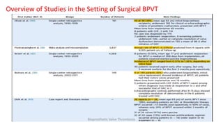
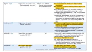
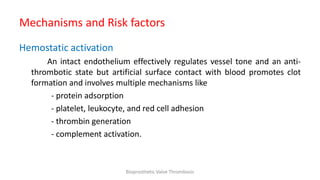
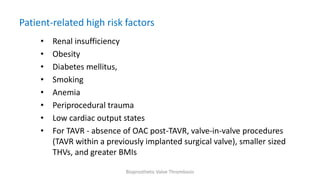
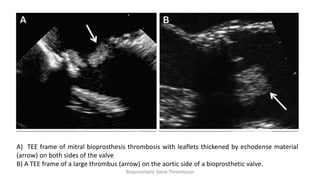
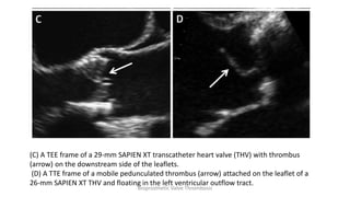
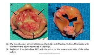
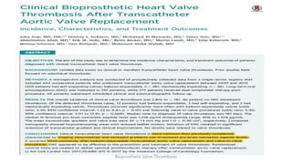
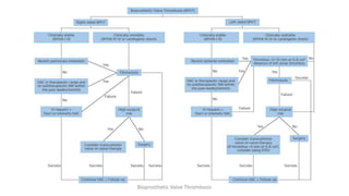
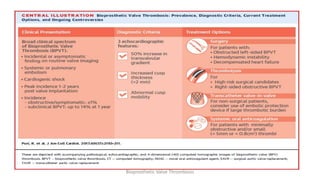
Bio Prosthetic Valve Thrombosis
Dr G. Raghu Kishore
Bioprosthetic Valve Thrombosis
Introduction
• Bioprosthetic valve (BPV) thrombosis is considered a relatively rare clinical entity in
the past.
• With advent of careful echo follow up, 4D CT diagnosis of sub clinical thrombosis
on both surgically or transcatheterly placed bioprosthetic valves(THV) has been
increasing.
• Debate continues among clinicians as to both the clinical relevance of subclinical
BPV thrombosis and the value of empirical oral anticoagulation following BPV
implantation.
• currently no systematic, prospective data exist regarding the optimal treatment
approach in THV recipients.
Bioprosthetic Valve Thrombosis
• Of the > 110,000 valve replacements undertaken annually within the United States
alone , there has been a gradual shift from mechanical to bioprosthetic valve (BPV)
implantations.
• Bioprostheses now account for nearly 80% of all surgical aortic valve replacements
(SAVRs) within the US
• This is because
a) changing demographics of valve recipients,
b) older, with high risk surgical profiles,
c) has greater bleeding risk because of the need for lifelong oral
anticoagulation (OAC) following valve replacement
Bioprosthetic Valve Thrombosis
• Although BPVs are less thrombogenic than their mechanical counterparts,
clinically apparent BPV thrombosis (BPVT) is a rare yet important clinical entity,
known to occur at all 4 valve locations.
• Its true incidence is probably greater than previously thought, and its clinical
relevance is likely to increase in conjunction with the rapidly expanding field of
THV technologies.
Bioprosthetic Valve Thrombosis
Types of BPV
• Human tissue valves - 2 categories –
- Allografts
- Autografts.
• Allografts are cadaveric valves that are cryopreserved in liquid nitrogen until use
and implanted without a stent.
• Auto grafts are composed of the patient’s own valve. The most common
autograft is the Ross procedure, during which the patient’s pulmonary valve is
transplanted to the aortic position, and an allograft valve is placed in the
pulmonary position.
• Clinical indications for human tissue valves are currently limited(i.e., women of
childbearing age, endocarditis).
Bioprosthetic Valve Thrombosis
Types of BPV
• Animal tissue valves – xenografts(Heterografts) - most widely used type of
BPV
Stented Vs stentless valves
Porcine Vs bovine.
• Valve leaflets are crosslinked with glutaraldehyde and mounted on a
metallic or polymer supporting stent.
• Pericardial valves consist of sheets of bovine pericardium mounted inside
or outside a supporting stent
Bioprosthetic Valve Thrombosis
• Stentless BPVs tend to be hemodynamically superior compared with stented BPVs.
• Sutureless valves have recently emerged as an option to reduce cross-clamp and
CBP duration, thereby improving surgical outcomes and facilitating minimally
invasive approaches.
• These valves seem also especially suitable for patients with small aortic annuli, to
optimize hemodynamic results
Bioprosthetic Valve Thrombosis
Types of BPVs
Bioprosthetic Valve Thrombosis
THVs
• Most THVs are stent-caged BPVs implanted using a catheter-based delivery system.
• First-generation devices have been the most widely used THVs in clinical practice
to date.
• The SAPIEN valve (Edwards Lifesciences, Irvine, California) and its current iterations
(SAPIEN XT and SAPIEN 3) consist of bovine pericardial leaflets sewed within a
balloon expandable cobalt-chromium stent, with the SAPIEN 3 THV being covered
by a polymer skirt at the bottom of the stent frame aimed to reduce paravalvular
leaks.
Bioprosthetic Valve Thrombosis
• THV leaflets are thinner than surgical BPV leaflets (0.25 mm vs. 0.40 mm
respectively)
• The CoreValve, CoreValve Evolut R and CoreValve Evolut PRO (Medtronic,
Minneapolis, Minnesota) are made of a self-expanding nitinol stent with porcine
pericardial leaflets
Bioprosthetic Valve Thrombosis
DEFINITION AND DIAGNOSIS OF BPVT
• The diagnosis of BPVT is challenging because of
- a lack of general awareness of this condition
- lack of a universal definition, and
- often confused with the term used as “valve deterioration’’
Valve thrombosis is considered any thrombosis unrelated to infection,
attached to or within close proximity of the valve per se, thus occluding the path
of blood flow, interfering with valve function, or sufficiently large so as to warrant
treatment
Bioprosthetic Valve Thrombosis
• Structural valve deterioration includes dysfunction or deterioration of the prosthetic
valve, exclusive of infection or thrombosis, as determined by reoperation, autopsy, or
clinical investigation.
• This usually involves wear and tear, fracture, calcification, leaflet tear, stent creep,
suture line disruption of valve components.
• Non structural dysfuntion relates to problems (excluding thrombus and infection)
indirectly involving valve components, while still resulting in a dysfunctional valve
(pannus, paravalvular leak, inappropriate sizing or positioning)
Bioprosthetic Valve Thrombosis
• In clinical practice, frequent confusion of these terms and definitions has invariably
led to underreporting of valve thrombosis.
• the majority of BPVTs have been reported in the aortic position, thus presenting a
major limitation in truly understanding data related to BPVT in other positions,
including those involving THVs.
• Egbe et al. demonstrated that the possibility of surgical BPVT was mentioned in
only 2 of 42 (5%) TTE and in 6 of 45 (13%) TEEs, although echo reports described
these BPVs as being “abnormal” in all instances
• Daniel et al. reported that if TTE is the first-line screening examination for
specifically detecting BPV abnormalities, it would identify BPVT in only 13% of
cases
Bioprosthetic Valve Thrombosis
Transesophageal echocardiography (TEE) is strongly
recommended when the clinical suspicion for BPV
abnormalities remains high, particularly for BPVs within
the mitral or tricuspid position
Bioprosthetic Valve Thrombosis
Incidence of BPVT
• The precise incidence of BPVT is difficult to ascertain, as most large prospective
registries failed to systematically report BPVTs
• In a recent meta-analysis involving SAVR recipients, early and late BPVT were only
reported in 3 and 13 of the 54 included studies, with rates of 0.34% and 0.04% per
year, respectively.
• Puvimanasinghe et al. also reported an extremely low 0.03% per year predicted rate
of BPVT using microsimulation models.
• Other investigators reported up to a 6% incidence of BPVT in retrospective analyses
of mitral valve recipients occurring at a mean followup of 12 months.
Bioprosthetic Valve Thrombosis
Overview of Studies in the Setting of Surgical BPVT
Bioprosthetic Valve Thrombosis
Mechanisms and Risk factors
Hemostatic activation
An intact endothelium effectively regulates vessel tone and an anti-
thrombotic state but artificial surface contact with blood promotes clot
formation and involves multiple mechanisms like
- protein adsorption
- platelet, leukocyte, and red cell adhesion
- thrombin generation
- complement activation.
Bioprosthetic Valve Thrombosis
Perturbations in blood flow
• Flow conditions around valve prostheses are associated with endothelial damage,
vascular remodeling, and thrombosis.
• High wall shear stress levels promote platelet activation, combined with flow
recirculation, stimulate localized thrombogenesis, within the BPV region and there
is a complex interplay between flow patterns and blood stasis
• Moderate or severe PPM occurs when the effective aortic valve area of a normal
functioning prosthetic valve in up to 44% of SAVR recipients.
• Wall shear rate, was significantly higher in those TAVR pts demonstrating PPM
compared with those without PPM although a direct link between PPM and BPVT
has yet to be elucidated,
Bioprosthetic Valve Thrombosis
Patient-related high risk factors
• Renal insufficiency
• Obesity
• Diabetes mellitus,
• Smoking
• Anemia
• Periprocedural trauma
• Low cardiac output states
• For TAVR - absence of OAC post-TAVR, valve-in-valve procedures
(TAVR within a previously implanted surgical valve), smaller sized
THVs, and greater BMIs
Bioprosthetic Valve Thrombosis
Clinical presentation
• The spectrum of BPVT can range from an incidental finding in
asymptomatic patients to overt syncope, acute HF, and , in its most severe
form, cardiogenic shock due to mechanical obstruction.
• Progressive dyspnea is the most common clinical presentation
• In some cases, patients may present with systemic or pulmonary
embolism from friable valve-associated thrombi owing to a process of
delayed thrombus organization
Bioprosthetic Valve Thrombosis
When to suspect BPV thrombosis…?
• Occurrence within 1–2 yrs of BPV implantation
• Presence of risk factors: paroxysmal AF, sub therapeutic INR, recent
withdrawal of OAC, history of TE event, depressed LVEF, known
hypercoagulable condition.
• New-onset acute HF, progressively worsening dyspnea
• New thromboembolic event
• Regression of HF symptoms with OAC
Bioprosthetic Valve Thrombosis
Echocardiographic features
• Direct visualization of valve thrombosis in rare cases
• 50% mean gradient increase compared with the initial post-operative evaluation
• Increased cusp thickness (>2 mm), especially on the downstream aspect of the
BPV (ventricular side for mitral and tricuspid prostheses, arterial side for aortic and
pulmonary prostheses)
• Abnormal/reduced leaflet mobility
• Regression of BPV abnormalities with OAC, usually within 1–3 months of OAC
initiation
Bioprosthetic Valve Thrombosis
A) TEE frame of mitral bioprosthesis thrombosis with leaflets thickened by echodense material
(arrow) on both sides of the valve
B) A TEE frame of a large thrombus (arrow) on the aortic side of a bioprosthetic valve.
Bioprosthetic Valve Thrombosis
(C) A TEE frame of a 29-mm SAPIEN XT transcatheter heart valve (THV) with thrombus
(arrow) on the downstream side of the leaflets.
(D) A TTE frame of a mobile pedunculated thrombus (arrow) attached on the leaflet of a
26-mm SAPIEN XT THV and floating in the left ventricular outflow tract.
Bioprosthetic Valve Thrombosis
(A) BPV thrombosis of a 25-mm Bicor prosthesis (St. Jude Medical, St. Paul, Minnesota) with
thrombi on the downstream side of the cusps.
(B) Explanted Sorin Mitraflow BPV with thrombus on the downstream side of the valve
leaflets
Bioprosthetic Valve Thrombosis
• Egbe et al. proposed a diagnostic model combining 3 echocardiographic predictors:
1) a 50% increase in the transvalvular gradient compared with baseline within 5
years of surgery, in the absence of a high cardiac output state
2) increased cusp thickness (>2 mm), especially on the downstream aspect of the
valve
3) abnormal cusp mobility.
• Applying this simple model to the 138 patients included in their study yielded rates of
72%, 90%, 78%, and 87% for sensitivity, specificity, and positive and negative predicted
values for diagnosing BPVT, respectively
Bioprosthetic Valve Thrombosis
CT features
• Reduced leaflet motion (RLM) on 4D CT
• HALT (hypoattenuated leaflet thickening)
Plays most important role in diagnosis of sub clinical BPV thrombosis
early in the course
Bioprosthetic Valve Thrombosis
Core Valve
Portico valve
Arrows depict hypoattenuating opacities during diastole and systole.
Leaflets with reduced motion aredepicted by wedge-shaped or semilunar opacities during the cardiac cycleBioprosthetic Valve Thrombosis
Studies of post SAVR/TAVR patients followed by 4D CT
Bioprosthetic Valve Thrombosis
THV Thrombosis
• The incidence of THV thrombosis is likely to be at least 0.6%, possibly more.
• Most cases occurred within the first year post–transcatheter aortic valve
replacement (TAVR), with a median onset of 6 months post-TAVR.
• The majority of patients with THV thrombosis (65%) presented with gradual onset
of dyspnea, with no reported or published cases to date of overt systemic
embolism.
• Nearly one-third of patients were asymptomatic.
• Direct echocardiographic visualization of thrombus was possible in only a minority
of cases, with almost all patients demonstrating a rising transaortic gradient from
baseline (>90% presented with a mean transaortic gradient >20 mm Hg).
Bioprosthetic Valve Thrombosis
• Morphological features demonstrable on echocardiography related to leaflet
thickening and reduced leaflet mobility.
• A preponderance of reported cases occurred following balloon expanding valve
implantation, with a large majority of THV thrombosis cases occurring despite the
use of DAPT.
• However, the prompt and empirical commencement of OAC was nearly universal
in restoring transaortic gradients to close to their baseline values, following a
median period of 40 days post-OAC commencement.
Bioprosthetic Valve Thrombosis
Treatment of BPVT
Treatment strategy for BPVT depends on
1. Mode of clinical presentation
2. Patient’s hemodynamic status
3. Presence or absence of BPV obstruction
4. Valve location
Bioprosthetic Valve Thrombosis
Conventional treatment options
– Surgery
– Fibrinolysis
– Anticoagulation
Bioprosthetic Valve Thrombosis
• Nonobstructive left-sided BPVTs
• Large (>5 mm)
• Mobile
• Pedunculated thrombi
• Intravenous heparin fails
For small (<5 mm) thrombi  MEDICAL THERAPY
SURGERY
Bioprosthetic Valve Thrombosis
Fibrinolysis
– Second line therapy reserved for patients with surgical contra-
indications
– acceptable for obstructed tricuspid or pulmonary BPVs
Bioprosthetic Valve Thrombosis
THV thrombosis
• THV thrombosis has been usually managed with OAC therapy, with significant
clinical and hemodynamic improvement and normalization of transvalvular
gradients following 2 months of OAC therapy in most of the cases.
• The duration of OAC following BPVT remains unclear and should be determined on
an individual basis, taking into consideration the risks for bleeding events versus
recurrent valve thrombosis.
• Long-term OAC therapy may be the preferred strategy, but in the event of OAC
cessation, antiplatelet therapy and frequent echocardiographic surveillance should
probably be recommended.
Bioprosthetic Valve Thrombosis
Prevention of BPVT
Post-procedural thromboprophylaxis
• Following surgical BPV implantation, 3 months of OAC is recommended
(irrespective of valve position), during which time appropriate endothelialization
of the BPV surface generally occurs.
• Following the initial 3-month period post-SAVR, guidelines tend to recommend
the indefinite continuation of at least aspirin.
• However, with respect to aortic BPVs, treatment guidelines remain inconclusive.
Bioprosthetic Valve Thrombosis
• STS Adult Cardiac Surgery Database, from > 25,600 patients > 65 yrs in 800 U.S.
hospitals, Brennan et al. showed lower death and embolic events in those
prescribed both aspirin and warfarin within the first 3 months post-SAVR (with
BPVs) compared with aspirin only, with the cost of an added bleeding risk.
• Interestingly, an aspirin-only versus warfarin only strategy was equally efficacious
in terms of reducing mortality and TE complications, outlining the synergistic
benefit of inhibiting platelets, as well as the coagulation cascade
• A similar benefit of warfarin was demonstrated within the first 3 months post–
surgical BPV implantation within a Danish national registry
Bioprosthetic Valve Thrombosis
• In a recent meta-analysis, Riaz et al. included 13 studies comprising 6,431 cases
versus 18,210 controls found that warfarin use post-SAVR using a BPV associated
with a significantly elevated the risk for bleeding (OR: 1.96; 95% CI: 1.25 to 3.08; p
<0.0001) compared with aspirin or placebo.
• With regard to composite primary outcome variables (risk for VTE, stroke, or TIA)
at 3 months, no significant difference with warfarin (OR: 1.13; 95% CI: 0.82 to
1.56; p = 0.67), and OAC use was not shown to improve clinical outcomes beyond
3 months post-SAVR (OR: 1.12; 95% CI: 0.80 to 1.58; p ¼ 0.79).
Bioprosthetic Valve Thrombosis
Peri- and post-procedural thromboprophylaxis in TAVR
• Periprocedurally, intravenous heparin anticoagulation 100 units/kg followed by
additional boluses to maintain an ACT of >250 s is the most common regime
• BRAVO-3 (Effect of Bivalirudin on Aortic Valve Intervention Outcomes 3) trial
randomizing more than 800 TAVR recipients to either bivalirudin or heparin, no
significant differences were observed in in-hospital major bleeding rates (6.9% vs.
9.0%, p = 0.27, respectively) or 30-day net adverse CV events(14.4% vs. 16.1%, p =
0.35)
Bioprosthetic Valve Thrombosis
• Unlike during primary PCI, these data likely suggest no added benefit of direct
thrombin inhibition over heparin for lowering bleeding rates during TAVR.
• Current U.S. guidelines recommend indefinite aspirin therapy and 3 to 6 months of
DAPT post-TAVR with aspirin and clopidogrel, whereas Canadian guidelines differ
slightly by recommending post-TAVR DAPT for 1 to 3 months
Bioprosthetic Valve Thrombosis
Current Antithrombotic Therapeutic Recommendations for BPV Implantation
Bioprosthetic Valve Thrombosis
Conclusion
• The advent of TAVR, and recent findings of subclinical leaflet thrombosis in both
transcatheter and surgically implanted BPVs, importance of BPV thrombosis is
increasing.
• Although symptomatic BPVT is considered a relatively rare phenomenon,
accumulating evidence suggests that this phenomenon may be more prevalent
than previously thought, particularly with THVs.
• Clinicians should have a heightened clinical awareness of this phenomenon,
especially in BPV recipients who present with dyspnea and/or rising trans-
prosthetic gradients at any time post implantation.
Bioprosthetic Valve Thrombosis
• This should prompt echocardiographic (and possibly computed tomographic)
imaging to evaluate bioprosthetic leaflet morphology and motion.
• In the absence of clinically overt BPV obstruction, OAC therapy is recommended,
with follow-up echocardiography scheduled 1 to 2 months following treatment
initiation.
• Although exquisitely sensitive in detecting leaflet anomalies, the role of routine 4D
CT post-BPV insertion remains uncertain until further research identifies clinical
utility.
• However, CT could play an important role in the diagnosis of BPVT in symptomatic
patients or those presenting with rising transprosthetic gradients.
Bioprosthetic Valve Thrombosis
• The role of routine OAC following surgical BPV implantation within the aortic
position remains unclear.
• Nevertheless, until prospective studies are undertaken, clinical guidelines
suggesting a limited period (1 to 3 months) of OAC with or without aspirin should
probably be adhered to.
• Following TAVR, a period of DAPT is also advisable. Thereafter, the precise long
term antithrombotic regimen should be dictated by patient-specific co morbidities
relating to both thromboembolic and bleeding risk.
Bioprosthetic Valve Thrombosis
• Future studies are required to better elucidate THV durability and identify novel
risk factors for BPVT, such as PPM, smaller-sized THVs, newer iteration trans
catheter procedures (i.e., valve-in-valve procedures), and THVs implanted in non
aortic positions.
• A range of clinical trials are currently under way that will be pivotal in informing
us of the role of novel OACs following TAVR.
Bioprosthetic Valve Thrombosis
THANK YOU
Bioprosthetic Valve Thrombosis

Bioprosthetic valve thrombosis

  • 1.
    Bio Prosthetic ValveThrombosis Dr G. Raghu Kishore Bioprosthetic Valve Thrombosis
  • 2.
    Introduction • Bioprosthetic valve(BPV) thrombosis is considered a relatively rare clinical entity in the past. • With advent of careful echo follow up, 4D CT diagnosis of sub clinical thrombosis on both surgically or transcatheterly placed bioprosthetic valves(THV) has been increasing. • Debate continues among clinicians as to both the clinical relevance of subclinical BPV thrombosis and the value of empirical oral anticoagulation following BPV implantation. • currently no systematic, prospective data exist regarding the optimal treatment approach in THV recipients. Bioprosthetic Valve Thrombosis
  • 3.
    • Of the> 110,000 valve replacements undertaken annually within the United States alone , there has been a gradual shift from mechanical to bioprosthetic valve (BPV) implantations. • Bioprostheses now account for nearly 80% of all surgical aortic valve replacements (SAVRs) within the US • This is because a) changing demographics of valve recipients, b) older, with high risk surgical profiles, c) has greater bleeding risk because of the need for lifelong oral anticoagulation (OAC) following valve replacement Bioprosthetic Valve Thrombosis
  • 4.
    • Although BPVsare less thrombogenic than their mechanical counterparts, clinically apparent BPV thrombosis (BPVT) is a rare yet important clinical entity, known to occur at all 4 valve locations. • Its true incidence is probably greater than previously thought, and its clinical relevance is likely to increase in conjunction with the rapidly expanding field of THV technologies. Bioprosthetic Valve Thrombosis
  • 5.
    Types of BPV •Human tissue valves - 2 categories – - Allografts - Autografts. • Allografts are cadaveric valves that are cryopreserved in liquid nitrogen until use and implanted without a stent. • Auto grafts are composed of the patient’s own valve. The most common autograft is the Ross procedure, during which the patient’s pulmonary valve is transplanted to the aortic position, and an allograft valve is placed in the pulmonary position. • Clinical indications for human tissue valves are currently limited(i.e., women of childbearing age, endocarditis). Bioprosthetic Valve Thrombosis
  • 6.
    Types of BPV •Animal tissue valves – xenografts(Heterografts) - most widely used type of BPV Stented Vs stentless valves Porcine Vs bovine. • Valve leaflets are crosslinked with glutaraldehyde and mounted on a metallic or polymer supporting stent. • Pericardial valves consist of sheets of bovine pericardium mounted inside or outside a supporting stent Bioprosthetic Valve Thrombosis
  • 7.
    • Stentless BPVstend to be hemodynamically superior compared with stented BPVs. • Sutureless valves have recently emerged as an option to reduce cross-clamp and CBP duration, thereby improving surgical outcomes and facilitating minimally invasive approaches. • These valves seem also especially suitable for patients with small aortic annuli, to optimize hemodynamic results Bioprosthetic Valve Thrombosis
  • 8.
  • 9.
  • 10.
  • 11.
    THVs • Most THVsare stent-caged BPVs implanted using a catheter-based delivery system. • First-generation devices have been the most widely used THVs in clinical practice to date. • The SAPIEN valve (Edwards Lifesciences, Irvine, California) and its current iterations (SAPIEN XT and SAPIEN 3) consist of bovine pericardial leaflets sewed within a balloon expandable cobalt-chromium stent, with the SAPIEN 3 THV being covered by a polymer skirt at the bottom of the stent frame aimed to reduce paravalvular leaks. Bioprosthetic Valve Thrombosis
  • 12.
    • THV leafletsare thinner than surgical BPV leaflets (0.25 mm vs. 0.40 mm respectively) • The CoreValve, CoreValve Evolut R and CoreValve Evolut PRO (Medtronic, Minneapolis, Minnesota) are made of a self-expanding nitinol stent with porcine pericardial leaflets Bioprosthetic Valve Thrombosis
  • 13.
    DEFINITION AND DIAGNOSISOF BPVT • The diagnosis of BPVT is challenging because of - a lack of general awareness of this condition - lack of a universal definition, and - often confused with the term used as “valve deterioration’’ Valve thrombosis is considered any thrombosis unrelated to infection, attached to or within close proximity of the valve per se, thus occluding the path of blood flow, interfering with valve function, or sufficiently large so as to warrant treatment Bioprosthetic Valve Thrombosis
  • 14.
    • Structural valvedeterioration includes dysfunction or deterioration of the prosthetic valve, exclusive of infection or thrombosis, as determined by reoperation, autopsy, or clinical investigation. • This usually involves wear and tear, fracture, calcification, leaflet tear, stent creep, suture line disruption of valve components. • Non structural dysfuntion relates to problems (excluding thrombus and infection) indirectly involving valve components, while still resulting in a dysfunctional valve (pannus, paravalvular leak, inappropriate sizing or positioning) Bioprosthetic Valve Thrombosis
  • 15.
    • In clinicalpractice, frequent confusion of these terms and definitions has invariably led to underreporting of valve thrombosis. • the majority of BPVTs have been reported in the aortic position, thus presenting a major limitation in truly understanding data related to BPVT in other positions, including those involving THVs. • Egbe et al. demonstrated that the possibility of surgical BPVT was mentioned in only 2 of 42 (5%) TTE and in 6 of 45 (13%) TEEs, although echo reports described these BPVs as being “abnormal” in all instances • Daniel et al. reported that if TTE is the first-line screening examination for specifically detecting BPV abnormalities, it would identify BPVT in only 13% of cases Bioprosthetic Valve Thrombosis
  • 16.
    Transesophageal echocardiography (TEE)is strongly recommended when the clinical suspicion for BPV abnormalities remains high, particularly for BPVs within the mitral or tricuspid position Bioprosthetic Valve Thrombosis
  • 17.
    Incidence of BPVT •The precise incidence of BPVT is difficult to ascertain, as most large prospective registries failed to systematically report BPVTs • In a recent meta-analysis involving SAVR recipients, early and late BPVT were only reported in 3 and 13 of the 54 included studies, with rates of 0.34% and 0.04% per year, respectively. • Puvimanasinghe et al. also reported an extremely low 0.03% per year predicted rate of BPVT using microsimulation models. • Other investigators reported up to a 6% incidence of BPVT in retrospective analyses of mitral valve recipients occurring at a mean followup of 12 months. Bioprosthetic Valve Thrombosis
  • 18.
    Overview of Studiesin the Setting of Surgical BPVT Bioprosthetic Valve Thrombosis
  • 19.
  • 20.
    Mechanisms and Riskfactors Hemostatic activation An intact endothelium effectively regulates vessel tone and an anti- thrombotic state but artificial surface contact with blood promotes clot formation and involves multiple mechanisms like - protein adsorption - platelet, leukocyte, and red cell adhesion - thrombin generation - complement activation. Bioprosthetic Valve Thrombosis
  • 21.
    Perturbations in bloodflow • Flow conditions around valve prostheses are associated with endothelial damage, vascular remodeling, and thrombosis. • High wall shear stress levels promote platelet activation, combined with flow recirculation, stimulate localized thrombogenesis, within the BPV region and there is a complex interplay between flow patterns and blood stasis • Moderate or severe PPM occurs when the effective aortic valve area of a normal functioning prosthetic valve in up to 44% of SAVR recipients. • Wall shear rate, was significantly higher in those TAVR pts demonstrating PPM compared with those without PPM although a direct link between PPM and BPVT has yet to be elucidated, Bioprosthetic Valve Thrombosis
  • 22.
    Patient-related high riskfactors • Renal insufficiency • Obesity • Diabetes mellitus, • Smoking • Anemia • Periprocedural trauma • Low cardiac output states • For TAVR - absence of OAC post-TAVR, valve-in-valve procedures (TAVR within a previously implanted surgical valve), smaller sized THVs, and greater BMIs Bioprosthetic Valve Thrombosis
  • 23.
    Clinical presentation • Thespectrum of BPVT can range from an incidental finding in asymptomatic patients to overt syncope, acute HF, and , in its most severe form, cardiogenic shock due to mechanical obstruction. • Progressive dyspnea is the most common clinical presentation • In some cases, patients may present with systemic or pulmonary embolism from friable valve-associated thrombi owing to a process of delayed thrombus organization Bioprosthetic Valve Thrombosis
  • 24.
    When to suspectBPV thrombosis…? • Occurrence within 1–2 yrs of BPV implantation • Presence of risk factors: paroxysmal AF, sub therapeutic INR, recent withdrawal of OAC, history of TE event, depressed LVEF, known hypercoagulable condition. • New-onset acute HF, progressively worsening dyspnea • New thromboembolic event • Regression of HF symptoms with OAC Bioprosthetic Valve Thrombosis
  • 25.
    Echocardiographic features • Directvisualization of valve thrombosis in rare cases • 50% mean gradient increase compared with the initial post-operative evaluation • Increased cusp thickness (>2 mm), especially on the downstream aspect of the BPV (ventricular side for mitral and tricuspid prostheses, arterial side for aortic and pulmonary prostheses) • Abnormal/reduced leaflet mobility • Regression of BPV abnormalities with OAC, usually within 1–3 months of OAC initiation Bioprosthetic Valve Thrombosis
  • 26.
    A) TEE frameof mitral bioprosthesis thrombosis with leaflets thickened by echodense material (arrow) on both sides of the valve B) A TEE frame of a large thrombus (arrow) on the aortic side of a bioprosthetic valve. Bioprosthetic Valve Thrombosis
  • 27.
    (C) A TEEframe of a 29-mm SAPIEN XT transcatheter heart valve (THV) with thrombus (arrow) on the downstream side of the leaflets. (D) A TTE frame of a mobile pedunculated thrombus (arrow) attached on the leaflet of a 26-mm SAPIEN XT THV and floating in the left ventricular outflow tract. Bioprosthetic Valve Thrombosis
  • 28.
    (A) BPV thrombosisof a 25-mm Bicor prosthesis (St. Jude Medical, St. Paul, Minnesota) with thrombi on the downstream side of the cusps. (B) Explanted Sorin Mitraflow BPV with thrombus on the downstream side of the valve leaflets Bioprosthetic Valve Thrombosis
  • 29.
    • Egbe etal. proposed a diagnostic model combining 3 echocardiographic predictors: 1) a 50% increase in the transvalvular gradient compared with baseline within 5 years of surgery, in the absence of a high cardiac output state 2) increased cusp thickness (>2 mm), especially on the downstream aspect of the valve 3) abnormal cusp mobility. • Applying this simple model to the 138 patients included in their study yielded rates of 72%, 90%, 78%, and 87% for sensitivity, specificity, and positive and negative predicted values for diagnosing BPVT, respectively Bioprosthetic Valve Thrombosis
  • 30.
  • 31.
  • 32.
  • 33.
    CT features • Reducedleaflet motion (RLM) on 4D CT • HALT (hypoattenuated leaflet thickening) Plays most important role in diagnosis of sub clinical BPV thrombosis early in the course Bioprosthetic Valve Thrombosis
  • 34.
    Core Valve Portico valve Arrowsdepict hypoattenuating opacities during diastole and systole. Leaflets with reduced motion aredepicted by wedge-shaped or semilunar opacities during the cardiac cycleBioprosthetic Valve Thrombosis
  • 35.
    Studies of postSAVR/TAVR patients followed by 4D CT Bioprosthetic Valve Thrombosis
  • 36.
    THV Thrombosis • Theincidence of THV thrombosis is likely to be at least 0.6%, possibly more. • Most cases occurred within the first year post–transcatheter aortic valve replacement (TAVR), with a median onset of 6 months post-TAVR. • The majority of patients with THV thrombosis (65%) presented with gradual onset of dyspnea, with no reported or published cases to date of overt systemic embolism. • Nearly one-third of patients were asymptomatic. • Direct echocardiographic visualization of thrombus was possible in only a minority of cases, with almost all patients demonstrating a rising transaortic gradient from baseline (>90% presented with a mean transaortic gradient >20 mm Hg). Bioprosthetic Valve Thrombosis
  • 37.
    • Morphological featuresdemonstrable on echocardiography related to leaflet thickening and reduced leaflet mobility. • A preponderance of reported cases occurred following balloon expanding valve implantation, with a large majority of THV thrombosis cases occurring despite the use of DAPT. • However, the prompt and empirical commencement of OAC was nearly universal in restoring transaortic gradients to close to their baseline values, following a median period of 40 days post-OAC commencement. Bioprosthetic Valve Thrombosis
  • 38.
  • 39.
    Treatment of BPVT Treatmentstrategy for BPVT depends on 1. Mode of clinical presentation 2. Patient’s hemodynamic status 3. Presence or absence of BPV obstruction 4. Valve location Bioprosthetic Valve Thrombosis
  • 40.
    Conventional treatment options –Surgery – Fibrinolysis – Anticoagulation Bioprosthetic Valve Thrombosis
  • 41.
    • Nonobstructive left-sidedBPVTs • Large (>5 mm) • Mobile • Pedunculated thrombi • Intravenous heparin fails For small (<5 mm) thrombi  MEDICAL THERAPY SURGERY Bioprosthetic Valve Thrombosis
  • 42.
    Fibrinolysis – Second linetherapy reserved for patients with surgical contra- indications – acceptable for obstructed tricuspid or pulmonary BPVs Bioprosthetic Valve Thrombosis
  • 43.
  • 44.
    THV thrombosis • THVthrombosis has been usually managed with OAC therapy, with significant clinical and hemodynamic improvement and normalization of transvalvular gradients following 2 months of OAC therapy in most of the cases. • The duration of OAC following BPVT remains unclear and should be determined on an individual basis, taking into consideration the risks for bleeding events versus recurrent valve thrombosis. • Long-term OAC therapy may be the preferred strategy, but in the event of OAC cessation, antiplatelet therapy and frequent echocardiographic surveillance should probably be recommended. Bioprosthetic Valve Thrombosis
  • 45.
    Prevention of BPVT Post-proceduralthromboprophylaxis • Following surgical BPV implantation, 3 months of OAC is recommended (irrespective of valve position), during which time appropriate endothelialization of the BPV surface generally occurs. • Following the initial 3-month period post-SAVR, guidelines tend to recommend the indefinite continuation of at least aspirin. • However, with respect to aortic BPVs, treatment guidelines remain inconclusive. Bioprosthetic Valve Thrombosis
  • 46.
    • STS AdultCardiac Surgery Database, from > 25,600 patients > 65 yrs in 800 U.S. hospitals, Brennan et al. showed lower death and embolic events in those prescribed both aspirin and warfarin within the first 3 months post-SAVR (with BPVs) compared with aspirin only, with the cost of an added bleeding risk. • Interestingly, an aspirin-only versus warfarin only strategy was equally efficacious in terms of reducing mortality and TE complications, outlining the synergistic benefit of inhibiting platelets, as well as the coagulation cascade • A similar benefit of warfarin was demonstrated within the first 3 months post– surgical BPV implantation within a Danish national registry Bioprosthetic Valve Thrombosis
  • 47.
    • In arecent meta-analysis, Riaz et al. included 13 studies comprising 6,431 cases versus 18,210 controls found that warfarin use post-SAVR using a BPV associated with a significantly elevated the risk for bleeding (OR: 1.96; 95% CI: 1.25 to 3.08; p <0.0001) compared with aspirin or placebo. • With regard to composite primary outcome variables (risk for VTE, stroke, or TIA) at 3 months, no significant difference with warfarin (OR: 1.13; 95% CI: 0.82 to 1.56; p = 0.67), and OAC use was not shown to improve clinical outcomes beyond 3 months post-SAVR (OR: 1.12; 95% CI: 0.80 to 1.58; p ¼ 0.79). Bioprosthetic Valve Thrombosis
  • 48.
    Peri- and post-proceduralthromboprophylaxis in TAVR • Periprocedurally, intravenous heparin anticoagulation 100 units/kg followed by additional boluses to maintain an ACT of >250 s is the most common regime • BRAVO-3 (Effect of Bivalirudin on Aortic Valve Intervention Outcomes 3) trial randomizing more than 800 TAVR recipients to either bivalirudin or heparin, no significant differences were observed in in-hospital major bleeding rates (6.9% vs. 9.0%, p = 0.27, respectively) or 30-day net adverse CV events(14.4% vs. 16.1%, p = 0.35) Bioprosthetic Valve Thrombosis
  • 49.
    • Unlike duringprimary PCI, these data likely suggest no added benefit of direct thrombin inhibition over heparin for lowering bleeding rates during TAVR. • Current U.S. guidelines recommend indefinite aspirin therapy and 3 to 6 months of DAPT post-TAVR with aspirin and clopidogrel, whereas Canadian guidelines differ slightly by recommending post-TAVR DAPT for 1 to 3 months Bioprosthetic Valve Thrombosis
  • 50.
    Current Antithrombotic TherapeuticRecommendations for BPV Implantation Bioprosthetic Valve Thrombosis
  • 51.
  • 52.
  • 53.
  • 54.
    Conclusion • The adventof TAVR, and recent findings of subclinical leaflet thrombosis in both transcatheter and surgically implanted BPVs, importance of BPV thrombosis is increasing. • Although symptomatic BPVT is considered a relatively rare phenomenon, accumulating evidence suggests that this phenomenon may be more prevalent than previously thought, particularly with THVs. • Clinicians should have a heightened clinical awareness of this phenomenon, especially in BPV recipients who present with dyspnea and/or rising trans- prosthetic gradients at any time post implantation. Bioprosthetic Valve Thrombosis
  • 55.
    • This shouldprompt echocardiographic (and possibly computed tomographic) imaging to evaluate bioprosthetic leaflet morphology and motion. • In the absence of clinically overt BPV obstruction, OAC therapy is recommended, with follow-up echocardiography scheduled 1 to 2 months following treatment initiation. • Although exquisitely sensitive in detecting leaflet anomalies, the role of routine 4D CT post-BPV insertion remains uncertain until further research identifies clinical utility. • However, CT could play an important role in the diagnosis of BPVT in symptomatic patients or those presenting with rising transprosthetic gradients. Bioprosthetic Valve Thrombosis
  • 56.
    • The roleof routine OAC following surgical BPV implantation within the aortic position remains unclear. • Nevertheless, until prospective studies are undertaken, clinical guidelines suggesting a limited period (1 to 3 months) of OAC with or without aspirin should probably be adhered to. • Following TAVR, a period of DAPT is also advisable. Thereafter, the precise long term antithrombotic regimen should be dictated by patient-specific co morbidities relating to both thromboembolic and bleeding risk. Bioprosthetic Valve Thrombosis
  • 57.
    • Future studiesare required to better elucidate THV durability and identify novel risk factors for BPVT, such as PPM, smaller-sized THVs, newer iteration trans catheter procedures (i.e., valve-in-valve procedures), and THVs implanted in non aortic positions. • A range of clinical trials are currently under way that will be pivotal in informing us of the role of novel OACs following TAVR. Bioprosthetic Valve Thrombosis
  • 58.