
Recommended
More Related Content
Similar to anatomy of the disc in spine anatomy complete
Similar to anatomy of the disc in spine anatomy complete (20)
Recently uploaded
Recently uploaded (20)
anatomy of the disc in spine anatomy complete
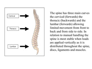
- 1. The spine has three main curves the cervical (forwards) the thoracic (backwards) and the lumbar (forwards) allowing limited movement from front to back and from side to side. In relation to manual handling the spine is most stable when loads are applied vertically as it is distributed throughout the spine, discs, ligaments and muscles.
- 2. The spine contains 24 individual vertebra (7 cervical, 12 thoracic, 5 lumbar) and nine fused to form the sacrum and coccyx (5 and 4 respectively).
- 3. The vertebrae are separated by a fibroelastic structure called the intervertebral disc. It acts to absorb loads transmitted through the bones, joints and ligaments. It contains an outer ring called the annulus fibrosis, much like a radial tire, and an inner core called the nucleus pulpolsus. The disc has no blood supply and therefore normally degenerates and dries out with time and aging. Like dried rubber the annulus can crack with flexion, and shear forces.
- 4. The vertebra are supported in position by ligaments and a pair of small apophyseal joints that slide together and protect the spine from excessive flexion and rotation. Strength is further given by muscles of the back, trunk and abdomen.
- 5. The vertebra are interspersed with intervertebral discs. Each of these discs which act as shock absorbers and allow flexibility. The discs contain a capsule of thick fluid called the nucleus pulposus is the translucent gel in the central area and is surrounded by a tough fibrous layer called the annulus fibrosus.
- 6. As the outer annulus dries out with age it can crack and let the inner nucleus pulposus extend outward. Note the disc is thinner in the posterior or back region. This is the area of most disc failures. When an individual bends forward the inner nucleus pulposus exerts pressure backwads. Therefore bending at the waist is a dangerous move for the aging disc. Most disc injuries involve bending or flexion at the waist.
- 7. Different phases of disc injury. Note that increased injury occurs with disruption, and tearing of the outer annulus fibrosis. The extent of injury can be consistent with extension of Nucleus pulposis.
- 8. As the outer annulus fails, the inner nucleus pulposis can migrate posteriorly. As the disc ages it can become easier to get cracks in the outer annulus weakening the wall of the disc. When the inner nucleus pulposis is exposed to the environment of the body it can be resorbed. Therefore disc herniations in the absence of neurologic deficit can be treated conservatively by Chiropractic manipulation.
- 11. MRI demonstrating a herniated disc in the cervical spine, or neck. Note the appearance of the normal discs. Can you identify the herniated disc based on the review of the previous slides?
- 13. Picture of a hernaited nucleus pulposis surgically removed.