Downloaded 764 times


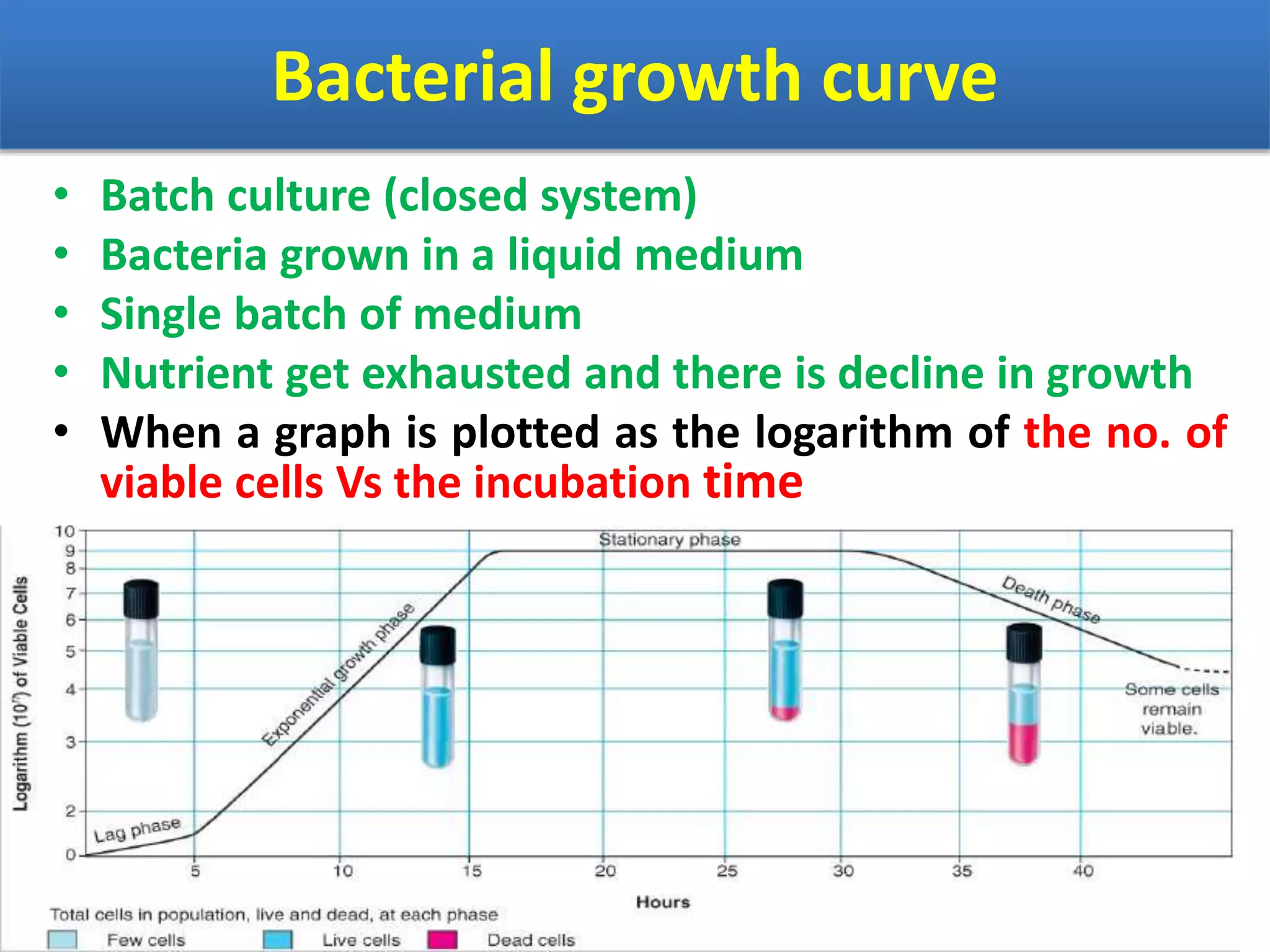


Microbial growth involves an increase in cell size or population numbers through cell division and reproduction. There are two levels of growth - increasing cell size through synthesis of new components, and increasing population size through cell division. Microbiologists study population growth curves, which typically have four phases: lag phase as cells adapt, exponential or log phase of rapid growth, stationary phase as resources are depleted, and death phase. Growth is measured by counting cell numbers directly under a microscope or using counting chambers, or indirectly by culturing cells on agar plates and counting colonies. Environmental factors like nutrients, oxygen, pH, and temperature affect microbial growth rates.