Name : SNEHASISADHIKARY
Student Code : BWU/BPA/23/018
Program Name : B.Sc. In Physician Assistant
Semester /Year : 4th
Sem – 2nd
Year
Department : Department of Allied Health Sciences
Course Name : Basic Radiology and ImagingTechnology
Course Code : BPAS403
Rgistration No. : 23013000477 of 2023-2024
Roll No. : 23010313017
Introduction to Ultrasonography :
Ultrasonic Waves and Imaging Principles
2.
Sound Waves
Soundwaves are mechanical
Require a medium for transmission
Sound is formed by the vibration or movement of
air or liquid.
When a sound is made the vibrations make the
surrounding particles in the air or liquid vibrate.
The sound makes the molecules compress and
expand bump into each other.
Sound waves with frequencies above the range of
human hearing i.e. 20Hz to 20KHz.
3.
Sound wavesand ultrasound waves follow the rules of
propagation and reflection similar to those that govern light
waves in that they can be
Reflection – to throw back the sound wave at the interface of two
materials.
Refraction – to bend the sound wave at the interface of two
materials , i.e. the waves are transmitted.
Absorption – attnuation of a sound wave by “relaxation” or
frictional processes in the medium that convert acoustic energy to
heat, i.e. some portion of the waves is not transmitted.
Speed of Sound :
Depends upon the compressibility of material.
Speed is low in air (331 m/s) < higher in soft tissue (1540m/s) <
highest in the bone (3360 m/s)
4.
Ultrasound Frequency
Thewavelength of ultrasound for diagnostic purposes should
be on the order of 1 mm or less.
The wavelength of sound decreases as frequently (MHz)
increases.
Diagnostic imaging typically uses frequencies between
20KHz to 1MHz.
5.
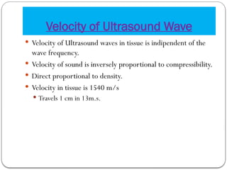
Velocity of UltrasoundWave
Velocity of Ultrasound waves in tissue is indipendent of the
wave frequency.
Velocity of sound is inversely proportional to compressibility.
Direct proportional to density.
Velocity in tissue is 1540 m/s
Travels 1 cm in 13m.s.
6.
Formation of UltrasoundWave
Medical ultrasound waves are generated by electrically
vibrating a transducer.
Like piston in a water
The wave length of ultrasound wave is distance between two
bands of compression and rarefaction.
7.
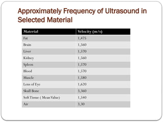
Approximately Frequency ofUltrasound in
Selected Material
Material Velocity (m/s)
Fat 1,475
Brain 1,560
Liver 1,570
Kidney 1,560
Spleen 1,570
Blood 1,570
Muscle 1,580
Lens of Eye 1,620
Skull Bone 3,360
SoftTissue ( MeanValue) 1,540
Air 3,30
8.
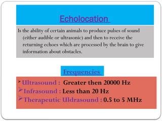
Echolocation
Is the abilityof certain animals to produce pulses of sound
(either audible or ultrasonic) and then to receive the
returning echoes which are processed by the brain to give
information about obstacles.
Frequencies
Ultrasound : Greater then 20000 Hz
Infrasound : Less than 20 Hz
Therapeutic Uldrasound : 0.5 to 5 MHz
9.
Ultrasound
Ultrasound orUltrasonography is a medical imaging
technique that uses high frequency sound waves and their
echoes.
An ultrasound test is a radiology technique, which uses high
– frequency sound waves to produce images of the organs
and structures of the body.
The technique is similar to the echolocation used by bats,
whales and dolphins, as well as SONAR used by submarines.
10.
In ultrasound, thefollowing events happen :
The sound waves are sent through body tissues with a device called a
transducer.
The transducer is placed directly on top of the skin, which has a gel
applied to the surface.
The sound waves that are sent by the transducer into the body and
hit a boundary of organs.
Some of the sound waves get reflected back to the probe, while
some travel on further until they reach another boundary and get
reflected.
The machine calculates the distance from the probe to the tissue or
organ (boundaries) and the time of the each echo’s return (usually
on the order of millionths of a second).
11.
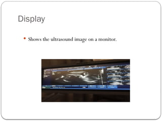
The machine displaysthe distances and intensities of the echoes
on the screen, forming a two dimensional image.
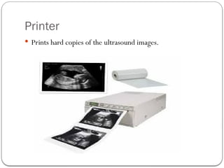
The echo images are then recorded on a plane film and can also
be recorded on videotape.
In ultrasound millions of pulses and echoes are sent and received
each second.The probe can be moved along the surface of the
body and angled to obtain various views.
After the ultrasound, the gel is easily wiped off.
The technical term for ultrasound testing and recording is
“sonography”.
12.
Principles of Ultrasonography
1.Piezoelectric effect: USG machines use piezoelectric
crystals that produce ultrasonic waves when an electric current
is applied.
2. Reflection and scattering: When ultrasonic waves
encounter tissues or organs, they reflect and scatter, creating
echoes.
3. Detection and conversion: The echoes are detected by
the transducer and converted into electrical signals.
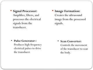
Signal Processor:
Amplifies,filters, and
processes the electrical
signals from the
transducer.
Image Formation:
Creates the ultrasound
image from the processed
signals.
• Pulse Generator :
Produces high-frequency
electrical pulses to drive
the transducer.
• Scan Converter:
Controls the movement
of the transducer to scan
the body.
16.
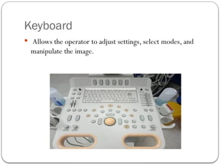
Keyboard
Allows theoperator to adjust settings, select modes, and
manipulate the image.
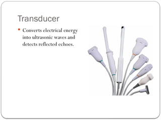
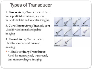
Types of Transducer
1.Linear ArrayTransducer: Used
for superficial structures, such as
musculoskeletal and vascular imaging.
2. Curvilinear ArrayTransducer:
Used for abdominal and pelvic
imaging.
3. Phased ArrayTransducer:
Used for cardiac and vascular
imaging.
4. EndocavitaryTransducer:
Used for transvaginal, transrectal,
and transesophageal imaging.
20.
Modes of USGImaging
1. B-Mode (Brightness Mode): Displays the ultrasound
image in shades of gray.
2. M-Mode (Motion Mode): Displays the movement of
structures over time.
3. Doppler Mode: Measures blood flow and velocity.
4.Colour Doppler Mode: Displays blood flow in color.
5. Power Doppler Mode: Sensitive to low-velocity blood
flow.
21.
Reference
Conclusions
https://doi.org/10.1016/j.sopen.2024.02.005
www.SlideShare.com
I would like to thank our Department of Allied Health Sciences and our
respected Subject teachers, to give me the opportunity to present this
interesting topic, it help us to gain the knowledge about the
Ultrasonography.
This presentation help us for better understanding the
ultrasound waves, ultrasonography machine principles and
the part of the machine.
Acknowledgement