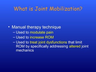
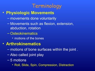
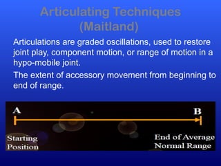
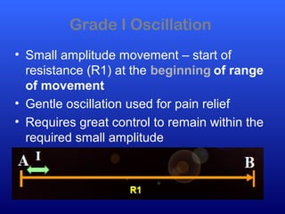
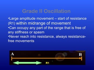
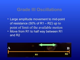
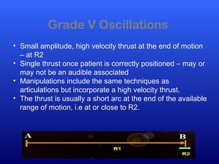
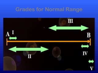
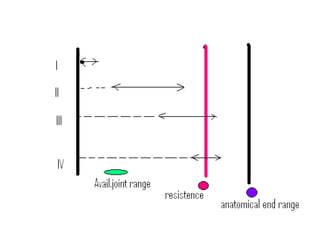
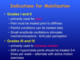
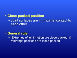
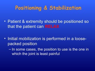
This document defines joint mobilization techniques and provides guidelines for their use. It describes mobilization as a manual therapy that uses passive joint movement to increase range of motion or decrease pain. Accessory joint movements like gliding and traction are explained. Precautions and contraindications for mobilization are outlined. A grading scale from I to V is presented to indicate the amplitude of oscillations used in different mobilization techniques.
Goniometry is the measuring of angles created by the bones of the body at the joints.1, 2, 3
The term goniometry is derived from two Greek words, gonia meaning angle and metron, meaning measure. 1, 2, 3, 4, 5,
System to measure the joint ranges in each plane of the joint is termed goniometry. 4
These measurements are done with instrument such as goniometer, a tape measure, inclinometers or by visual estimate.
Joint mobilization refers to a technique of manual therapy by which a therapist applies a brief stretch of 30s or less through traction and gliding along a joint surface.
a detailed description on theory behind Strength duration curve, along with procedure for plotting SD Curve and measuring the Rheobase and Chronaxie of the plotted graph.
Co-ordination Exercise,Definitions,Nervous control,Motor pathway,Cerebral cortex,Kinesthetic sensation,Causes of Incoordination,Flaccidity
Spasticity ,Cerebellar ataxia,Loss of kinesthetic sensation,Types of coordination,Posterior column,Test for Incoordination.
this PPT contains all the detailed information about walking aids including types, measurements, advantages & disadvantages, gait training with specific aid, etc.
This Presentation is about Mitchell relaxation technique also known a physiological relaxation technique Mitchell’s physiological relaxation technique is based on reciprocal inhibition and involves diaphragmatic breathing and a series of ordered isotonic contractions.
The manual muscle testing procedure was described in this power point, indications, contraindications, limitations of MMT was included. the MMT grading system (scale) was explained well in this PPT.
this PPT contain detailed kinetics & kinematics of ankle joint & all joints of foot complex, muscles of ankle & foot complex, plantar arches & weight distribution during standing.
Basic Introduction about Joint Mobilisation and Manipulation, This article gives clear notes for the students to understand the Mobilisation techniques.
Brian Mulligan described novel concept of the simultaneous application of therapist applied accessory mobilizations and patient generated active movements
A great presentation on joint mobilisation and manual therapy for sports and massage therapist's. Presentation from our workshop event at the St John Street clinic on 12th December 2015.
Ylinen Jari, Vorwort Leon Chaitow
Das Lern- und Nachschlagewerk über klinisch erforschte und bewährte Dehntechniken für Therapie und Sport. Mit über 200 Zeichnungen und Muskelbeschreibungen. Mehr als 160 Farbfotos zeigen die Lagerung des Patienten, die Positionierung der Hände des Therapeuten und die Dehnrichtung.
Sowohl inhaltlich und erst recht aus pädagogischer Sicht erfüllt dieses Lehrbuch in höchster Kompetenz alle fachlichen Ansprüche zu diesem komplexen Thema. Und genau das ist die Stärke dieses in zwei Teile klar gegliederten Lehr- und Arbeitsbuches: Es beschreibt Fakten aus Basis wissenschaftlicher Erkenntnisse sowie deren Lücken, ergänzt durch praktische Erfahrungen. Der zweite Teil ist perfekt! Klar gegliedert von Kopf bis Fuß, toll bebildert und prägnant beschrieben. Physiopraxis.
Buch: 296 Seiten.
Größe: 27,4 x 22 x 1,6 cm
In der folgenden Abbildung zu, wenn Sie darauf klicken.
Urban & Fischer bei Elsevier, die ersete Auflage 2009
Medirehabook, zweiten Auflage geändert 2013
ISBN 9789519209081
Bestellungen per E-mail : info@medirehab.com
oder durch www.amazon.de
Ardian dan Dicky
pemrograman game unity (roll ball)
Absen : 06 & 21
SMK DARMA SISWA 1 SIDOARJO
TEKNIK KOMPUTER DAN JARINGAN
UJI KOMPETENSI KEJURUAN TAHUN 2015
This presentation will give an basic insights about the spinal mobilisation and various manual therapy techniques used on Lumbar spine especially Maitland & Mulligan techniques.
SELF- MOBILIZATION ( AUTO MOBILIZATION)-
Self stretching techniques that specifically used joint traction and glides that directs the stretch force to the joint force.
MOBILIZATION WITH MOVEMENT (MWM)- Concurrent application of a sustained accessory mobilization applied by a clinician and an active physiological movement to end range applied by the patient.
Applied in a pain free direction
Osteoarthritis of the Knee Joint is a quite common condition found in Indian Population. This presentation is made to understand how this condition affects patients and what are the different Physiotherapy measures to make the patient functionally independent.
In this article or theoretical interface we can read and learn about principle of joint mobilization, joint mobility . After the mobilization we can find in it a good way to learn its parts and types , as it as leg joint mobilization, hand joint mobilization or somewhere else mobilization. Joint mobilization is a part of our daily physique. In joint mobilization our body daily moves in a way and a good mobility need to movement. In this article or theoretical interface we can read and learn about principle of joint mobilization, joint mobility . After the mobilization we can find in it a good way to learn its parts and types , as it as leg joint mobilization, hand joint mobilization or somewhere else mobilization. Joint mobilization is a part of our daily physique. In joint mobilization our body daily moves in a way and a good mobility need to movement. In this article or theoretical interface we can read and learn about principle of joint mobilization, joint mobility . After the mobilization we can find in it a good way to learn its parts and types , as it as leg joint mobilization, hand joint mobilization or somewhere else mobilization. Joint mobilization is a part of our daily physique. In joint mobilization our body daily moves in a way and a good mobility need to movement. In this article or theoretical interface we can read and learn about principle of joint mobilization, joint mobility . After the mobilization we can find in it a good way to learn its parts and types , as it as leg joint mobilization, hand joint mobilization or somewhere else mobilization. Joint mobilization is a part of our daily physique. In joint mobilization our body daily moves in a way and a good mobility need to movement. In this article or theoretical interface we can read and learn about principle of joint mobilization, joint mobility . After the mobilization we can find in it a good way to learn its parts and types , as it as leg joint mobilization, hand joint mobilization or somewhere else mobilization. Joint mobilization is a part of our daily physique. In joint mobilization our body daily moves in a way and a good mobility need to movement. In this article or theoretical interface we can read and learn about principle of joint mobilization, joint mobility . After the mobilization we can find in it a good way to learn its parts and types , as it as leg joint mobilization, hand joint mobilization or somewhere else mobilization. Joint mobilization is a part of our daily physique. In joint mobilization our body daily moves in a way and a good mobility need to movement. In this article or theoretical interface we can read and learn about principle of joint mobilization, joint mobility . After the mobilization we can find in it a good way to learn its parts and types , as it as leg joint mobilization, hand joint mobilization or somewhere else mobilization. Joint mobilization is a part of our daily physique. In joint mobilization our body daily moves in a way that's
- Video recording of this lecture in English language: https://youtu.be/lK81BzxMqdo
- Video recording of this lecture in Arabic language: https://youtu.be/Ve4P0COk9OI
- Link to download the book free: https://nephrotube.blogspot.com/p/nephrotube-nephrology-books.html
- Link to NephroTube website: www.NephroTube.com
- Link to NephroTube social media accounts: https://nephrotube.blogspot.com/p/join-nephrotube-on-social-media.html
The prostate is an exocrine gland of the male mammalian reproductive system
It is a walnut-sized gland that forms part of the male reproductive system and is located in front of the rectum and just below the urinary bladder
Function is to store and secrete a clear, slightly alkaline fluid that constitutes 10-30% of the volume of the seminal fluid that along with the spermatozoa, constitutes semen
A healthy human prostate measures (4cm-vertical, by 3cm-horizontal, 2cm ant-post ).
It surrounds the urethra just below the urinary bladder. It has anterior, median, posterior and two lateral lobes
It’s work is regulated by androgens which are responsible for male sex characteristics
Generalised disease of the prostate due to hormonal derangement which leads to non malignant enlargement of the gland (increase in the number of epithelial cells and stromal tissue)to cause compression of the urethra leading to symptoms (LUTS
Anti ulcer drugs and their Advance pharmacology ||
Anti-ulcer drugs are medications used to prevent and treat ulcers in the stomach and upper part of the small intestine (duodenal ulcers). These ulcers are often caused by an imbalance between stomach acid and the mucosal lining, which protects the stomach lining.
||Scope: Overview of various classes of anti-ulcer drugs, their mechanisms of action, indications, side effects, and clinical considerations.
Ozempic: Preoperative Management of Patients on GLP-1 Receptor Agonists Saeid Safari
Preoperative Management of Patients on GLP-1 Receptor Agonists like Ozempic and Semiglutide
ASA GUIDELINE
NYSORA Guideline
2 Case Reports of Gastric Ultrasound
Explore natural remedies for syphilis treatment in Singapore. Discover alternative therapies, herbal remedies, and lifestyle changes that may complement conventional treatments. Learn about holistic approaches to managing syphilis symptoms and supporting overall health.
These lecture slides, by Dr Sidra Arshad, offer a quick overview of physiological basis of a normal electrocardiogram.
Learning objectives:
1. Define an electrocardiogram (ECG) and electrocardiography
2. Describe how dipoles generated by the heart produce the waveforms of the ECG
3. Describe the components of a normal electrocardiogram of a typical bipolar leads (limb II)
4. Differentiate between intervals and segments
5. Enlist some common indications for obtaining an ECG
Study Resources:
1. Chapter 11, Guyton and Hall Textbook of Medical Physiology, 14th edition
2. Chapter 9, Human Physiology - From Cells to Systems, Lauralee Sherwood, 9th edition
3. Chapter 29, Ganong’s Review of Medical Physiology, 26th edition
4. Electrocardiogram, StatPearls - https://www.ncbi.nlm.nih.gov/books/NBK549803/
5. ECG in Medical Practice by ABM Abdullah, 4th edition
6. ECG Basics, http://www.nataliescasebook.com/tag/e-c-g-basics
Ethanol (CH3CH2OH), or beverage alcohol, is a two-carbon alcohol
that is rapidly distributed in the body and brain. Ethanol alters many
neurochemical systems and has rewarding and addictive properties. It
is the oldest recreational drug and likely contributes to more morbidity,
mortality, and public health costs than all illicit drugs combined. The
5th edition of the Diagnostic and Statistical Manual of Mental Disorders
(DSM-5) integrates alcohol abuse and alcohol dependence into a single
disorder called alcohol use disorder (AUD), with mild, moderate,
and severe subclassifications (American Psychiatric Association, 2013).
In the DSM-5, all types of substance abuse and dependence have been
combined into a single substance use disorder (SUD) on a continuum
from mild to severe. A diagnosis of AUD requires that at least two of
the 11 DSM-5 behaviors be present within a 12-month period (mild
AUD: 2–3 criteria; moderate AUD: 4–5 criteria; severe AUD: 6–11 criteria).
The four main behavioral effects of AUD are impaired control over
drinking, negative social consequences, risky use, and altered physiological
effects (tolerance, withdrawal). This chapter presents an overview
of the prevalence and harmful consequences of AUD in the U.S.,
the systemic nature of the disease, neurocircuitry and stages of AUD,
comorbidities, fetal alcohol spectrum disorders, genetic risk factors, and
pharmacotherapies for AUD.
Title: Sense of Smell
Presenter: Dr. Faiza, Assistant Professor of Physiology
Qualifications:
MBBS (Best Graduate, AIMC Lahore)
FCPS Physiology
ICMT, CHPE, DHPE (STMU)
MPH (GC University, Faisalabad)
MBA (Virtual University of Pakistan)
Learning Objectives:
Describe the primary categories of smells and the concept of odor blindness.
Explain the structure and location of the olfactory membrane and mucosa, including the types and roles of cells involved in olfaction.
Describe the pathway and mechanisms of olfactory signal transmission from the olfactory receptors to the brain.
Illustrate the biochemical cascade triggered by odorant binding to olfactory receptors, including the role of G-proteins and second messengers in generating an action potential.
Identify different types of olfactory disorders such as anosmia, hyposmia, hyperosmia, and dysosmia, including their potential causes.
Key Topics:
Olfactory Genes:
3% of the human genome accounts for olfactory genes.
400 genes for odorant receptors.
Olfactory Membrane:
Located in the superior part of the nasal cavity.
Medially: Folds downward along the superior septum.
Laterally: Folds over the superior turbinate and upper surface of the middle turbinate.
Total surface area: 5-10 square centimeters.
Olfactory Mucosa:
Olfactory Cells: Bipolar nerve cells derived from the CNS (100 million), with 4-25 olfactory cilia per cell.
Sustentacular Cells: Produce mucus and maintain ionic and molecular environment.
Basal Cells: Replace worn-out olfactory cells with an average lifespan of 1-2 months.
Bowman’s Gland: Secretes mucus.
Stimulation of Olfactory Cells:
Odorant dissolves in mucus and attaches to receptors on olfactory cilia.
Involves a cascade effect through G-proteins and second messengers, leading to depolarization and action potential generation in the olfactory nerve.
Quality of a Good Odorant:
Small (3-20 Carbon atoms), volatile, water-soluble, and lipid-soluble.
Facilitated by odorant-binding proteins in mucus.
Membrane Potential and Action Potential:
Resting membrane potential: -55mV.
Action potential frequency in the olfactory nerve increases with odorant strength.
Adaptation Towards the Sense of Smell:
Rapid adaptation within the first second, with further slow adaptation.
Psychological adaptation greater than receptor adaptation, involving feedback inhibition from the central nervous system.
Primary Sensations of Smell:
Camphoraceous, Musky, Floral, Pepperminty, Ethereal, Pungent, Putrid.
Odor Detection Threshold:
Examples: Hydrogen sulfide (0.0005 ppm), Methyl-mercaptan (0.002 ppm).
Some toxic substances are odorless at lethal concentrations.
Characteristics of Smell:
Odor blindness for single substances due to lack of appropriate receptor protein.
Behavioral and emotional influences of smell.
Transmission of Olfactory Signals:
From olfactory cells to glomeruli in the olfactory bulb, involving lateral inhibition.
Primitive, less old, and new olfactory systems with different path
micro teaching on communication m.sc nursing.pdfAnurag Sharma
Microteaching is a unique model of practice teaching. It is a viable instrument for the. desired change in the teaching behavior or the behavior potential which, in specified types of real. classroom situations, tends to facilitate the achievement of specified types of objectives.
3. Objectives
At the end of this lecture students will be
able to
• Define mobilization, Self-Mobilization, Mobilization with
Movement, physiologic movements, accessory
movements, arthrokinematics, muscle energy, thrust,
convex & concave surface,
• Describe Joint Shapes & Arthrokinematics
• Explain Convex-Concave & Concave-Convex Rule
• Describe Effects of Joint Mobilization
• Enumerate precautions & Contraindications for
Mobilization
• Describe Maitland Joint Mobilization Grading Scale
4. What is Joint Mobilization?
• Manual therapy technique
– Used to modulate pain
– Used to increase ROM
– Used to treat joint dysfunctions that limit
ROM by specifically addressing altered joint
mechanics
5. • Factors that may alter joint mechanics:
– Pain & Muscle guarding
– Joint hypomobility
– Joint effusion
– Contractures or adhesions in the joint
capsules or supporting ligaments
– Malalignment or subluxation of bony surfaces
6. Terminology
• Mobilization
– passive joint movement for increasing ROM or
decreasing pain
– Applied to joints & related soft tissues at varying speeds
& amplitudes using physiologic or accessory motions
– Force is light enough that patient’s can stop the
movement
• Manipulation
– passive joint movement for increasing joint
mobility
– Incorporates a sudden, forceful thrust that is
beyond the patient’s control
7. Terminology
• Self-Mobilization (Auto-mobilization)
– self-stretching techniques that specifically
use joint traction or glides that direct the
stretch force to the joint capsule
• Mobilization with Movement (MWM)
– Concurrent application of a sustained
accessory mobilization applied by a clinician,
Physiotherapist to end range and
physiological movement applied by the
patient
– Applied in a pain-free direction
8. Terminology
• Physiologic Movements
– movements done voluntarily
– Movements such as flexion, extension,
abduction, rotation
– Osteokinematics
• motions of the bones
• Arthrokinematics
– motions of bone surfaces within the joint .
– Also called joint play
– 5 motions
• Roll, Slide, Spin, Compression, Distraction
9. Accessory Movements
– Movements within the joint & surrounding tissues that
are necessary for normal ROM, but can not be
voluntarily performed
– Component motions
• motions that accompany active motion, but are not
under voluntary control
• Ex: Upward rotation of scapula & rotation of clavicle
that occur with shoulder flexion
– Joint play
• motions that occur within the joint
• Determined by joint capsule’s laxity
• Can be demonstrated passively, but not performed
actively
10. Terminology
• Muscle energy
– use an active contraction of deep muscles
that attach near the joint & whose line of pull
can cause the desired accessory motion
– Clinician stabilizes segment on which the
distal aspect of the muscle attaches;
command for an isometric contraction of the
muscle is given, which causes the
accessory movement of the joint
11. Terminology
• Thrust
– high-velocity, short-amplitude motion that the
patient can not prevent
– Performed at end of pathologic limit of the joint
(snap adhesions, stimulate joint receptors)
• Concave
– hollowed or rounded inward
• Convex
– curved or rounded outward
12. Relationship Between Physiological &
Accessory Motion
• Biomechanics of joint motion
– Physiological motion
• Result of concentric or eccentric active muscle
contractions
• flexion, extension, abduction, adduction or rotation
– Accessory Motion
• Motion of articular surfaces relative to one another
• Generally associated with physiological movement
• Necessary for full range of physiological motion to
occur
• Ligament & joint capsule involvement in motion
13. Joint Shapes
• Ovoid
– one surface is convex,
other surface is concave
– E.g. hip joint
• Sellar (saddle)
– one surface is concave in
one direction & convex in
the other, with the
opposing surface convex &
concave respectively
– Subtalar joint
14. Basic concepts of joint motion :
Arthrokinematics
Types of joint motion
• 5 types of joint arthrokinematics
– Roll
– Slide
– Spin
– Compression
– Distraction
• Joint motion usually often involves a
combination of rolling, sliding & spinning
15. Roll
• A series of points on one articulating
surface come into contact with a series of
points on another surface
– ball rolling on ground
– Example: Femoral condyles rolling on tibial plateau
– Roll occurs in direction of movement
– Occurs on incongruent (unequal) surfaces
– Usually occurs in combination with sliding or spinning
16. Slide
• Specific point on one surface comes into
contact with a series of points on another
surface
• Surfaces are congruent
• When a passive mobilization technique is
applied to produce a slide in the joint –
referred to as a GLIDE.
• Combined rolling-sliding in a joint
– The more congruent the surfaces are, the more
sliding there is
– The more incongruent the joint surfaces are, the
more rolling there is
17. Spin
• Occurs when one bone rotates around a
stationary longitudinal mechanical axis
– Same point on the moving surface creates an
arc of a circle as the bone spins
• Example: Shoulder with flexion/extension,
the hip with flexion/extension, and Radial
head at the humeroradial joint during
pronation/supination
18. • Compression
– Decrease in space between two joint surfaces
– Adds stability to a joint
– Normal reaction of a joint to muscle
contraction
• Distraction
– Two surfaces are pulled apart
– Often used in combination with joint
mobilizations to increase stretch of capsule.
19. Convex-Concave & Concave-Convex Rule
• Basic application of correct
mobilization techniques
• One joint surface is MOBILE & one
is STABLE
• Concave-convex rule: concave
joint surfaces slide in the SAME
direction as the bone movement
(convex is STABLE)
– If concave joint is moving on
stationary convex surface – glide
occurs in same direction as roll
20. Convex-concave rule: convex joint surfaces
slide in the OPPOSITE direction of the bone
movement (concave is STABLE)
If convex surface in moving on stationary
concave surface – gliding occurs in opposite
direction to roll
21.
22. Effects of Joint Mobilization
• Neurophysiological effects
– Stimulates mechanoreceptors to pain
– Affect muscle spasm & muscle guarding –
nociceptive stimulation
– Increase in awareness of position & motion
because of afferent nerve impulses
• Nutritional effects
– Distraction or small gliding movements – cause
synovial fluid movement
– Movement can improve nutrient exchange
23. • Mechanical effects
– Improve mobility of hypo-mobile joints
(adhesions & thickened Connective tissue
from immobilization – loosens)
– Maintains extensibility & tensile strength of
articular tissues
24. Contraindications for
Mobilization
• Avoid the following:
– Inflammatory arthritis
– Malignancy
– Tuberculosis
– Osteoporosis
– Ligamentous rupture
– Herniated disks with nerve
compression
– Bone disease
– Neurological involvement
– Bone fracture
– Congenital bone
deformities
– Vascular disorders
– Joint effusion
• May use I & II
mobilizations to relieve
pain
26. Articulating Techniques
(Maitland)
Articulations are graded oscillations, used to restore
joint play, component motion, or range of motion in a
hypo-mobile joint.
The extent of accessory movement from beginning to
end of range.
28. Grade I Oscillation
• Small amplitude movement – start of
resistance (R1) at the beginning of range
of movement
• Gentle oscillation used for pain relief
• Requires great control to remain within the
required small amplitude
29. Grade II Oscillation
•Large amplitude movement – start of resistance
(R1) within midrange of movement
•Can occupy any part of the range that is free of
any stiffness or spasm
•Never reach into resistance, always resistance-
free movements
30. Grade III Oscillations
• Large amplitude movement to mid-point
of resistance (50% of R1 – R2) up to
point of limit of the available motion
• Move from R1 to half way between R1
and R2
31. Grade IV Oscillations
• Small amplitude movement to the mid-point
of resistance– between R1 and R2 at very
end range of movement
• Oscillatory movement often stretching into
stiffness or spasm
32. Grade V Oscillations
• Small amplitude, high velocity thrust at the end of motion
– at R2
• Single thrust once patient is correctly positioned – may or
may not be an audible associated
• Manipulations include the same techniques as
articulations but incorporate a high velocity thrust.
• The thrust is usually a short arc at the end of the available
range of motion, i.e at or close to R2.
35. Indications for Mobilization
• Grades I and II
– primarily used for pain
– Pain must be treated prior to stiffness
– Painful conditions can be treated daily
– Small amplitude oscillations stimulate
mechanoreceptors - limit pain perception
• Grades III and IV
– primarily used to increase motion
– Stiff or hypomobile joints should be treated 3-4
times per week – alternate with active motion
exercises
36. Joint Positions
• Resting position
– Maximum joint play - position in which joint
capsule and ligaments are most relaxed
– Evaluation and treatment position utilized with
hypomobile joints
• Loose-packed position
– Articulating surfaces are maximally separated
– Joint will exhibit greatest amount of joint play
– Position used for both traction and joint
mobilization
37. • Close-packed position
–Joint surfaces are in maximal contact to
each other
• General rule:
– Extremes of joint motion are close-packed, &
midrange positions are loose-packed.
38. Joint Mobilization Application
• All joint mobilizations follow the convex-
concave rule
• Patient should be relaxed
• Explain purpose of treatment & sensations
to expect to patient
• Evaluate BEFORE & AFTER treatment
goniometry
39. • Stop the treatment if it is too painful for the
patient
• Use proper body mechanics
• Use gravity to assist the mobilization
technique if possible
• Begin & end treatments with Grade I or II
oscillations
40. Positioning & Stabilization
• Patient & extremity should be positioned so
that the patient can RELAX
• Initial mobilization is performed in a loose-
packed position
– In some cases, the position to use is the one in
which the joint is least painful
41. • Firmly & comfortably stabilize one joint
segment, usually the proximal bone
– Hand, belt, assistant
– Prevents unwanted stress & makes the
stretch force more specific & effective
42. Treatment Force & Direction of
Movement
• Treatment force is applied as close to the
opposing joint surface as possible
• The larger the contact surface is, the more
comfortable the procedure will be (e.g. use
flat surface of the hand instead of forcing
with the thumb)
• Direction of movement during treatment is
either PARALLEL or PERPENDICULAR to
the treatment plane
43. Treatment Direction
• Treatment plane lies on the
concave articulating surface,
perpendicular to a line from
the center of the convex
articulating surface
• Joint traction techniques are
applied perpendicular to the
treatment plane
– Entire bone is moved so that the
joint surfaces are separated
44.
45. • Gliding techniques are applied parallel to
the treatment plane
• Glide in the direction in which the slide
would normally occur for the desired
motion
• Direction of sliding is easily determined by
using the convex-concave rule. The entire
bone is moved so that there is gliding of
one joint surface on the other.
• The bone should not be used as a lever; it
should have no arcing motion (swing) that
would cause rolling and thus compression
of the joint surfaces.
46. • When using grade III gliding techniques, a
grade I distraction should be used
• If gliding in the restricted direction is too
painful, begin gliding mobilizations in the
painless direction then progress to gliding
in restricted direction when not as painful
47. • Reevaluate the joint response the next
day or have the patient report at the
next visit
– If increased pain, reduce amplitude of
oscillations
– If joint is the same or better, perform either
of the following:
• Repeat the same maneuver if goal is to
maintain joint play
• Progress to sustained grade III traction or
glides if the goal is to increase joint play