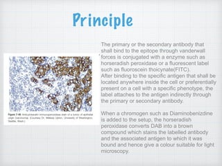
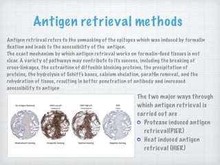
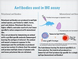
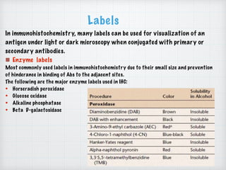
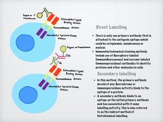
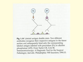
Immunohistochemistry is a technique that uses antibodies to identify antigens in cells of a tissue sample. It relies on the principle of antibodies binding specifically to antigens in cells. The primary antibody binds to the antigen of interest, while the secondary antibody is conjugated to an enzyme or fluorescent label for visualization. A chromogen is used to produce a colored precipitate that can be seen under the microscope. Immunohistochemistry has many applications in pathology for tumor diagnosis and classification by identifying cellular markers of differentiation. It allows identification of cell lineages and tumor types through characteristic protein expression patterns revealed by specific antibody staining.
Immunohistochemistry (IHC) is the localization of a known antigen in tissues by utilizing antibodies directed towards that (specific) antigen. In this presentation, we will introduce the procedure of IHC and the troubleshooting solutions.
How To Optimize Your Immunohistochemistry ExperimentProteintech Group
Immunohistochemistry allows the visualization of proteins in tissue while retaining its microstructure. This guide includes general protocols, technical tips and troubleshooting.
Immunohistochemistry (IHC) is the process of detecting antigens (e.g. proteins) in cells of a tissue section by exploiting the principle of antibodies binding specifically to antigens in biological tissues.
https://www.creative-bioarray.com/protocol/immunohistochemistry-protocol.htm
Basics of immunohistochemistry, steps of histochemistry, primary and secondary antibody use as protein locator and different fixatives with their advantages
This immunohistochemistry presentation discusses assay principles, a general protocol and tips and hints for simplifying your staining procedures.
To view the webinar recording please visit: http://www.innovabiosciences.com/bioconjugation-and-immunoassay-webinars/immunohistochemistry-introduction.html
EXPLORING THE CONCEPT OF MULTIPLEXING IN IMMUNOHISTOCHEMISTRYChelsea Osayande
Conventional immunohistochemistry (IHC) is commonly used as a diagnostic technique in the field of tissue pathology but suffers from certain limitations. The most critical of these is that this technique only permits the labelling of a single marker per tissue section. This results in missed opportunities to gain important prognostic and diagnostic information from patient samples.
Multiplex Immunohistochemistry/Immunofluorescence (mIHC/IF) technologies, which allow the simultaneous detection of multiple markers on a single tissue section, have been introduced and adopted in both research and clinical settings in response to increased demand for improved techniques.
A number of highly multiplexed tissue imaging technologies have also emerged, permitting comprehensive studies of cell composition, functional state and cell-cell interactions which suggest improved diagnostic benefit.
Immunohistochemistry (IHC) is a highly sensitive method that allows the localization of antigen within a cell or a tissue with high resolution. The method is based on the use of a primary antibody that specifically binds to its complementary antigen. The bound antibody may then be visualized by a variety of methods such as colorimetric end points.
Antibodies are compelling proteins that are essential to the immune system and extremely powerful in biotechnology applications; existing as major players in our defence against external agents (viruses, bacteria, etc.), they are also widely used as tools for research, diagnosis and treatments.
Immunohistochemistry (IHC) combines histological, immunological, and biochemical techniques for the identification of specific tissue components by means of a particular antigen/antibody reaction tagged with a visible label
Demystifying Immunohistochemistry A Step-by-Step Guide to IHC Staining Proced...ihc-prs
Immunohistochemistry (IHC) staining is a powerful technique in the field of pathology that allows researchers and clinicians to visualize specific proteins within tissue samples. This procedure is essential for understanding the molecular composition of cells and tissues, aiding in the diagnosis and classification of diseases. Let's delve into the step-by-step process of IHC staining to demystify this invaluable tool.
Western Blotting: Techniques, Applications, and Innovations | The Lifescience...The Lifesciences Magazine
Innovations and Advancements in Western Blotting: 1. Automated Western Blotting 2. Multiplexing Capabilities 3. Improved Detection Sensitivity 4. Digital Imaging and Quantification
Unveiling the Power of Immunohistochemistry Decoding the Procedure and Antibo...ihc-prs
Immunohistochemistry (IHC) stands as a cornerstone technique in the realm of pathology, offering insights into the localization and expression of proteins within tissue samples. This indispensable method not only aids in diagnosing diseases but also contributes to unraveling the intricate mechanisms underlying various pathological conditions. Let's delve into the procedure of IHC and explore the diverse range of antibodies pivotal in its execution.
We are engaged in manufacturing, supplying and exporting of Laboratory Filtration Products (syringe filters, membrane filters, glass fiber filters and filter papers). These filtration products are precisely manufactured by making use of superior quality raw material and ultra-modern techniques by the experience team. Our offered products are widely used in various laboratories for various filtration works in worldwide. http://www.axivasichem.com/syringe-filters.aspx
This ppt file represents a simple overview on what is antibody validation & how to validate an antibody before performing any research.
Used references are also included.
OTECHNOLOGY IS CHALLENGING SUBJECT TO TEACH AND UNDERSTAND ALSO .....THEIR INTERESTING PART IS TO LEARN ABOUT MICROBIAL GENETICS AND THEIR METHODS OF GENE TRANSFER
Richard's entangled aventures in wonderlandRichard Gill
Since the loophole-free Bell experiments of 2020 and the Nobel prizes in physics of 2022, critics of Bell's work have retreated to the fortress of super-determinism. Now, super-determinism is a derogatory word - it just means "determinism". Palmer, Hance and Hossenfelder argue that quantum mechanics and determinism are not incompatible, using a sophisticated mathematical construction based on a subtle thinning of allowed states and measurements in quantum mechanics, such that what is left appears to make Bell's argument fail, without altering the empirical predictions of quantum mechanics. I think however that it is a smoke screen, and the slogan "lost in math" comes to my mind. I will discuss some other recent disproofs of Bell's theorem using the language of causality based on causal graphs. Causal thinking is also central to law and justice. I will mention surprising connections to my work on serial killer nurse cases, in particular the Dutch case of Lucia de Berk and the current UK case of Lucy Letby.
Comparing Evolved Extractive Text Summary Scores of Bidirectional Encoder Rep...University of Maribor
Slides from:
11th International Conference on Electrical, Electronics and Computer Engineering (IcETRAN), Niš, 3-6 June 2024
Track: Artificial Intelligence
https://www.etran.rs/2024/en/home-english/
Nutraceutical market, scope and growth: Herbal drug technologyLokesh Patil
As consumer awareness of health and wellness rises, the nutraceutical market—which includes goods like functional meals, drinks, and dietary supplements that provide health advantages beyond basic nutrition—is growing significantly. As healthcare expenses rise, the population ages, and people want natural and preventative health solutions more and more, this industry is increasing quickly. Further driving market expansion are product formulation innovations and the use of cutting-edge technology for customized nutrition. With its worldwide reach, the nutraceutical industry is expected to keep growing and provide significant chances for research and investment in a number of categories, including vitamins, minerals, probiotics, and herbal supplements.
Multi-source connectivity as the driver of solar wind variability in the heli...Sérgio Sacani
The ambient solar wind that flls the heliosphere originates from multiple
sources in the solar corona and is highly structured. It is often described
as high-speed, relatively homogeneous, plasma streams from coronal
holes and slow-speed, highly variable, streams whose source regions are
under debate. A key goal of ESA/NASA’s Solar Orbiter mission is to identify
solar wind sources and understand what drives the complexity seen in the
heliosphere. By combining magnetic feld modelling and spectroscopic
techniques with high-resolution observations and measurements, we show
that the solar wind variability detected in situ by Solar Orbiter in March
2022 is driven by spatio-temporal changes in the magnetic connectivity to
multiple sources in the solar atmosphere. The magnetic feld footpoints
connected to the spacecraft moved from the boundaries of a coronal hole
to one active region (12961) and then across to another region (12957). This
is refected in the in situ measurements, which show the transition from fast
to highly Alfvénic then to slow solar wind that is disrupted by the arrival of
a coronal mass ejection. Our results describe solar wind variability at 0.5 au
but are applicable to near-Earth observatories.
This pdf is about the Schizophrenia.
For more details visit on YouTube; @SELF-EXPLANATORY;
https://www.youtube.com/channel/UCAiarMZDNhe1A3Rnpr_WkzA/videos
Thanks...!
Professional air quality monitoring systems provide immediate, on-site data for analysis, compliance, and decision-making.
Monitor common gases, weather parameters, particulates.
2. Introduction
Immunohistochemistry is a technique for identifying cellular or tissue
constituents by means of antigen-antibody interactions. The site of antibody
binding to a particular antigenic determinant ie a epitope could be either direct
labelling of the antibody or by the use of secondary labelling method.
3. Principle
The primary or the secondary antibody that
shall bind to the epitope through vanderwall
forces is conjugated with a enzyme such as
horseradish peroxidase or a fluorescent label
such as fluoroscein thoicynate(FITC).
After binding to the specific antigen that shall be
located anywhere inside the cell or preferentially
present on a cell with a specific phenotype, the
label attaches to the antigen indirectly through
the primary or secondary antibody.
When a chromogen such as Diaminobenizdine
is added to the setup, the horseradish
peroxidase converts DAB into a brown
compound which stains the labelled antibody
and the associated antigen to which it was
bound and hence give a colour suitable for light
microscopy.
4. Obtaining a sample
• For IHC, sample could be either
obtained from a biopsy or a surgical
procedure. The most routine in
obtaining a sample is through Fine
Needle Aspiration(FNA).
• In this technique, a thin (23-25) gauge,
hollow needle is inserted into the mass
for sampling of cells.
• Fine needle aspiration biopsies are very
safe minor surgical procedures. Often
a major surgical biopsy can be avoided
by performing a needle aspiration
biopsy instead, eliminating the need
for hospitalisation.
5. Aspiration techniques
Suction FNAC Capillary method
• In this method, the needle is
passed into the lesion and
negative pressure is applied,
usually by virtue of a syringe
attached to the needle
• Used predominantly, also known
as the non aspiration technique
• The needle is passed into the
lesion and multiple fast jabbing
movements in and out of the
lesion as well as in different
directions are performed.
8. Fixation
The purpose of fixation is to preserve tissue so that it is resistant to undergoing further
changes, e.g. by the action of tissue enzymes or microorganisms.
Fixation of tissues is necessary:
a. To adequately preserve cellular components, including soluble and structural proteins.
b. To prevent autolysis and displacement of cell constituents, including Ags and enzymes.
c. To stabilize cellular materials against deleterious effects of subsequent procedures
d. To facilitate conventional staining and immunostaining.
However, the effects of fixation,
including protein-protein and
protein-nucleic acid cross linking and
calcium ion bonding, mask or damage
epitopes through alteration of the
protein 3 dimensional structure.
These changes can often be reversed
by any of the multiple antigen
retrieval methods.
9. Prevention of false positives
Sometimes normal and neoplasticism cells have endogenous enzyme activity
such as peroxidase or alkaline phosphatase. This leads to the generation of false
results as the chromogen specific as a substrate for the enzyme stains the
antigen when associated with a primary or secondary antibody, but also stains
the tissue under inspection diffusely due to the presence of endogenous enzymes.
To counteract this, endogenous enzymes need to be blocked before addition of the
conjugated Ab- chromogen complex. The most frequently used method is pre-
incubation of the sections in absolute methanol containing hydrogen peroxide.
A. Before blocking endogenous enzyme
activity
B. After blocking endogenous
enzyme activity
10. Antigen retrieval methods
Antigen retrieval refers to the unmasking of the epitopes which was induced by formalin
fixation and leads to the accessibility of the antigen.
The exact mechanism by which antigen retrieval works on formalin-fixed tissues is not
clear. A variety of pathways may contribute to its success, including the breaking of
cross-linkages, the extraction of diffusible blocking proteins, the precipitation of
proteins, the hydrolysis of Schiff's bases, calcium chelation, paraffin removal, and the
rehydration of tissue, resulting in better penetration of antibody and increased
accessibility to antigen
The two major ways through
which antigen retrieval is
carried out are
Protease induced antigen
retrieval(PIER)
Heat induced antigen
retrieval (HIER)
11. Several heating methods can be used to achieve the
AR results(HIER):
Water baths
Microwave ovens
Pressure cookers
Steamers
Antigen retrieval methods
12. Antibodies used in IHC assay
Polyclonal antibodies Monoclonal antibod
1. Purified immunogen is injected in mice
2. B lymphocytes are harvested f
3. B lymphocytes who have a limited lifespan are fused with ever dividing
non senescent myeloma cells to produce a hybridoma
The hybridomas having the desired specificity is
then selected. The hybrid cell produced is a
immortal cell that produces Igs specific for a single
epitope.(monoclonal antibodies)
Polyclonal antibodies are produced in multiple
animal species, particularly rabbit, horse,
goat and chicken. Polyclonal Abs have a
higher affinity and wide reactivity but lower
specificity when compared with monoclonal
Abs.
They are produced by immunizing an animal
with a purified specific molecule (immunogen)
bearing the antigen of interest. The animal
will mount a humoral response to the
immunogen and the antibodies so produced
can be harvested. It is likely that the animal
will produce multiple clones of plasma cells
and hence polyclonal Abs are obtained.
13. Labels
In immunohistochemistry, many labels can be used for visualization of an
antigen under light or dark microscopy when conjugated with primary or
secondary antibodies.
Enzyme labels
Most commonly used labels in immunohistochemistry due to their small size and prevention
of hinderance in binding of Abs to the adjacent sites.
The following are the major enzyme labels used in IHC:
• Horseradish peroxidase
• Glucose oxidase
• Alkaline phosphatase
• Beta D-galactosidase
14. Fluorescent labels
More commonly used in frozen sections.
Fluorophore linked antibodies emit a specific colour when excited by correct wavelengths of
light.
The benefit of using fluorophores is that they provide an easy method for performing double-
labelling experiments where several antibodies towards multiple targets are assayed in the
same sample. The secondary antibodies need to be targeted towards different primary
antibodies and also coupled to different fluorophores. The different secondary antibodies are
then observed separately by exciting them sequentially with different wavelengths. These
different excitation results are saved as separate images or colour channels and may later
be overlaid to infer protein co-localizations.
The various fluorochromes
commonly used are:
• Fluoroscein thiocynate(FITC)
• TRITC(rhodamine)
• Texas red
• R-phytoerythrin(PE)
15. Direct Labelling
Secondary labelling
• There is only one primary antibody that is
attached to the antigenic epitope which
could be cytoplasmic, membranous or
nuclear.
• Immunohistochemical staining methods
include use of fluorophore-labeled
(immunofluorescence) and enzyme-labeled
(immunoperoxidase) antibodies to identify
proteins and other molecules in cells
• In this method, the primary antibody
devoid of any fluorochrome or
immunoperoxidase activity binds to the
epitope of a protein.
• A secondary antibody binds to an
epitope on the initial primary antibody
and has associated with it some
labelling activity. This is also referred
to as the indirect method of
histochemical labelling.
16.
17.
18. Procedure at SMIH
• Deparafinization of tissue sections taken on polylysine coated slides
• Quenching endogenous peroxidase
• Antigen retrieval
• Serum incubation
• Binding of primary antibody
• Binding with enzyme labelled secondary antibody
• Addition of chromogen substrate DAB
• Counterstain, dehydrate coverslipping
• Interpretation of pattern of staining and integration of IHC in correct
clinical setting
19. Application of IHC in diagnostics and theranostics.
• IHC is a major tool in the armentarium of a pathologist to confirm or
differentiate between tumors
• Certain differentiation markers present in the cytoplasm, nucleus or
the cell membrane serve as epitopes for antibody labelling. These
markers are localised in certain cell lines such as epithelial,
mesenchymal, hematopoetic, neuroendocrine, melanocytes to name a
few. It is exciting to see that these markers can serve as strong
diagnostic tools and panels for differentiating between different
tumour phenotype.
20. Markers of differentiation
1. Cytokeratins
• Cytokeratins are the the most important markers
used for the diagnosis of epithelial neoplasms.
• Cytokeratins are intermediate filament proteins
building an intracytoplasmic network between the
nucleus and cell membrane of epithelial cells
• Cytokeratins are a complex family composed of
more than 20 isotypes and divided into two
groups which usually heterodimerize with each
other:
• Type I(acidic group) including cytokeratins 9-20
• Type II(basic group) including cytokeratins 1-8
Keratins are also divided into high molecular weight
keratins(HMWK) and low molecular weight
keratins(LMWK)
22. CK8/18= simple one layered non polar
epithelial cells
Gastrointestinal epithelium additionally
expresses CK19
Goblet cells and mature enterocytes: CK20
Biliary,pancreatic ducts, renal collecting
tubules: CK7(and transitional epithelium)
Basal layer: CK5/14
Intermediate and superficial layers:
1. Non cornified: 4/13
2. Cornified: 1/10
Transitional epithelium- additional CK20 expression along with 8,18,19,7
Mesothelium- CK8,18,19,7(simple epithelia) and stratified epithelial CK5,14
1) HMWK- expressed in squamous cell carcinomas
2) LMWK- expressed in glandular epithelium and visceral parenchyma
Simple epithelium
Stratified epithelium
23.
24.
25. 2. Epithelial membrane antigen: LMWK equivalent(marker of glandular
epithelium)
3. Carcinoembryonic antigen: marker of glandular epithelium
4. p63: HMWK equivalent: squamous and urothelial epithelium
5. Vimentin: vimentin is a 57kDa protein, a member of the Type III family of
intermediate filaments, expressed in all mesenchymal cells forming an
important part of the cytoskeleton of these cells.
6. S100 protein: structurally similar to calmodulin however differ from it
in terms of cell specific expression pattern. S100 proteins are involved in
regulation of protein phosphorylation, transcription factors, Ca++
homeostasis, the dynamics of cytoskeleton constituents, cell growth and
differentiation.
It can be found in 100% of melanomas and schwannomas.
7. CD45/leukocyte common antigen: family of high molecular mass integral
membrane glycoprotein molecules expressed on all hematopoietic cells
except mature red cells and their immediate progenitors,
megakaryocytes, and platelets.
Specific marker for lymphatic and hemopoetic tumours.