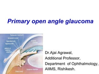
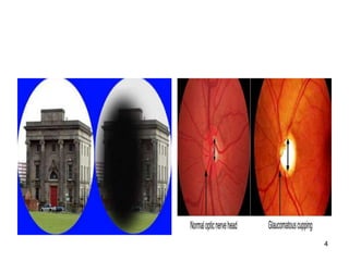
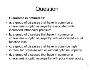
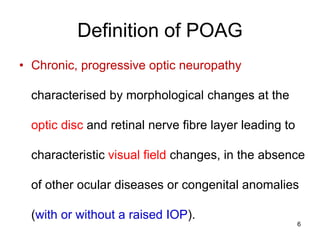
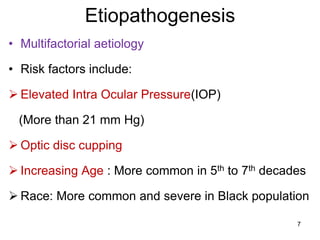
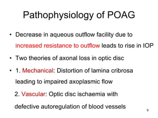
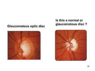
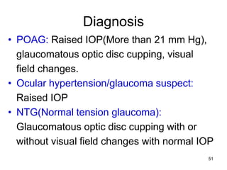
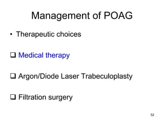
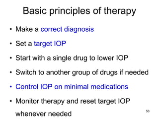
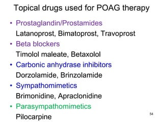
The Most Attractive Hyderabad Call Girls Kothapet 𖠋 6297143586 𖠋 Will You Mis...
Primary open angle glaucoma presentation
1. Primary open angle glaucoma
Dr.Ajai Agrawal,
Additional Professor,
Department of Ophthalmology,
AIIMS, Rishikesh.
2. Acknowledgement
• Kanski’s Clinical Ophthalmology (8th Edition).
• Becker- Schaffer’s Diagnosis and therapy of The
Glaucomas (8th Edition).
• Comprehensive Ophthalmology (A.K.Khurana)
(7th Edition).
2
3. Learning Objectives
• At the end of this class the students shall be able to :
• Define primary open angle glaucoma(POAG).
• Comprehend the pathophysiology and
risk factors of POAG.
• Understand the clinical features of POAG.
• Understand the fundamentals of managing
primary open angle glaucoma 3
5. Question
Glaucoma is defined as:
• a. a group of diseases that have in common a
characteristic optic neuropathy associated with
increased intraocular pressure.
• b. a group of diseases that have in common a
characteristic optic neuropathy with associated visual
function loss.
• c. a group of diseases that have in common high
intraocular pressure with or without optic neuropathy.
• d. a group of diseases that have in common a
characteristic optic neuropathy with poor visual acuity.
5
6. Definition of POAG
• Chronic, progressive optic neuropathy
characterised by morphological changes at the
optic disc and retinal nerve fibre layer leading to
characteristic visual field changes, in the absence
of other ocular diseases or congenital anomalies
(with or without a raised IOP).
6
7. Etiopathogenesis
• Multifactorial aetiology
• Risk factors include:
Elevated Intra Ocular Pressure(IOP)
(More than 21 mm Hg)
Optic disc cupping
Increasing Age : More common in 5th to 7th decades
Race: More common and severe in Black population
7
8. Etiopathogenesis
Heredity/ Family History: Risk of about 10% in
siblings; 4% in off springs
Diabetes
Systemic Hypertension
Myopia
Thin central corneas
Steroid usage
??Migraine, Cigarette smoking 8
9. Pathophysiology of POAG
• Decrease in aqueous outflow facility due to
increased resistance to outflow leads to rise in IOP
• Two theories of axonal loss in optic disc
• 1. Mechanical: Distortion of lamina cribrosa
leading to impaired axoplasmic flow
2. Vascular: Optic disc ischaemia with
defective autoregulation of blood vessels 9
10. FORMATION OF AQUEOUS HUMOR
CILIARY PROCESSES
-approx. 70-80 radial folds in the pars plicata which form
the site of aqueous production.
-Zonular fibers attach primarily in the valleys of the ciliary
processes and also along the pars plana
12. Formation of aqueous humor
• Diffusion and ultrafiltration are both
passive mechanisms so no active cellular
participation occurs.
• Active secretion is an active process.
• Rate of formation of aqueous humor in a
healthy human eye is-
2 - 3 microlitres/minute
12
14. Differences between
aqueous humor & plasma
-Marked deficit of
proteins
-Marked excess of
Ascorbate
-Excess of Lactate
-Excess of Chloride &
certain amino acids
AQUEOUS PLASMA
0.024 7.0
gm/dl gm/dl
1.06 0.04
micromol/ml micromol/ml
4.5 1.9
micromol/ml micromol/ml
14
15. Functions of aqueous humor
*Maintaining IOP :
-important for early ocular development &
maintaining global integrity throughout life.
*Serves as a vascular system for the avascular
structures of the eye: cornea, lens & TM.
- by providing substrates & nutrients & removing
metabolites.
15
16. Functions of aqueous humor
*Delivering high concentration of Ascorbate:
- scavenges free radicals & protects against UV
rays & other radiations.
*Local paracrine signaling & immune responses.
*Colourless & transparent medium as part of eye’s
optical system.
16
19. Major amount of aqueous humor leaves the
eye by
BULK FLUID FLOW
i.e. fluid flows along normal pressure
gradient through non-energy dependent
process
19
24. • Veins from the anterior part of ciliary body form the
Ciliary venous plexus
Anterior ciliary veins & Episcleral veins
communicate with Schlemm’s canal
24
28. Trans-corneal outflow
- Aqueous humor from anterior chamber
goes into tear film through cornea.
- Very little aqueous passes through this
pathway.
- Total volume of fluid transferred is limited
by high hydraulic resistance of the cornea.
28
29. Clinical features of POAG
Symptoms
• Usually asymptomatic in early cases
• Mild headache and eye ache
• Frequent changes in presbyopic glasses
• Delayed dark adaptation
• Loss of peripheral vision
• Loss of central vision(late cases)
29
30. Signs of POAG
• Normal anterior segment
• Pupil reaction to light may be sluggish(in
advanced cases only)
• Elevated IOP(More than 21 mm Hg) with
diurnal variation more than 5-8 mmHg
• Optic disc changes (Progressive,
asymmetric)
• Visual field defects
30
32. Optic disc changes in glaucoma
• Early changes
o Retinal nerve fibre layer atrophy
o Vertically oval cup
o Asymmetry of the cups(More than 0.2
difference)
o Large cup(CD more than 0.6)
o Splinter haemorrhages
32
33. Advanced glaucomatous disc changes
• Marked cupping (More than 0.7)
• Thinning of NRR (Neuroretinal rim)
• Lamellar dot sign
• Vascular alterations
o Nasal shifting of retinal vessels
o Bayonetting sign(convoluted path due to NRR
loss)
o Baring of circumlinear vessels and overpass
vessels
• Glaucomatous optic atrophy
33
39. Visual field defects in glaucoma
• Arcuate nerve fibres in the superior and
inferior temporal portions of the optic disc:
Most sensitive to damage
• Macular fibres : Most resistant to damage
CENTRAL VISION IS PRESERVED TILL
THE LAST IN GLAUCOMA
39
40. Progression of field defects
• Isopter contraction: Generalised field
constriction
• Baring of blind spot : Non specific
(Exclusion of blind spot from central field)
• Paracentral scotoma: Wing shaped and
occurs above or below the blind spot in the
Bjerrum’s area(10-25 degrees from
fixation)
Is the earliest clinically significant defect
40
41. Progression of field defects
• Seidel’s scotoma: sickle shaped
Due to joining of blind spot and
paracentral scotoma
• Bjerrum’s/Arcuate scotoma:
Extension of Seidel’s scotoma to reach the
horizontal line.
• Double arcuate/ring scotoma
41
42. Progression of field defects
• Roenne’s central nasal step:
Sharp right angled defect at the horizontal
meridian when arcuate scotomas run in
different arcs
• Peripheral field defects
• Advanced defects
Residual Tubular vision
Temporal island of vision
42
43. Quantification of visual field defects
• Visual field analyzer
Kinetic perimeter
Static perimeter (automated)
Testing more than once is required before
final interpretation
43
50. Diagnostic work up/Investigations
• Tonometry
• Goniscopy: Open angles
• Perimetry: To detect visual field defects
• Slit lamp examination: To rule out causes
of secondary open angle glaucoma
• Fundus examination to document optic
disc changes
• Diurnal variation testing
• Provocative testing: Water drinking test
50
51. Diagnosis
• POAG: Raised IOP(More than 21 mm Hg),
glaucomatous optic disc cupping, visual
field changes.
• Ocular hypertension/glaucoma suspect:
Raised IOP
• NTG(Normal tension glaucoma):
Glaucomatous optic disc cupping with or
without visual field changes with normal IOP
51
52. Management of POAG
• Therapeutic choices
Medical therapy
Argon/Diode Laser Trabeculoplasty
Filtration surgery
52
53. Basic principles of therapy
• Make a correct diagnosis
• Set a target IOP
• Start with a single drug to lower IOP
• Switch to another group of drugs if needed
• Control IOP on minimal medications
• Monitor therapy and reset target IOP
whenever needed
53
55. Systemic drugs used for POAG therapy
• Used rarely, for short term control of IOP
• Oral carbonic anhydrase inhibitors
Acetazolamide, Methazolamide
55
56. Laser treatment
• Indications
Target IOP not achieved with medical
therapy
Non compliance of medical therapy
Argon/ Diode Laser Trabeculoplasty (ALT)
Selective Laser Trabeculoplasty (SLT)
56
57. Surgical therapy
• Indications
Target IOP not achieved with maximal
tolerated medical therapy and laser
trabeculoplasty
Non compliance of medical therapy
Non availability of laser therapy
Advanced glaucoma
57
58. Surgical therapy
• Filtration surgery : Trabeculectomy
• Modified trabeculectomy :
Use of antifibrotic agents
Mitomycin/5FU
• Aqueous drainage devices:
Ahmed glaucoma valve
In cases with no/poor visual potential:
Cycloablative therapy with laser/cryotherapy
58
59. Conclusion
• Primary open angle glaucoma is a
progressive optic neuropathy with
characteristic optic disc and visual field
changes.
• Increased resistance to aqueous outflow
leads to rise in IOP.
• Aim of management is to reduce IOP to
minimize damage to optic disc and
resultant visual field defects.
59
60. Question
Which of the following is not a risk factor
for the development of primary open
angle glaucoma?
a. positive family history.
b. advanced age.
c. increased IOP.
d. increased corneal thickness.
60
61. Question
You have been referred a case of open
angle glaucoma. Which of the following
would be an important point in
diagnosing the case?
a. Shallow anterior chamber
b. Optic disc cupping
c. Narrow angle
d. Visual acuity and refractive error
61