SlideShare a Scribd company logo
1 of 86
DR. ARNAV SINGH SAROYA
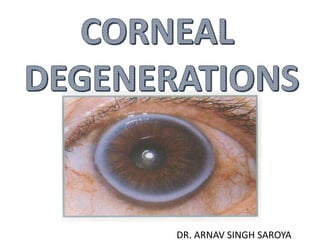
CORNEAL DEGENERATION
Corneal degenerations refers to the conditions in which
the normal cells undergo degenerative changes under the
influence of age or some pathologic conditions.
• Non-familial, late onset
•Asymmetric, unilateral, central or peripheral
•Changes to the tissue caused by inflammation, age, or
systemic disease.
•Characterized by a deposition of material, a thinning of
tissue, or vascularization
DIFFERENCE BETWEEN
DYSTROPHY DEGENERATION
• Age Early Late
• Heredity AD/AR None
• Laterality B/L U/L(B/L)
• Location Central Peripheral
• Corneal layers Discrete Not discrete
• Systemic diseases no common
• Characteristic Well defined Poorly defined
• Vascularity avascular correspond with vascularity
CLASSIFICATION
•1.Depending upon etiology
•INVOLUTIONAL NON-
(age related) INVOLUTIONAL (pathological)
1.Arcus senilis 1. Band keratopathy
2.Limbal girdle of Vogt 2. Amyloid degeneration
3.Crocodile shagreen 3. Lipid degeneration
4.Cornea Farinata 4. Salzmann's nodular
5.Hassal – Henle bodies 5. Terrien’s marginal
6.Furrow degeneration 6. Spheroidal
2. Depending upon location
I. Axial Corneal Degenerations
a) Fatty Degenerations
b)Hyaline Degenerations
c) Amyloidosis
d)Calcific Degenerations (Band Keratopathy)
e)Salzmann’s Nodular Degeneration
•II. Peripheral Degenerations
a) Arcus Senilis
b) Vogt’s White Limbal Girdle
c) Hassall – Henle Bodies
d) Terriens’s Marginal Degeneration
e) Mooren’s Ulcer
f) Pellucid Marginal Degeneration
g) Furrow Degeneration (Senile Marginal
Degeneration)
ARCUS SENILIS & ARCUS JUVENILIS
Gerontoxon in the aged & Anterior Embryotoxon in the young.
Area : Peripheral cornea (starts inferiorly and progress superiorly to encircle
entire circumference)
Pathology : Lipid deposition
Prevalence
Increases with age
Men > Women
Age: >40yrs
Blacks affected at a younger age than Whites
Arcus senilis
• Innocuous and extremely common in elderly
• Occasionally associated with hyperlipoproteinaemia
• Bilateral, circumferential bands
of lipid deposits
• Diffuse central and sharp
peripheral border
• Peripheral border separated
from limbus by clear zone
• Clear zone may be thinned
( senile furrow)
Slit lamp examination:
• Best in Sclerotic scatter or Broad tangential view.
• Sharp peripheral border – ending at the edge of Bowman’s
layer with a lucent zone to the limbus.
• Diffuse central edge.
• Lipid deposition in the stroma – near Bowman’s layer > the
Descemet's membrane –.
Clear zone (furrow in slit beam)
Arcus
senilis
Histochemical
• Cholesterol, Cholesterol esters, phospholipids, neutral
Glycerides.
• Experimental studies: Vascular origin – in the form of low
density lipoproteins (LDL) cross the capillary wall.
• Bilateral symmetric & progresses slowly
• Lucid interval – due to vessel’s ability to reabsorb the lipid in
this area/ descemet's ends here.
• Stains for lipids- Oil red O & Sudan black.
Histopathology
• Has an hourglass appearance
• lipid is extracellular .
• advanced stages – involves
stromal lamellae.
Significance :
• No visual problem hence no treatment required.
• <40yrs with arcus – risk of Coronary artery disease –
evaluated for Hyperlipoproteinemia
• Increased levels of beta lipoproteins rich in cholesterol.
• Diseases – Nephrotic syndrome, Hypothyroidism,
Obstructive jaundice, Diabetic ketoacidosis.
• Seen in Lecithin cholesterol acyltransferase (LCAT) deficiency
Differential diagnosis
• Peripheral mosaic crocodile shagreen.
• Limbal girdle of Vogt.
• Residual scars from peripheral corneal hypersensitivity
(catarrhal) ulcer.
LIPID DEGENERATIONS
Primary :
• Rare – B/L, no disorders of lipid metabolism.
• Fatty acids – cholesterol, triglyceride, phospholipids
• Etiology – altered metabolic activity of
Keratocytes, increased vascular
permeability
• Clinical manifestation –
cosmetic or decrease vision.
LIPID DEGENERATIONS
Secondary lipid degeneration:
Located in superficial and deep stroma at areas of
vascularization.
Common – vascularised cornea – MC associated with HERPES
SIMPLEX AND HERPES ZOSTER, Interstitial keratitis, trauma,
corneal hydrops, corneal ulcers.
• Etiopathogenesis – increased permeability of vessels – or
decreased ability to remove lipid.
• Onset sudden – rapid decrease in vision.
Secondary lipid degeneration:
• Shape :
1.Sea fan with feathery edges – areas of inactive
neovascularization
2.Discoid lesion – active neovascularization
Lipid keratopathy
• Usually unilateral stromal deposits
without vascularization
• Rare, occurs spontaneously in
avascular cornea
• Unilateral stromal deposits with
vascularization
• Common, secondary to previous
disciform keratitis
Treatment - coagulation of feeder
vessels and/or keratoplasty
Treatment - keratoplasty, if severe
Primary Secondary
Secondary lipid degeneration
TREATMENT
1.Medical control of inflammatory disease.
2. Argon laser photocoagulation-feeder vessels.
3. Needle point cautery-feeder vessels.
4. Keratoplasty
5.Lipid might resolve with Subconjunctival Bevacizumab.
Band Shaped Keratopathy
TYPES : Calcific and Non – calcific forms
Clinically : begins at the periphery – 3 & 9 o’clock
• May also begin centrally
• Typical peripheral form – sharply demarcated peripheral
edge – separated from limbus by lucent zone.
• Small holes in the deposit represent areas where the corneal
nerves penetrate Bowman’s MEMBRANE.
•Early – opacity gray – becomes white & chalky – lucent
holes – penetrating corneal nerves.
•Lesion subepithelial –
Histopathology
•Fine basophilic granules –
Bowman’s layer.
Granules coalesce.
•Hyaline – like material – deposited in subepithelial
tissue( reduplication of Bowman’s layer)
• Calcific-Hydroxyapatite deposits of calcium in epithelium, Bowman`s
layer, and superficial stroma.
• Non-Calcified-Depositions of Urates- Brown colored.
• Can develop in glaucoma patients who use medications with
phenylmercuric nitrate or mercury.
• Fibrous pannus –
associated with calcification
• Overlying epithelium
atrophic
• Intracellular calcium
deposits.
• Extracellular only with local
disease or renal failure.
Physiology:
•Alteration of corneal metabolism – increased tissue
pH – precipitation of calcium, evaporation of tears –
carbon dioxide release – rise in pH.
Evaluation:
•History – medical workup – ocular examination
Signs & symptoms:
•Decrease vision ,foreign body sensation ,
photophobia, tearing
BAND KERATOPATHY (ADVANCED)
TREATMENT
If vision affected or eye uncomfortable-
• A) Chelation.
-Forceps, diamond burr
-No. 15 blade, spatula
-EDTA- 0.5-1.5%
• B) Excimer laser keratectomy.
• C) BCL, NSAIDS, antibiotic, steroids
LIMBAL GIRDLE OF VOGT
Girdle - crescentic yellow – white band – interpalpebral
limbus.
• B/L ,Symmetric , subepithelial ? STROMAL
• Nasal limbus > Temporal > inferior
Two types :
Type I – white band with holes “Swiss cheese pattern” –
central border sharp – represents early calcific band
keratopathy- lucent zone
Type II – true limbal girdle – chalky band without holes.
LIMBAL GIRDLE OF VOGT
•Incidence increases with age
> 20yrs
•Histopathology :lesion subepithelial
- overlying epithelial atrophy
- destruction & calcification of
Bowman
- Elastoid degeneration
•No treatment is required
CROCODILE SHAGREEN
• Usually bilateral
• Polygonal stromal opacities separated by
clear space
• Most frequently involve anterior stroma
(anterior crocodile shagreen)
• Occasionally involve posterior stroma
(posterior crocodile shagreen)
CROCODILE SHAGREEN
(Mosaic Keratopathy)
• Anterior Crocodile Shagreen-
 B/L symmetrical, asymptomatic.
 Level of Bowman's layer.
 Pattern-way stromal collagen fibrils inserts into
Bowman's layer OR breaks in bowman after trauma.
 Senile change, keratoconus+ hard contact lens, trauma,
BSK, hypotony
CROCODILE SHAGREEN
•Posterior crocodile shagreen-
• In central posterior stroma.
• B/L, does not disrupt descemet or endothelium,
asymptomatic.
• Histologically- irregular alignment of stromal lamellae with
a “Saw-Tooth Pattern
• Age related only
• Should be distinguished from central cloudy dystrophy of
Francois.
Crocodile shagreen-Mosaic pattern
CORNEA FARINATA
• Asymptomatic
• Tiny opacities
• Bilateral in deep corneal stroma.
• Flour – dust appearance, gray brown to white
• Histopathology – vacuoles filled with lipofuscin like substance
Corneal farinata- Flour like opacities
SPHEROIDAL DEGENERATION
•Corneal elastosis,Labrador keratopathy, Climatic droplet
keratopathy, Bietti nodular dystrophy, Fisherman’s
keratitis, chronic actinic keratopathy, Keratinoid
degeneration , Proteinaceous degeneration.
Classification:
• Type I (primary) : bilateral without ocular pathology
•Type II(secondary) : in association with ocular pathology
Clinically :
•Located in the anterior stroma ,replacing Bowman’s
layer.
•Clear to yellow gold spherules – sub epithelium within
Bowman’s or in superficial corneal stroma.
•Size : 0.1mm to 0.4mm
•Early stages – interpalpebral zone – 3 and 9 o’clock
•Progression – tend to darken with age – light yellow to
brownish yellow.
Etiologic factors:
•Ultraviolet radiation
•Micro trauma – sand, wind, dust, drying.
Incidence: Men > Women
Clinical system for grading
• Grade I : involvement of medial and lateral interpalpebral
strips.
• Grade II : central cornea affected – no visual problems.
• Grade III : central cornea affected with reduced vision.
• Grade IV : elevated nodules present with gr-III findings.
Deposits located superficial and deep to Bowman's
layer & in anterior stroma
Source of degenerative material
•Primary degeneration shows irregular
collagen from abnormal fibroblasts.
• Secondary degeneration shows protein
deposits from interaction of UV light
and plasma proteins.
Signs & symptoms
•Progressive – as long as exposed to causative
factors
•Initially asymptomatic – advanced stages – visual
deterioration.
•Advanced lesions – nodular – break through the
epithelium – foreign body sensation & irritation.
TREATMENT
a) Protection against UV damage-sunglasses.
b) Superficial keratectomy-improve vision.
c) Lamellar keratoplasty.
Salzmann Nodular Degeneration
•Degenerative process that follows episodes of
keratitis.
•Commonly associated – Phytencular disease, vernal
keratoconjunctivitis, trachoma, measles, scarlet
fever, interstitial keratitis, Thygeson`s superficial
punctate keratitis.
•Bilateral or may present unilateral.
•Commonly associated with meibomian gland
dysfunction.
Classification
1. Asymptomatic – isolated paracentral or
peripheral lesions.
2. Symptomatic – with irritation and foreign body
sensation.
3. Nodules – overlying the pupil, with reduction of
visual acuity.
Clinical examination
• Women > men
• Grayish-white to blue nodular lesions, 0.5-2mm
diameter.
• Nodules annular in location – mid periphery.
• Adjacent to corneal scarring or pannus, epithelial iron
line may outline the base of lesion.
• Progression is slow, Not Vascularized.
Histopathological:
• Dense collagen plaques with hyalinization – between
epithelium and Bowman’s layer.
• Bowman’s- absent, epithelium- atrophic
C/F:
• Generally asymptomatic
• Epithelial erosions- lacrimation, photophobia, irritation
Treatment:
• Lubrication and control of cause
• Superficial nodules – Excimer laser .
• Nodule in visual axis – decreased vision – Superficial
keratectomy, Penetrating keratectomy
• Surface flattened with diamond burr
• Mitomycin C
Salzmann nodular degeneration
AMYLOID DEGENERATION
• Group of proteins with starch like staining characteristics
• Amorphous extracellular substance- stains with Congo red and
thioflavin T.
• β-pleated sheet configuration of the protein organized into fibrils
Polymorphic Amyloid Degeneration
•After age 50
•Polymorphic punctate or filamentous opacities in
the central cornea
•Throughout the stroma( typically posterior)
•Gray on direct illumination
•Retroillumination- clear and crystalline
•Bilaterally and asymptomatic
AMYLOID
FURROW DEGENERATION
• Thinning can develop in the lucid interval between an
arcus & the limbus
• Noninflammatory
• Asymptomatic
• Intact epithelium
TERRIEN’S MARGINAL CORNEAL
DEGENERATION
• Peripheral inflammatory/non-inflammatory thinning of
cornea.
• Rare disorder – unknown etiology.
• Seen at any age – most common between 20 – 40 yrs.
• Men > Women : 3:1
• Bilateral & Symmetric
Clinical examination
• Begins superonasally – fine punctate opacities – anterior
stroma
• Fine superficial vascularization – leading to the lesion
• Stroma progressively thins
• Gutter formation between opacity and limbus.
• Peripheral edge slopes – central steeper.
• Degeneration due to excess Lipid deposition at advancing
edge or the inability to metabolize lipids.
Types:
Quiescent type : common – older patients –
asymptomatic – produces no pain.
Inflammatory type : younger : recurrent episodes of
inflammation, episcleritis, scleritis resembles Fuch’s
superficial marginal keratitis
Histopathology:
•Fibrillar degeneration of collagen
•Fibrous tissue seen
•Cholesterol crystal deposition
•Sparse inflammatory cells
• Bowman’s- fragmented/ absent
• Epithelium- intact
Course of disease:
• Can progress to high against-the-rule or oblique astigmatism.
• Disease progression is slow.
• Visual deterioration d/t increasing corneal astigmatism.
• Perforation may occur spontaneously or secondary to minor
trauma.
• Pseudopterygium may develop in long standing cases.
TREATMENT:
• Astigmatism- Gas permeable Scleral contact lenses.
• lamellar or eccentric penetrating grafts.
• Crescent shaped excision of gutter with suturing of the
healthier margins if possible-results are unsatisfactory.
• Peripheral corneal thinning in terrian
• marginal degeneration
• Pseudopterygium in
• Terrian marginal degeneration
PELLUCID MARGINAL DEGENERATION
• uncommon form of corneal thinning and ectasia
• Seen in inferior cornea – between 4 – 8o’clock
• Stroma – clear, epithelized, nonvascularized.
• Thinned Area 1 – 2mm.
• Age 20 – 40yrs.
• Maybe progressive.
• Hydrops and corneal scarring can develop.
• Histology : localized loss of Bowman’s layer.
TREATMENT:
a) Spectacles-Fails.
b) Gas permeable scleral contact lens.
c) Surgical:
1. Central lamellar keratoplasty.
2. Wedge resection of diseased tissue, large
penetrating keratoplasty, crescentic lamellar
keratoplasty, and thermokeratoplasty.
Coats White Ring
• A less than 1mm circle area of gray-
white dot in the superficial stroma.
• The ring stains for iron
• Is an iron deposit of fibrotic remnants
of a metallic foreign body.
• Prevention and treatment –
The lesions do not progress unless
there is associated inflammation.
Patients should not be treated with
steroids and anti inflammatory
medication.
Peripheral cornea guttae
• Hassall-Henle bodies
• Small, wartlike excrescences
• Peripheral portion of Descemet's membrane
• Small, dark dimples within the endothelial
mosaic
• localized overproduction of basement
membrane by endothelial cells
Metabolic disorders
Systemic
consultatio
n
1. Cystinosis (AR, cysteine deposition,
pediatric renal failure)
2. Mucopolysacharidosis MPS (GAG,
pigmentary retinopathy, optic atrophy, facial
coarseness, cardiac, skeletal)
3. Wilson (copper, deccease cerruloplasmin, kayser
fleischer ring, sunflower cat, liver basal ganglia,
Gonio, penecillamine)
4. Fabry (X linked, decrease alpha galactosidase,
vortex keratopathy, spoke shaped post cat,
IRON LINES
Corneal Degenerations - Dr Arnav Saroya

More Related Content

What's hot

Corneal degenerations
Corneal degenerationsCorneal degenerations
Corneal degenerationsdrkvasantha
 
Pigment dispersion syndrome
Pigment dispersion syndromePigment dispersion syndrome
Pigment dispersion syndromeSSSIHMS-PG
 
Corneal topography
Corneal topographyCorneal topography
Corneal topographySatish Jeria
 
Peripheral ulcerative keratitis (puk)
Peripheral ulcerative keratitis (puk)Peripheral ulcerative keratitis (puk)
Peripheral ulcerative keratitis (puk)Desta Genete
 
Biometry: Iol calculation
Biometry: Iol calculation Biometry: Iol calculation
Biometry: Iol calculation Noor Munirah Aab
 
Congenital corneal disorders
Congenital corneal disordersCongenital corneal disorders
Congenital corneal disorderssneha_thaps
 
Optic nerve head evaluation in glaucoma
Optic nerve head evaluation in glaucomaOptic nerve head evaluation in glaucoma
Optic nerve head evaluation in glaucomaDr Laltanpuia Chhangte
 
Branch retinal vein occlusion (BRVO)
Branch retinal vein occlusion (BRVO)Branch retinal vein occlusion (BRVO)
Branch retinal vein occlusion (BRVO)NIKHIL GOTMARE
 
Indirect ophthalmoscopy
Indirect ophthalmoscopy Indirect ophthalmoscopy
Indirect ophthalmoscopy Shruti Laddha
 
Retinal vascular occlusions
Retinal vascular occlusions Retinal vascular occlusions
Retinal vascular occlusions Pooja Kandula
 
Difference between follicles &amp; papillae.
Difference between follicles &amp; papillae.Difference between follicles &amp; papillae.
Difference between follicles &amp; papillae.Kape John
 

What's hot (20)

Corneal drawings
Corneal drawingsCorneal drawings
Corneal drawings
 
Corneal degenerations
Corneal degenerationsCorneal degenerations
Corneal degenerations
 
Pigment dispersion syndrome
Pigment dispersion syndromePigment dispersion syndrome
Pigment dispersion syndrome
 
Corneal Dystrophies
Corneal DystrophiesCorneal Dystrophies
Corneal Dystrophies
 
Corneal topography
Corneal topographyCorneal topography
Corneal topography
 
Peripheral ulcerative keratitis (puk)
Peripheral ulcerative keratitis (puk)Peripheral ulcerative keratitis (puk)
Peripheral ulcerative keratitis (puk)
 
Retinal Vein Occlusion
Retinal Vein OcclusionRetinal Vein Occlusion
Retinal Vein Occlusion
 
Biometry: Iol calculation
Biometry: Iol calculation Biometry: Iol calculation
Biometry: Iol calculation
 
A scan biometry
A scan biometryA scan biometry
A scan biometry
 
Evaluation of squint
Evaluation of squint Evaluation of squint
Evaluation of squint
 
Congenital corneal disorders
Congenital corneal disordersCongenital corneal disorders
Congenital corneal disorders
 
Optic nerve head evaluation in glaucoma
Optic nerve head evaluation in glaucomaOptic nerve head evaluation in glaucoma
Optic nerve head evaluation in glaucoma
 
Pediatric contact lens
Pediatric contact lensPediatric contact lens
Pediatric contact lens
 
Vitreous
VitreousVitreous
Vitreous
 
Branch retinal vein occlusion (BRVO)
Branch retinal vein occlusion (BRVO)Branch retinal vein occlusion (BRVO)
Branch retinal vein occlusion (BRVO)
 
Corneal dystrophy
Corneal dystrophy Corneal dystrophy
Corneal dystrophy
 
Indirect ophthalmoscopy
Indirect ophthalmoscopy Indirect ophthalmoscopy
Indirect ophthalmoscopy
 
Corneal topography
Corneal topographyCorneal topography
Corneal topography
 
Retinal vascular occlusions
Retinal vascular occlusions Retinal vascular occlusions
Retinal vascular occlusions
 
Difference between follicles &amp; papillae.
Difference between follicles &amp; papillae.Difference between follicles &amp; papillae.
Difference between follicles &amp; papillae.
 

Similar to Corneal Degenerations - Dr Arnav Saroya

Corneal degeneration &amp; depos
Corneal degeneration &amp; deposCorneal degeneration &amp; depos
Corneal degeneration &amp; deposNiwar Ameen
 
CORNEAL_DYSTROPHIES_Autosaved_DHBbwn.pptx
CORNEAL_DYSTROPHIES_Autosaved_DHBbwn.pptxCORNEAL_DYSTROPHIES_Autosaved_DHBbwn.pptx
CORNEAL_DYSTROPHIES_Autosaved_DHBbwn.pptxDHIR EYE HOSPITAL
 
Eye in Metabolic Disorders
Eye in Metabolic DisordersEye in Metabolic Disorders
Eye in Metabolic DisordersSarmila Acharya
 
Peripheral corneal diseases
Peripheral corneal diseasesPeripheral corneal diseases
Peripheral corneal diseasesSadhwini Harish
 
Soft tissue calcifications of the oral cavity
Soft tissue calcifications of the oral cavitySoft tissue calcifications of the oral cavity
Soft tissue calcifications of the oral cavityNarmathaN2
 
premalignant lesions& conditions.pptx
premalignant lesions& conditions.pptxpremalignant lesions& conditions.pptx
premalignant lesions& conditions.pptxDrNonithaS
 
Corneal degeneration dystrophies
Corneal degeneration dystrophiesCorneal degeneration dystrophies
Corneal degeneration dystrophiesSamhaa Mohammed
 
OCULAR SARCOIDOSIS
OCULAR SARCOIDOSISOCULAR SARCOIDOSIS
OCULAR SARCOIDOSISSSSIHMS-PG
 
Oral lesions with dyskeratosis
Oral lesions with dyskeratosisOral lesions with dyskeratosis
Oral lesions with dyskeratosisYousef Abosaif
 
Keratoconus, lenticonus, lentiglobus , microspherophakia &amp;
Keratoconus, lenticonus, lentiglobus , microspherophakia &amp;Keratoconus, lenticonus, lentiglobus , microspherophakia &amp;
Keratoconus, lenticonus, lentiglobus , microspherophakia &amp;Vinitkumar MJ
 
Vascular disorders of retina
Vascular disorders of retinaVascular disorders of retina
Vascular disorders of retinaHaris Khan
 

Similar to Corneal Degenerations - Dr Arnav Saroya (20)

Corneal degeneration &amp; depos
Corneal degeneration &amp; deposCorneal degeneration &amp; depos
Corneal degeneration &amp; depos
 
Corneal degeneration
Corneal degeneration Corneal degeneration
Corneal degeneration
 
CORNEAL_DYSTROPHIES_Autosaved_DHBbwn.pptx
CORNEAL_DYSTROPHIES_Autosaved_DHBbwn.pptxCORNEAL_DYSTROPHIES_Autosaved_DHBbwn.pptx
CORNEAL_DYSTROPHIES_Autosaved_DHBbwn.pptx
 
Coats ppt
Coats pptCoats ppt
Coats ppt
 
16Corneal Degenerations.ppt
16Corneal Degenerations.ppt16Corneal Degenerations.ppt
16Corneal Degenerations.ppt
 
Eye in Metabolic Disorders
Eye in Metabolic DisordersEye in Metabolic Disorders
Eye in Metabolic Disorders
 
Peripheral corneal diseases
Peripheral corneal diseasesPeripheral corneal diseases
Peripheral corneal diseases
 
Soft tissue calcifications of the oral cavity
Soft tissue calcifications of the oral cavitySoft tissue calcifications of the oral cavity
Soft tissue calcifications of the oral cavity
 
premalignant lesions& conditions.pptx
premalignant lesions& conditions.pptxpremalignant lesions& conditions.pptx
premalignant lesions& conditions.pptx
 
Corneal degeneration dystrophies
Corneal degeneration dystrophiesCorneal degeneration dystrophies
Corneal degeneration dystrophies
 
CORNEAL DYSTROPHIES
CORNEAL DYSTROPHIESCORNEAL DYSTROPHIES
CORNEAL DYSTROPHIES
 
ocular Sarcoidosis
ocular Sarcoidosisocular Sarcoidosis
ocular Sarcoidosis
 
OCULAR SARCOIDOSIS
OCULAR SARCOIDOSISOCULAR SARCOIDOSIS
OCULAR SARCOIDOSIS
 
Disorders of the eye
Disorders of the eyeDisorders of the eye
Disorders of the eye
 
Oral lesions with dyskeratosis
Oral lesions with dyskeratosisOral lesions with dyskeratosis
Oral lesions with dyskeratosis
 
Keratoconus, lenticonus, lentiglobus , microspherophakia &amp;
Keratoconus, lenticonus, lentiglobus , microspherophakia &amp;Keratoconus, lenticonus, lentiglobus , microspherophakia &amp;
Keratoconus, lenticonus, lentiglobus , microspherophakia &amp;
 
Vascular disorders of retina
Vascular disorders of retinaVascular disorders of retina
Vascular disorders of retina
 
Ophthalmology Revision
Ophthalmology RevisionOphthalmology Revision
Ophthalmology Revision
 
Dry eyes
Dry eyesDry eyes
Dry eyes
 
Cornea 2
Cornea 2Cornea 2
Cornea 2
 

Recently uploaded

VIP Call Girls Tirunelveli Aaradhya 8250192130 Independent Escort Service Tir...
VIP Call Girls Tirunelveli Aaradhya 8250192130 Independent Escort Service Tir...VIP Call Girls Tirunelveli Aaradhya 8250192130 Independent Escort Service Tir...
VIP Call Girls Tirunelveli Aaradhya 8250192130 Independent Escort Service Tir...narwatsonia7
 
VIP Mumbai Call Girls Hiranandani Gardens Just Call 9920874524 with A/C Room ...
VIP Mumbai Call Girls Hiranandani Gardens Just Call 9920874524 with A/C Room ...VIP Mumbai Call Girls Hiranandani Gardens Just Call 9920874524 with A/C Room ...
VIP Mumbai Call Girls Hiranandani Gardens Just Call 9920874524 with A/C Room ...Garima Khatri
 
VIP Call Girls Pune Sanjana 9907093804 Short 1500 Night 6000 Best call girls ...
VIP Call Girls Pune Sanjana 9907093804 Short 1500 Night 6000 Best call girls ...VIP Call Girls Pune Sanjana 9907093804 Short 1500 Night 6000 Best call girls ...
VIP Call Girls Pune Sanjana 9907093804 Short 1500 Night 6000 Best call girls ...Miss joya
 
Kesar Bagh Call Girl Price 9548273370 , Lucknow Call Girls Service
Kesar Bagh Call Girl Price 9548273370 , Lucknow Call Girls ServiceKesar Bagh Call Girl Price 9548273370 , Lucknow Call Girls Service
Kesar Bagh Call Girl Price 9548273370 , Lucknow Call Girls Servicemakika9823
 
Bangalore Call Girls Marathahalli 📞 9907093804 High Profile Service 100% Safe
Bangalore Call Girls Marathahalli 📞 9907093804 High Profile Service 100% SafeBangalore Call Girls Marathahalli 📞 9907093804 High Profile Service 100% Safe
Bangalore Call Girls Marathahalli 📞 9907093804 High Profile Service 100% Safenarwatsonia7
 
(Rocky) Jaipur Call Girl - 9521753030 Escorts Service 50% Off with Cash ON De...
(Rocky) Jaipur Call Girl - 9521753030 Escorts Service 50% Off with Cash ON De...(Rocky) Jaipur Call Girl - 9521753030 Escorts Service 50% Off with Cash ON De...
(Rocky) Jaipur Call Girl - 9521753030 Escorts Service 50% Off with Cash ON De...indiancallgirl4rent
 
Russian Escorts Girls Nehru Place ZINATHI 🔝9711199012 ☪ 24/7 Call Girls Delhi
Russian Escorts Girls  Nehru Place ZINATHI 🔝9711199012 ☪ 24/7 Call Girls DelhiRussian Escorts Girls  Nehru Place ZINATHI 🔝9711199012 ☪ 24/7 Call Girls Delhi
Russian Escorts Girls Nehru Place ZINATHI 🔝9711199012 ☪ 24/7 Call Girls DelhiAlinaDevecerski
 
Bangalore Call Girls Hebbal Kempapura Number 7001035870 Meetin With Bangalor...
Bangalore Call Girls Hebbal Kempapura Number 7001035870  Meetin With Bangalor...Bangalore Call Girls Hebbal Kempapura Number 7001035870  Meetin With Bangalor...
Bangalore Call Girls Hebbal Kempapura Number 7001035870 Meetin With Bangalor...narwatsonia7
 
Call Girls Service Surat Samaira ❤️🍑 8250192130 👄 Independent Escort Service ...
Call Girls Service Surat Samaira ❤️🍑 8250192130 👄 Independent Escort Service ...Call Girls Service Surat Samaira ❤️🍑 8250192130 👄 Independent Escort Service ...
Call Girls Service Surat Samaira ❤️🍑 8250192130 👄 Independent Escort Service ...CALL GIRLS
 
Call Girls Colaba Mumbai ❤️ 9920874524 👈 Cash on Delivery
Call Girls Colaba Mumbai ❤️ 9920874524 👈 Cash on DeliveryCall Girls Colaba Mumbai ❤️ 9920874524 👈 Cash on Delivery
Call Girls Colaba Mumbai ❤️ 9920874524 👈 Cash on Deliverynehamumbai
 
Call Girls Horamavu WhatsApp Number 7001035870 Meeting With Bangalore Escorts
Call Girls Horamavu WhatsApp Number 7001035870 Meeting With Bangalore EscortsCall Girls Horamavu WhatsApp Number 7001035870 Meeting With Bangalore Escorts
Call Girls Horamavu WhatsApp Number 7001035870 Meeting With Bangalore Escortsvidya singh
 
Call Girl Number in Vashi Mumbai📲 9833363713 💞 Full Night Enjoy
Call Girl Number in Vashi Mumbai📲 9833363713 💞 Full Night EnjoyCall Girl Number in Vashi Mumbai📲 9833363713 💞 Full Night Enjoy
Call Girl Number in Vashi Mumbai📲 9833363713 💞 Full Night Enjoybabeytanya
 
Aspirin presentation slides by Dr. Rewas Ali
Aspirin presentation slides by Dr. Rewas AliAspirin presentation slides by Dr. Rewas Ali
Aspirin presentation slides by Dr. Rewas AliRewAs ALI
 
CALL ON ➥9907093804 🔝 Call Girls Baramati ( Pune) Girls Service
CALL ON ➥9907093804 🔝 Call Girls Baramati ( Pune)  Girls ServiceCALL ON ➥9907093804 🔝 Call Girls Baramati ( Pune)  Girls Service
CALL ON ➥9907093804 🔝 Call Girls Baramati ( Pune) Girls ServiceMiss joya
 
Call Girl Number in Panvel Mumbai📲 9833363713 💞 Full Night Enjoy
Call Girl Number in Panvel Mumbai📲 9833363713 💞 Full Night EnjoyCall Girl Number in Panvel Mumbai📲 9833363713 💞 Full Night Enjoy
Call Girl Number in Panvel Mumbai📲 9833363713 💞 Full Night Enjoybabeytanya
 
Bangalore Call Girls Majestic 📞 9907093804 High Profile Service 100% Safe
Bangalore Call Girls Majestic 📞 9907093804 High Profile Service 100% SafeBangalore Call Girls Majestic 📞 9907093804 High Profile Service 100% Safe
Bangalore Call Girls Majestic 📞 9907093804 High Profile Service 100% Safenarwatsonia7
 
Russian Call Girls in Pune Tanvi 9907093804 Short 1500 Night 6000 Best call g...
Russian Call Girls in Pune Tanvi 9907093804 Short 1500 Night 6000 Best call g...Russian Call Girls in Pune Tanvi 9907093804 Short 1500 Night 6000 Best call g...
Russian Call Girls in Pune Tanvi 9907093804 Short 1500 Night 6000 Best call g...Miss joya
 
Call Girls Service Pune Vaishnavi 9907093804 Short 1500 Night 6000 Best call ...
Call Girls Service Pune Vaishnavi 9907093804 Short 1500 Night 6000 Best call ...Call Girls Service Pune Vaishnavi 9907093804 Short 1500 Night 6000 Best call ...
Call Girls Service Pune Vaishnavi 9907093804 Short 1500 Night 6000 Best call ...Miss joya
 

Recently uploaded (20)

VIP Call Girls Tirunelveli Aaradhya 8250192130 Independent Escort Service Tir...
VIP Call Girls Tirunelveli Aaradhya 8250192130 Independent Escort Service Tir...VIP Call Girls Tirunelveli Aaradhya 8250192130 Independent Escort Service Tir...
VIP Call Girls Tirunelveli Aaradhya 8250192130 Independent Escort Service Tir...
 
VIP Mumbai Call Girls Hiranandani Gardens Just Call 9920874524 with A/C Room ...
VIP Mumbai Call Girls Hiranandani Gardens Just Call 9920874524 with A/C Room ...VIP Mumbai Call Girls Hiranandani Gardens Just Call 9920874524 with A/C Room ...
VIP Mumbai Call Girls Hiranandani Gardens Just Call 9920874524 with A/C Room ...
 
Russian Call Girls in Delhi Tanvi ➡️ 9711199012 💋📞 Independent Escort Service...
Russian Call Girls in Delhi Tanvi ➡️ 9711199012 💋📞 Independent Escort Service...Russian Call Girls in Delhi Tanvi ➡️ 9711199012 💋📞 Independent Escort Service...
Russian Call Girls in Delhi Tanvi ➡️ 9711199012 💋📞 Independent Escort Service...
 
VIP Call Girls Pune Sanjana 9907093804 Short 1500 Night 6000 Best call girls ...
VIP Call Girls Pune Sanjana 9907093804 Short 1500 Night 6000 Best call girls ...VIP Call Girls Pune Sanjana 9907093804 Short 1500 Night 6000 Best call girls ...
VIP Call Girls Pune Sanjana 9907093804 Short 1500 Night 6000 Best call girls ...
 
Kesar Bagh Call Girl Price 9548273370 , Lucknow Call Girls Service
Kesar Bagh Call Girl Price 9548273370 , Lucknow Call Girls ServiceKesar Bagh Call Girl Price 9548273370 , Lucknow Call Girls Service
Kesar Bagh Call Girl Price 9548273370 , Lucknow Call Girls Service
 
Bangalore Call Girls Marathahalli 📞 9907093804 High Profile Service 100% Safe
Bangalore Call Girls Marathahalli 📞 9907093804 High Profile Service 100% SafeBangalore Call Girls Marathahalli 📞 9907093804 High Profile Service 100% Safe
Bangalore Call Girls Marathahalli 📞 9907093804 High Profile Service 100% Safe
 
(Rocky) Jaipur Call Girl - 9521753030 Escorts Service 50% Off with Cash ON De...
(Rocky) Jaipur Call Girl - 9521753030 Escorts Service 50% Off with Cash ON De...(Rocky) Jaipur Call Girl - 9521753030 Escorts Service 50% Off with Cash ON De...
(Rocky) Jaipur Call Girl - 9521753030 Escorts Service 50% Off with Cash ON De...
 
Russian Escorts Girls Nehru Place ZINATHI 🔝9711199012 ☪ 24/7 Call Girls Delhi
Russian Escorts Girls  Nehru Place ZINATHI 🔝9711199012 ☪ 24/7 Call Girls DelhiRussian Escorts Girls  Nehru Place ZINATHI 🔝9711199012 ☪ 24/7 Call Girls Delhi
Russian Escorts Girls Nehru Place ZINATHI 🔝9711199012 ☪ 24/7 Call Girls Delhi
 
Bangalore Call Girls Hebbal Kempapura Number 7001035870 Meetin With Bangalor...
Bangalore Call Girls Hebbal Kempapura Number 7001035870  Meetin With Bangalor...Bangalore Call Girls Hebbal Kempapura Number 7001035870  Meetin With Bangalor...
Bangalore Call Girls Hebbal Kempapura Number 7001035870 Meetin With Bangalor...
 
Call Girls Service Surat Samaira ❤️🍑 8250192130 👄 Independent Escort Service ...
Call Girls Service Surat Samaira ❤️🍑 8250192130 👄 Independent Escort Service ...Call Girls Service Surat Samaira ❤️🍑 8250192130 👄 Independent Escort Service ...
Call Girls Service Surat Samaira ❤️🍑 8250192130 👄 Independent Escort Service ...
 
Call Girls Colaba Mumbai ❤️ 9920874524 👈 Cash on Delivery
Call Girls Colaba Mumbai ❤️ 9920874524 👈 Cash on DeliveryCall Girls Colaba Mumbai ❤️ 9920874524 👈 Cash on Delivery
Call Girls Colaba Mumbai ❤️ 9920874524 👈 Cash on Delivery
 
Call Girls Horamavu WhatsApp Number 7001035870 Meeting With Bangalore Escorts
Call Girls Horamavu WhatsApp Number 7001035870 Meeting With Bangalore EscortsCall Girls Horamavu WhatsApp Number 7001035870 Meeting With Bangalore Escorts
Call Girls Horamavu WhatsApp Number 7001035870 Meeting With Bangalore Escorts
 
Call Girl Number in Vashi Mumbai📲 9833363713 💞 Full Night Enjoy
Call Girl Number in Vashi Mumbai📲 9833363713 💞 Full Night EnjoyCall Girl Number in Vashi Mumbai📲 9833363713 💞 Full Night Enjoy
Call Girl Number in Vashi Mumbai📲 9833363713 💞 Full Night Enjoy
 
Aspirin presentation slides by Dr. Rewas Ali
Aspirin presentation slides by Dr. Rewas AliAspirin presentation slides by Dr. Rewas Ali
Aspirin presentation slides by Dr. Rewas Ali
 
CALL ON ➥9907093804 🔝 Call Girls Baramati ( Pune) Girls Service
CALL ON ➥9907093804 🔝 Call Girls Baramati ( Pune)  Girls ServiceCALL ON ➥9907093804 🔝 Call Girls Baramati ( Pune)  Girls Service
CALL ON ➥9907093804 🔝 Call Girls Baramati ( Pune) Girls Service
 
Call Girl Number in Panvel Mumbai📲 9833363713 💞 Full Night Enjoy
Call Girl Number in Panvel Mumbai📲 9833363713 💞 Full Night EnjoyCall Girl Number in Panvel Mumbai📲 9833363713 💞 Full Night Enjoy
Call Girl Number in Panvel Mumbai📲 9833363713 💞 Full Night Enjoy
 
Bangalore Call Girls Majestic 📞 9907093804 High Profile Service 100% Safe
Bangalore Call Girls Majestic 📞 9907093804 High Profile Service 100% SafeBangalore Call Girls Majestic 📞 9907093804 High Profile Service 100% Safe
Bangalore Call Girls Majestic 📞 9907093804 High Profile Service 100% Safe
 
Escort Service Call Girls In Sarita Vihar,, 99530°56974 Delhi NCR
Escort Service Call Girls In Sarita Vihar,, 99530°56974 Delhi NCREscort Service Call Girls In Sarita Vihar,, 99530°56974 Delhi NCR
Escort Service Call Girls In Sarita Vihar,, 99530°56974 Delhi NCR
 
Russian Call Girls in Pune Tanvi 9907093804 Short 1500 Night 6000 Best call g...
Russian Call Girls in Pune Tanvi 9907093804 Short 1500 Night 6000 Best call g...Russian Call Girls in Pune Tanvi 9907093804 Short 1500 Night 6000 Best call g...
Russian Call Girls in Pune Tanvi 9907093804 Short 1500 Night 6000 Best call g...
 
Call Girls Service Pune Vaishnavi 9907093804 Short 1500 Night 6000 Best call ...
Call Girls Service Pune Vaishnavi 9907093804 Short 1500 Night 6000 Best call ...Call Girls Service Pune Vaishnavi 9907093804 Short 1500 Night 6000 Best call ...
Call Girls Service Pune Vaishnavi 9907093804 Short 1500 Night 6000 Best call ...
 

Corneal Degenerations - Dr Arnav Saroya

  • 2. CORNEAL DEGENERATION Corneal degenerations refers to the conditions in which the normal cells undergo degenerative changes under the influence of age or some pathologic conditions. • Non-familial, late onset •Asymmetric, unilateral, central or peripheral •Changes to the tissue caused by inflammation, age, or systemic disease. •Characterized by a deposition of material, a thinning of tissue, or vascularization
  • 3. DIFFERENCE BETWEEN DYSTROPHY DEGENERATION • Age Early Late • Heredity AD/AR None • Laterality B/L U/L(B/L) • Location Central Peripheral • Corneal layers Discrete Not discrete • Systemic diseases no common • Characteristic Well defined Poorly defined • Vascularity avascular correspond with vascularity
  • 4. CLASSIFICATION •1.Depending upon etiology •INVOLUTIONAL NON- (age related) INVOLUTIONAL (pathological) 1.Arcus senilis 1. Band keratopathy 2.Limbal girdle of Vogt 2. Amyloid degeneration 3.Crocodile shagreen 3. Lipid degeneration 4.Cornea Farinata 4. Salzmann's nodular 5.Hassal – Henle bodies 5. Terrien’s marginal 6.Furrow degeneration 6. Spheroidal
  • 5. 2. Depending upon location I. Axial Corneal Degenerations a) Fatty Degenerations b)Hyaline Degenerations c) Amyloidosis d)Calcific Degenerations (Band Keratopathy) e)Salzmann’s Nodular Degeneration
  • 6. •II. Peripheral Degenerations a) Arcus Senilis b) Vogt’s White Limbal Girdle c) Hassall – Henle Bodies d) Terriens’s Marginal Degeneration e) Mooren’s Ulcer f) Pellucid Marginal Degeneration g) Furrow Degeneration (Senile Marginal Degeneration)
  • 7. ARCUS SENILIS & ARCUS JUVENILIS Gerontoxon in the aged & Anterior Embryotoxon in the young. Area : Peripheral cornea (starts inferiorly and progress superiorly to encircle entire circumference) Pathology : Lipid deposition Prevalence Increases with age Men > Women Age: >40yrs Blacks affected at a younger age than Whites
  • 8. Arcus senilis • Innocuous and extremely common in elderly • Occasionally associated with hyperlipoproteinaemia • Bilateral, circumferential bands of lipid deposits • Diffuse central and sharp peripheral border • Peripheral border separated from limbus by clear zone • Clear zone may be thinned ( senile furrow)
  • 9. Slit lamp examination: • Best in Sclerotic scatter or Broad tangential view. • Sharp peripheral border – ending at the edge of Bowman’s layer with a lucent zone to the limbus. • Diffuse central edge. • Lipid deposition in the stroma – near Bowman’s layer > the Descemet's membrane –.
  • 10. Clear zone (furrow in slit beam) Arcus senilis
  • 11. Histochemical • Cholesterol, Cholesterol esters, phospholipids, neutral Glycerides. • Experimental studies: Vascular origin – in the form of low density lipoproteins (LDL) cross the capillary wall. • Bilateral symmetric & progresses slowly • Lucid interval – due to vessel’s ability to reabsorb the lipid in this area/ descemet's ends here. • Stains for lipids- Oil red O & Sudan black.
  • 12. Histopathology • Has an hourglass appearance • lipid is extracellular . • advanced stages – involves stromal lamellae.
  • 13. Significance : • No visual problem hence no treatment required. • <40yrs with arcus – risk of Coronary artery disease – evaluated for Hyperlipoproteinemia • Increased levels of beta lipoproteins rich in cholesterol. • Diseases – Nephrotic syndrome, Hypothyroidism, Obstructive jaundice, Diabetic ketoacidosis. • Seen in Lecithin cholesterol acyltransferase (LCAT) deficiency
  • 14. Differential diagnosis • Peripheral mosaic crocodile shagreen. • Limbal girdle of Vogt. • Residual scars from peripheral corneal hypersensitivity (catarrhal) ulcer.
  • 15.
  • 16. LIPID DEGENERATIONS Primary : • Rare – B/L, no disorders of lipid metabolism. • Fatty acids – cholesterol, triglyceride, phospholipids • Etiology – altered metabolic activity of Keratocytes, increased vascular permeability • Clinical manifestation – cosmetic or decrease vision.
  • 17. LIPID DEGENERATIONS Secondary lipid degeneration: Located in superficial and deep stroma at areas of vascularization. Common – vascularised cornea – MC associated with HERPES SIMPLEX AND HERPES ZOSTER, Interstitial keratitis, trauma, corneal hydrops, corneal ulcers. • Etiopathogenesis – increased permeability of vessels – or decreased ability to remove lipid. • Onset sudden – rapid decrease in vision.
  • 18. Secondary lipid degeneration: • Shape : 1.Sea fan with feathery edges – areas of inactive neovascularization 2.Discoid lesion – active neovascularization
  • 19. Lipid keratopathy • Usually unilateral stromal deposits without vascularization • Rare, occurs spontaneously in avascular cornea • Unilateral stromal deposits with vascularization • Common, secondary to previous disciform keratitis Treatment - coagulation of feeder vessels and/or keratoplasty Treatment - keratoplasty, if severe Primary Secondary
  • 20.
  • 21.
  • 23. TREATMENT 1.Medical control of inflammatory disease. 2. Argon laser photocoagulation-feeder vessels. 3. Needle point cautery-feeder vessels. 4. Keratoplasty 5.Lipid might resolve with Subconjunctival Bevacizumab.
  • 24. Band Shaped Keratopathy TYPES : Calcific and Non – calcific forms Clinically : begins at the periphery – 3 & 9 o’clock • May also begin centrally • Typical peripheral form – sharply demarcated peripheral edge – separated from limbus by lucent zone. • Small holes in the deposit represent areas where the corneal nerves penetrate Bowman’s MEMBRANE.
  • 25.
  • 26. •Early – opacity gray – becomes white & chalky – lucent holes – penetrating corneal nerves. •Lesion subepithelial – Histopathology •Fine basophilic granules – Bowman’s layer. Granules coalesce. •Hyaline – like material – deposited in subepithelial tissue( reduplication of Bowman’s layer)
  • 27. • Calcific-Hydroxyapatite deposits of calcium in epithelium, Bowman`s layer, and superficial stroma. • Non-Calcified-Depositions of Urates- Brown colored. • Can develop in glaucoma patients who use medications with phenylmercuric nitrate or mercury.
  • 28. • Fibrous pannus – associated with calcification • Overlying epithelium atrophic • Intracellular calcium deposits. • Extracellular only with local disease or renal failure.
  • 29. Physiology: •Alteration of corneal metabolism – increased tissue pH – precipitation of calcium, evaporation of tears – carbon dioxide release – rise in pH. Evaluation: •History – medical workup – ocular examination Signs & symptoms: •Decrease vision ,foreign body sensation , photophobia, tearing
  • 30.
  • 32. TREATMENT If vision affected or eye uncomfortable- • A) Chelation. -Forceps, diamond burr -No. 15 blade, spatula -EDTA- 0.5-1.5% • B) Excimer laser keratectomy. • C) BCL, NSAIDS, antibiotic, steroids
  • 33. LIMBAL GIRDLE OF VOGT Girdle - crescentic yellow – white band – interpalpebral limbus. • B/L ,Symmetric , subepithelial ? STROMAL • Nasal limbus > Temporal > inferior Two types : Type I – white band with holes “Swiss cheese pattern” – central border sharp – represents early calcific band keratopathy- lucent zone Type II – true limbal girdle – chalky band without holes.
  • 34.
  • 35.
  • 36. LIMBAL GIRDLE OF VOGT •Incidence increases with age > 20yrs •Histopathology :lesion subepithelial - overlying epithelial atrophy - destruction & calcification of Bowman - Elastoid degeneration •No treatment is required
  • 37. CROCODILE SHAGREEN • Usually bilateral • Polygonal stromal opacities separated by clear space • Most frequently involve anterior stroma (anterior crocodile shagreen) • Occasionally involve posterior stroma (posterior crocodile shagreen)
  • 38. CROCODILE SHAGREEN (Mosaic Keratopathy) • Anterior Crocodile Shagreen-  B/L symmetrical, asymptomatic.  Level of Bowman's layer.  Pattern-way stromal collagen fibrils inserts into Bowman's layer OR breaks in bowman after trauma.  Senile change, keratoconus+ hard contact lens, trauma, BSK, hypotony
  • 39. CROCODILE SHAGREEN •Posterior crocodile shagreen- • In central posterior stroma. • B/L, does not disrupt descemet or endothelium, asymptomatic. • Histologically- irregular alignment of stromal lamellae with a “Saw-Tooth Pattern • Age related only • Should be distinguished from central cloudy dystrophy of Francois.
  • 40.
  • 42. CORNEA FARINATA • Asymptomatic • Tiny opacities • Bilateral in deep corneal stroma. • Flour – dust appearance, gray brown to white • Histopathology – vacuoles filled with lipofuscin like substance
  • 43. Corneal farinata- Flour like opacities
  • 44. SPHEROIDAL DEGENERATION •Corneal elastosis,Labrador keratopathy, Climatic droplet keratopathy, Bietti nodular dystrophy, Fisherman’s keratitis, chronic actinic keratopathy, Keratinoid degeneration , Proteinaceous degeneration. Classification: • Type I (primary) : bilateral without ocular pathology •Type II(secondary) : in association with ocular pathology
  • 45. Clinically : •Located in the anterior stroma ,replacing Bowman’s layer. •Clear to yellow gold spherules – sub epithelium within Bowman’s or in superficial corneal stroma. •Size : 0.1mm to 0.4mm •Early stages – interpalpebral zone – 3 and 9 o’clock •Progression – tend to darken with age – light yellow to brownish yellow.
  • 46. Etiologic factors: •Ultraviolet radiation •Micro trauma – sand, wind, dust, drying. Incidence: Men > Women
  • 47.
  • 48. Clinical system for grading • Grade I : involvement of medial and lateral interpalpebral strips. • Grade II : central cornea affected – no visual problems. • Grade III : central cornea affected with reduced vision. • Grade IV : elevated nodules present with gr-III findings.
  • 49.
  • 50.
  • 51.
  • 52. Deposits located superficial and deep to Bowman's layer & in anterior stroma
  • 53. Source of degenerative material •Primary degeneration shows irregular collagen from abnormal fibroblasts. • Secondary degeneration shows protein deposits from interaction of UV light and plasma proteins.
  • 54. Signs & symptoms •Progressive – as long as exposed to causative factors •Initially asymptomatic – advanced stages – visual deterioration. •Advanced lesions – nodular – break through the epithelium – foreign body sensation & irritation.
  • 55. TREATMENT a) Protection against UV damage-sunglasses. b) Superficial keratectomy-improve vision. c) Lamellar keratoplasty.
  • 56. Salzmann Nodular Degeneration •Degenerative process that follows episodes of keratitis. •Commonly associated – Phytencular disease, vernal keratoconjunctivitis, trachoma, measles, scarlet fever, interstitial keratitis, Thygeson`s superficial punctate keratitis. •Bilateral or may present unilateral. •Commonly associated with meibomian gland dysfunction.
  • 57. Classification 1. Asymptomatic – isolated paracentral or peripheral lesions. 2. Symptomatic – with irritation and foreign body sensation. 3. Nodules – overlying the pupil, with reduction of visual acuity.
  • 58. Clinical examination • Women > men • Grayish-white to blue nodular lesions, 0.5-2mm diameter. • Nodules annular in location – mid periphery. • Adjacent to corneal scarring or pannus, epithelial iron line may outline the base of lesion. • Progression is slow, Not Vascularized.
  • 59.
  • 60. Histopathological: • Dense collagen plaques with hyalinization – between epithelium and Bowman’s layer. • Bowman’s- absent, epithelium- atrophic C/F: • Generally asymptomatic • Epithelial erosions- lacrimation, photophobia, irritation Treatment: • Lubrication and control of cause • Superficial nodules – Excimer laser . • Nodule in visual axis – decreased vision – Superficial keratectomy, Penetrating keratectomy • Surface flattened with diamond burr • Mitomycin C
  • 62. AMYLOID DEGENERATION • Group of proteins with starch like staining characteristics • Amorphous extracellular substance- stains with Congo red and thioflavin T. • β-pleated sheet configuration of the protein organized into fibrils
  • 63. Polymorphic Amyloid Degeneration •After age 50 •Polymorphic punctate or filamentous opacities in the central cornea •Throughout the stroma( typically posterior) •Gray on direct illumination •Retroillumination- clear and crystalline •Bilaterally and asymptomatic
  • 65. FURROW DEGENERATION • Thinning can develop in the lucid interval between an arcus & the limbus • Noninflammatory • Asymptomatic • Intact epithelium
  • 66.
  • 67. TERRIEN’S MARGINAL CORNEAL DEGENERATION • Peripheral inflammatory/non-inflammatory thinning of cornea. • Rare disorder – unknown etiology. • Seen at any age – most common between 20 – 40 yrs. • Men > Women : 3:1 • Bilateral & Symmetric
  • 68. Clinical examination • Begins superonasally – fine punctate opacities – anterior stroma • Fine superficial vascularization – leading to the lesion • Stroma progressively thins • Gutter formation between opacity and limbus. • Peripheral edge slopes – central steeper. • Degeneration due to excess Lipid deposition at advancing edge or the inability to metabolize lipids.
  • 69.
  • 70. Types: Quiescent type : common – older patients – asymptomatic – produces no pain. Inflammatory type : younger : recurrent episodes of inflammation, episcleritis, scleritis resembles Fuch’s superficial marginal keratitis Histopathology: •Fibrillar degeneration of collagen •Fibrous tissue seen •Cholesterol crystal deposition •Sparse inflammatory cells • Bowman’s- fragmented/ absent • Epithelium- intact
  • 71. Course of disease: • Can progress to high against-the-rule or oblique astigmatism. • Disease progression is slow. • Visual deterioration d/t increasing corneal astigmatism. • Perforation may occur spontaneously or secondary to minor trauma. • Pseudopterygium may develop in long standing cases.
  • 72. TREATMENT: • Astigmatism- Gas permeable Scleral contact lenses. • lamellar or eccentric penetrating grafts. • Crescent shaped excision of gutter with suturing of the healthier margins if possible-results are unsatisfactory.
  • 73.
  • 74. • Peripheral corneal thinning in terrian • marginal degeneration • Pseudopterygium in • Terrian marginal degeneration
  • 75. PELLUCID MARGINAL DEGENERATION • uncommon form of corneal thinning and ectasia • Seen in inferior cornea – between 4 – 8o’clock • Stroma – clear, epithelized, nonvascularized. • Thinned Area 1 – 2mm. • Age 20 – 40yrs. • Maybe progressive. • Hydrops and corneal scarring can develop.
  • 76. • Histology : localized loss of Bowman’s layer.
  • 77.
  • 78. TREATMENT: a) Spectacles-Fails. b) Gas permeable scleral contact lens. c) Surgical: 1. Central lamellar keratoplasty. 2. Wedge resection of diseased tissue, large penetrating keratoplasty, crescentic lamellar keratoplasty, and thermokeratoplasty.
  • 79. Coats White Ring • A less than 1mm circle area of gray- white dot in the superficial stroma. • The ring stains for iron • Is an iron deposit of fibrotic remnants of a metallic foreign body. • Prevention and treatment – The lesions do not progress unless there is associated inflammation. Patients should not be treated with steroids and anti inflammatory medication.
  • 80. Peripheral cornea guttae • Hassall-Henle bodies • Small, wartlike excrescences • Peripheral portion of Descemet's membrane • Small, dark dimples within the endothelial mosaic • localized overproduction of basement membrane by endothelial cells
  • 81. Metabolic disorders Systemic consultatio n 1. Cystinosis (AR, cysteine deposition, pediatric renal failure) 2. Mucopolysacharidosis MPS (GAG, pigmentary retinopathy, optic atrophy, facial coarseness, cardiac, skeletal) 3. Wilson (copper, deccease cerruloplasmin, kayser fleischer ring, sunflower cat, liver basal ganglia, Gonio, penecillamine) 4. Fabry (X linked, decrease alpha galactosidase, vortex keratopathy, spoke shaped post cat,
  • 82.

Editor's Notes

  1. 2