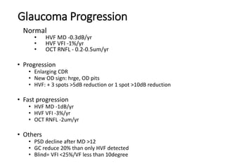
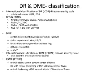
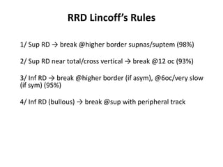
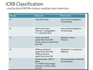
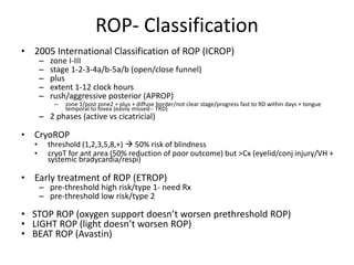
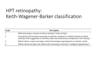
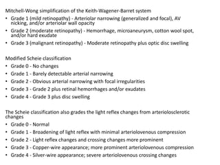
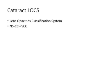
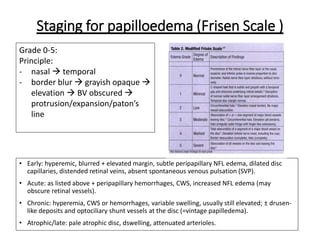
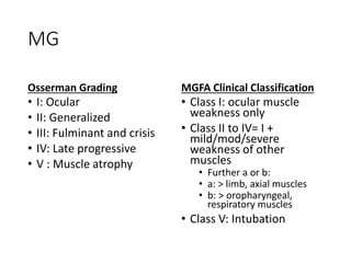
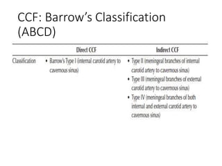
Classifications-Grading-Staging
@ Ophthalmology
Dummy Survival Guide
Dr Yong Meng Hsien 2021
Discussion Approach (Anatomy):
• Cornea & external eye diseases
• Glaucoma, angle and IOP
• Uveitis
• Lens and cataract
• Vitreo-retina
• Neuro-ophthalmology
• Orbit & Oculoplastic
• Trauma
General Classifications
• Risk factors
• E.g. modifiable/not, ocular/systemic
• Pathogenesis
• E.g. Ischemic/occlusive/haemorrhagic, necrotic/fibrotic
• Aetiology/causes
• Clinical features/Severity/Progression/Complications
• Investigation/tests
• E.g. ocular/systemic, invasive/not
• Management
• E.g. conservative/medical/laser/surgical
• E.g. factors to be considered
• Primary vs Secondary
• Systemic vs Ocular
• Congenital vs Acquired
• Anatomical approach
• Etiological approach (VITAMINS, VEINS, VINDICATE)
• Most common, important. life/sight threatening
Corneal & External Eye Diseases
• Keratoconus
• Corneal dystrophies
• Dry eye
• VKC
• HSV keratitis
• Prognosis for corneal transplant
• Trachoma
• GPC
Global Consensus on Keratoconus and
Ectatic Diseases 2015
• Currently there is no clinically adequate
classification system for keratoconus
• Currently, there is no consistent or clear definition
of ectasia progression
Ref + Astig + Topo + Km + CCT
Amsler-Krumeich KC Classification
Belin ABCD KC Staging
• Ant & Back radius of
curvature (3mm),
• Corneal thickness
(minimal)
• Distance VA (DCVA)
Pentacam for KC
• K Max & ABCD & Thinnest pachy  progression
• Indices  colour code, KISA (60-100), IS (1.4-1.9)
• BAD D (enhanced ectasia)  screening & colour code
(change/different: ant 5-7 & post 12-16 )
• Covis biomechanical: CBI & TBI
• Asphericity/Q value: normal -0.01 to -0.8
• Q-val <0/-ve/oblate = normal or KC or post hyperop LVC
• Q-val >0/+ve/prolate = post myopic LVC
• Q-val=0/sphere
IC3D 2015 (International Classification of Corneal
Dystrophies)
- Genetic (chromosomal/gene)
- Clinical (pattern/anatomical layer)
- Pathology (histo/biochem/confocal)
DEWS 2017 (International Dry Eye Workshop)
Classification: Aqueous deficiency VS evaporative
• Aq deficiency
– Sjogren VS Non Sjogren
• Evaporative
– Intrinsic VS Extrinsic
Dry Eye Severity Grading Scheme
• Symptoms + Signs (conj/cornea/tear/lid & gland) + Tests (TBUT/Schimer)
VKC- classification
• Limbal/bulbar vs palbebral vs mixed
• Chronic vs intermittent
• VKC Grade 0-4 (various)
• Mild/mod/severe (just noticeable/most of the
day/interrupt daily routine) & vision threatening
• Papillae Grading 0-4 (size: 0.3-1-3mm, location:
tarsal-limbus)
• Shield ulcer Grading 1-3 (content: clear-debris-
plaque, treatment response/re-epithelised)
HSV Keratitis
Brightbill’s Classification:
Corneal Graft Prognosis
• Grade I to V (excellent to poor)
• Underlying pathology + secondary complication
Trachoma: WHO classification & SAFE strategy
SAFE strategy: Surgery for in-turned eyelashes, Antibiotics for active disease, Face washing (or
promotion of facial cleanliness), and Environmental improvement to reduce transmission.
Uveitis
Uveitis
• Classification
• AC cells & flare
• Vitreous cells and haze
• Course & progression
• Sarcoidosis
• level of diagnostic certainty
• Staging of pulmonary sarcoidosis
• Phases of FFA
• Immunomodulators
• Hypersensitivity reactions
SUN Anatomical Classification
Uveitis SUN / NIH / MUST
International Workshop on Ocular Sarcoidosis (IWOS)
• 7signs:
– (1) mutton-fat keratic precipitates (KPs)/small granulomatous KPs and/or iris nodules
(Koeppe/Busacca),
– (2) trabecular meshwork (TM) nodules and/or tent-shaped peripheral anterior synechiae (PAS),
– (3) vitreous opacities displaying snowballs/strings of pearls,
– (4) multiple chorioretinal peripheral lesions (active and/or atrophic),
– (5) nodular and/or segmental peri-phlebitis (± candlewax drippings) and/or retinal macroaneurism in
an inflamed eye,
– 6) optic disc nodule(s)/granuloma(s) and/or solitary choroidal nodule, and
– (7) bilaterality
• 5labs/imaging Ix:
– (1) negative tuberculin skin test in a BCG-vaccinated patient or in a patient having had a positive
tuberculin skin test previously,
– (2) elevated serum angiotensin converting enzyme (ACE) levels and/or elevated serum lysozyme,
– (3) chest x-ray revealing bilateral hilar lymphadenopathy (BHL),
– (4) abnormal liver enzyme tests, and
– (5) chest CT scan in patients with a negative chest x-ray result.
• Four levels of certainty for the diagnosis of ocular sarcoidosis (diagnostic criteria) were
recommended in patients in whom other possible causes of uveitis had been excluded:
– (1) biopsy-supported diagnosis with a compatible uveitis was labeled as definite ocular sarcoidosis;
– (2) if biopsy was not done but chest x-ray was positive showing BHL associated with a compatible
uveitis, the condition was labeled as presumed ocular sarcoidosis;
– (3) if biopsy was not done and the chest x-ray did not show BHL but there were 3 of the above
intraocular signs and 2 positive laboratory tests, the condition was labeled as probable ocular
sarcoidosis; and
– (4) if lung biopsy was done and the result was negative but at least 4 of the above signs and 2
positive laboratory investigations were present, the condition was labeled as possible ocular
sarcoidosis
Pulmonary Sarcoidosis
• Stage 1: Bilateral hilar lymphadenopathy
• Stage 2: Bilateral hilar lymphadenopathy and
reticulo-nodular parenchymal infiltrates
• Stage 3: Reticulo-nodular parenchymal infiltrates
alone
• Stage 4: Progressive pulmonary fibrosis
Phases of FFA
Type V (Stimulating Ab)- grave’s, MG
ImmunoModulator (IMT)
• Stimulant (IF/IL)
• Immunosuppressant
– Corticosteroid
– Antimetabolite (AZT/MTX/MPM)
– T cell/calcineurin inhibitor (CSA/Tacro-Siro-limus)
– Alkylating agents (cyclophosphamide/chlorambucil)
– Biologic (TNF-a AG, IL-1/IL-6 AG, IFN-a, IVIg)
Glaucoma & Angle
Glaucoma & Angle
• Angle classification
• Stages of primary angle closure
• Stages of glaucoma (severity)
• Glaucoma progression
• Classification of topical antiglaucoma
• Grading of shallow AC
Angle
Shaffer Spaeth
Scheie
• Narrow slit beam
• Perpendicular to the most peripheral part of the cornea
• Slit beam angle of about 60°
• Compare the thickness of the cornea & visible aqueous
Becker Goniogram
Means of drawing gonioscopic findings
Allow description of the variable anatomy of an angle within a quadrant
Record synechiae, tumors, foreign bodies, and so on
Stages of primary angle closure
Glaucoma- Stage
Hodapp Parrish Anderson
– mild/mod/advanced/severe
– By MD: 6-12-20
– By points of depression: 25%-50%-75%
Glaucoma Progression
Normal
• HVF MD -0.3dB/yr
• HVF VFI -1%/yr
• OCT RNFL - 0.2-0.5um/yr
• Progression
• Enlarging CDR
• New OD sign: hrge, OD pits
• HVF: + 3 spots >5dB reduction or 1 spot >10dB reduction
• Fast progression
• HVF MD -1dB/yr
• HVF VFI -3%/yr
• OCT RNFL -2um/yr
• Others
• PSD decline after MD >12
• GC reduce 20% than only HVF detected
• Blind= VFI <25%/VF less than 10degree
Topical Antiglaucoma Classification
Vitreo-retinal
Vitreo-retinal
• DR
• DME
• AMD
• Geographical atrophy
• RD & PVR
• Macular hole
• Retinoblastoma
• Sickle cell retinopathy
• Hypertensive retinopathy
• Best disease
• Choroidal melanoma
DR & DME- classification
• International classification of DR (ICDR) disease severity scale
• mild-mod-severe NDPR, PDR
• DRS & ETDRS
• NPDR severe/very severe, PDR early/high risk
• NVD >/= 1/3 DD
• NVD <1/3 DD with VH/PRH
• NVE >/= ½ DD with VH/PRH
• DME
– center involvement: CMT (center 1mm) >250um
– vision impairment: VA =/< 6/9
– focal: micro-aneurysm with circinate ring
– diffuse: cystoid ME
– +- VMT
• International classification of DME (ICDME) disease severity scale
• DME absent vs present (mild-mod-severe)
• CSME (ETDRS)
– retinal edema within 500um center of fovea
– HE with retinal thickening within 500um center of fovea
– retinal thickening >1DD located within 1DD center of fovea
ARMD- Type & Classification
• Dry vs wet- 90 vs 10% (blindness opposite)
• Normal/normal aging/early/intermediate/late AMD
– drusen size + pigmentary changes → CNV/GA
• Dry- drusen/GA
• Drusen- size (drupelets/63/125=vein diameter at OD margin), morphology
(hard/soft/confluent), +- dystrophic calcification +- pigmentary changes
• PED- x 4 (drusenoid/serous/hrge/FVC)
• Wet- CNV/PED/RAP/PCV
• CNV (FFA/MPS study)
– classic (20%, well defined and early, predominant/minimal), (location:
extra/juxta/subfovea, 200um vs foveola)
– occult (80%, FV PED/LLUS)
• CNV (ICG)
– Hot spot <1DD (less common, for laser)
– Plaque >1DD (more common, poor natural history)
– Combination (rare)
• Wet (CNV)- type 1/2 (subRPE/subretina) or type 3 (RCA with RAP)
• CNV (active/not)- +fluid/hrge/leak on FFA/enlarging CNV membrane/deteriorating
vision
• Variant- RAP x 3 stages (1-3: IRN/SRN/RCA), IPCV
AMD Classification & AREDS Staging
GA New OCT Classification
• New classification (OCT)
• Outer retinal atrophy (ORA) only or with RPE (RORA)
• Complete vs incomplete
• cRORA = GA
• iRORA = early GA
• cORA
• iORA
PVR
Others PVR classification: RSC (1-4, a-c for 3-4), silicone study (1-6, 1-3 post, 4-6 ant)
RRD Lincoff’s Rules
1/ Sup RD → break @higher border supnas/suptem (98%)
2/ Sup RD near total/cross vertical → break @12 oc (93%)
3/ Inf RD → break @higher border (if asym), @6oc/very slow
(if sym) (95%)
4/ Inf RD (bullous) → break @sup with peripheral track
Macular Hole- Classification
• Gass VS IVTS
– Stage 0/VMA
• oblique VMA at parafovea with normal fovea
– Stage 1a/VMT
• Yellow spot
• Flattened foveal depression + cyst-like schisis (inner changes)
– Stage 1b/VMT
• Yellow ring
• 1a with centrifugal displacement of photoreceptor (outer changes)
– Stage 2/small-medium FTMH with VMT
• <250/<400um hole/red spot
– Stage 3/large FTMH without VMT (no complete PVD yet)
• >400um hole/red spot base with yellow dots +- operculum
– Stage 4/FTMH (any size) without VMT (complete PVD)
• Weiss ring with red spot +- ERM
ICRB Classification
- size/location/SRF/SR-vitreous seed/ant-post extension -
ROP- Classification
• 2005 International Classification of ROP (ICROP)
– zone I-III
– stage 1-2-3-4a/b-5a/b (open/close funnel)
– plus
– extent 1-12 clock hours
– rush/aggressive posterior (APROP)
– zone 1/post zone2 + plus + diffuse border/not clear stage/progress fast to RD within days + tongue
temporal to fovea (easily missed-- TRD)
– 2 phases (active vs cicatricial)
• CryoROP
• threshold (1,2,3,5,8,+)  50% risk of blindness
• cryoT for ant area (50% reduction of poor outcome) but >Cx (eyelid/conj injury/VH +
systemic bradycardia/respi)
• Early treatment of ROP (ETROP)
– pre-threshold high risk/type 1- need Rx
– pre-threshold low risk/type 2
• STOP ROP (oxygen support doesn’t worsen prethreshold ROP)
• LIGHT ROP (light doesn’t worsen ROP)
• BEAT ROP (Avastin)
Goldberg staging:
Proliferative Sickle Cell Retinopathy
HPT retinopathy:
Keith-Wagener-Barker classification
Mitchell-Wong simplification of the Keith-Wagener-Barret system​
• Grade 1 (mild retinopathy) - Arteriolar narrowing (generalized and focal), AV
nicking, and/or arteriolar wall opacity
• Grade 2 (moderate retinopathy) - Hemorrhage, microaneurysm, cotton wool spot,
and/or hard exudate
• Grade 3 (malignant retinopathy) - Moderate retinopathy plus optic disc swelling
Modified Scheie classification
• Grade 0 - No changes
• Grade 1 - Barely detectable arterial narrowing
• Grade 2 - Obvious arterial narrowing with focal irregularities
• Grade 3 - Grade 2 plus retinal hemorrhages and/or exudates
• Grade 4 - Grade 3 plus disc swelling
The Scheie classification also grades the light reflex changes from arteriolosclerotic
changes
• Grade 0 - Normal
• Grade 1 - Broadening of light reflex with minimal arteriolovenous compression
• Grade 2 - Light reflex changes and crossing changes more prominent
• Grade 3 - Copper-wire appearance; more prominent arteriolovenous compression
• Grade 4 - Silver-wire appearance; severe arteriolovenous crossing changes
Staging of Best disease
Collaborative Ocular melanoma Study (COMS)
• Small (1-3mm thick, <5mm long) @1989
–natural history/observe: 30% +growth in 5yr
–mortality: 6% 5yr, 15% 8yr → photoCoag
• Medium (3-8mm thick, <16mm long) @1998
–enucleation VS brachyT (iodine 125 gold
plaque/85Gy)
–survival: same (82% @5yr) → brachyT
• Large (8mm thick, >16mm long) @1994
–enucleation alone VS pre-radiation (ext
beam/20Gy)
–survival: same (60% @5yr) → enucleation
Staging of Lyme Disease
High myopia
META-PM (meta-analysis for pathologic myopia) international classification
• for myopic maculopathy
• OCT and colour fundus photography
• retinal changes + choroid, Bruch’s membrane and the RPE
• “no myopic retinal degenerative lesion” (Category 0), “tessellated fundus”
(Category 1), “diffuse chorioretinal atrophy” (Category 2), “patchy chorioretinal
atrophy” (Category 3), and “macular atrophy” (Category 4).
• “plus” lesions: lacquer cracks, myopic choroidal neovascularisation, and Fuchs
spot.
• Posterior staphyloma was considered as a further important sign
ATN classification
• three factors: atrophy, traction and neovascularisation.
Lens & Cataract
Cataract LOCS
• Lens Opacities Classification System
• NS-CC-PSCC
Neuro-Ophthalmology
Neuro-Ophthalmology
• RAPD
• Papilloedema
• MG
• CCF
• Duane syndrome
• Facial nerve palsy (severity)
RAPD
Grading (Bell et al 1993)
• Grade I: Weak constriction then greater dilatation
• Grade II: No initial constriction, stall, then dilatation
• Grade III: Immediate dilatation
• Grade IV: Immediate dilatation following prolonged illumination
of the good eye for six seconds
• Grade V: Immediate dilatation with no secondary constriction
Staging for papilloedema (Frisen Scale )
Grade 0-5:
Principle:
- nasal  temporal
- border blur  grayish opaque 
elevation  BV obscured 
protrusion/expansion/paton’s
line
• Early: hyperemic, blurred + elevated margin, subtle peripapillary NFL edema, dilated disc
capillaries, distended retinal veins, absent spontaneous venous pulsation (SVP).
• Acute: as listed above + peripapillary hemorrhages, CWS, increased NFL edema (may
obscure retinal vessels).
• Chronic: hyperemia, CWS or hemorrhages, variable swelling, usually still elevated; ± drusen-
like deposits and optociliary shunt vessels at the disc (=vintage papilledema).
• Atrophic/late: pale atrophic disc, dswelling, attenuated arterioles.
MG
Osserman Grading
• I: Ocular
• II: Generalized
• III: Fulminant and crisis
• IV: Late progressive
• V : Muscle atrophy
MGFA Clinical Classification
• Class I: ocular muscle
weakness only
• Class II to IV= I +
mild/mod/severe
weakness of other
muscles
• Further a or b:
• a: > limb, axial muscles
• b: > oropharyngeal,
respiratory muscles
• Class V: Intubation
CCF: Barrow’s Classification
(ABCD)
Duane Syndrome
Facial Nerve Palsy Severity
Oculoplastic & Orbit
• TED
• Orbital cellulitis
TED- Severity & Activity
• Mourits clinical activity score (CAS)
• PRSI: pain/red/swelling/impaired function-2/2/4/2 (=/>3/7 
4/10)
• Last 3 score for follow up (1-3mth): proptosis >2mm, VA
1line/more, diplopia >8 degree
• Mourits inflammatory index
• Conj edema3/conj red 1/lid edema 3/lid red1/pain rest1/move1
• NOSPECS (severity only)
• Modifield: SPECS further grade 0/a/b/c (Werner’s modified)
• VISA (vision/inflam/strabismus/appear: 1/10/6/3)
• Severe: Global severity grade (20pt)
• Active: I (=/>4 ), S (=/>12degree or fluctuate)
• EUGOGO
• Activity (CAS PRSI), severity (RSI + eyelid + cornea)
• mild (soft tissue/corneal SPK/no EOM)
• mod-severe (lid retraction 2mm, proptosis 3mm, EOM),
• sight threatening (ON/corneal bdown/frozen eye/choroidal fold)
Orbital Cellulitis: Chandler’s Classification
Ophthalmic Trauma
Ophthalmic Trauma
• Ocular trauma scoring
• Mid-facial fracture
• Chemical injury
• Hyphaema
Ocular Trauma Terminology
• Ocular Trauma Classification Group 1997 & Ocular
Trauma Score (OTS) 2002
• Birmingham Eye Trauma Terminology System
(BETTS)
OTS
• scores range from 1 (most severe injury and worst prognosis at 6 months follow-up) to 5 (least
severe injury and least poor prognosis at 6 months). Each score is associated with a range of
predicted post-injury visual acuities. It has a predictive accuracy of approximately 80%, which
means that the OTS will be accurate 4 out of 5 times
BETTS
Closed-globe injuries
• contusion (blunt trauma without break in eyewall)
• lamellar laceration (partial-thickness wound of the eyewall)
• superficial foreign bodies
Open-globe injuries
• rupture (blunt trauma with break in eyewall)
• laceration (full-thickness wound of the eyewall, caused by a sharp
object)
• intraocular foreign bodies, penetrating or perforating
• penetrating injury (entrance break; no exit break in eyewall)
• perforating injury (both entrance and exit breaks in eyewall)
Midface fracture
• Midface/Le Fort # (extended maxillary #)
I: low transverse maxillary (above teeth)
II: pyramidal maxillary-nasal/lacrimal/medial orbital
floor/rim
III: facial skeleton-base of skull detachM (orbital
floor/med/lat walls)
Chemical injury
End.
Thank you

Classification Staging Grading in Ophthalmology

  • 1.
  • 2.
    Discussion Approach (Anatomy): •Cornea & external eye diseases • Glaucoma, angle and IOP • Uveitis • Lens and cataract • Vitreo-retina • Neuro-ophthalmology • Orbit & Oculoplastic • Trauma
  • 3.
    General Classifications • Riskfactors • E.g. modifiable/not, ocular/systemic • Pathogenesis • E.g. Ischemic/occlusive/haemorrhagic, necrotic/fibrotic • Aetiology/causes • Clinical features/Severity/Progression/Complications • Investigation/tests • E.g. ocular/systemic, invasive/not • Management • E.g. conservative/medical/laser/surgical • E.g. factors to be considered • Primary vs Secondary • Systemic vs Ocular • Congenital vs Acquired • Anatomical approach • Etiological approach (VITAMINS, VEINS, VINDICATE) • Most common, important. life/sight threatening
  • 4.
    Corneal & ExternalEye Diseases
  • 5.
    Corneal & ExternalEye Diseases • Keratoconus • Corneal dystrophies • Dry eye • VKC • HSV keratitis • Prognosis for corneal transplant • Trachoma • GPC
  • 6.
    Global Consensus onKeratoconus and Ectatic Diseases 2015 • Currently there is no clinically adequate classification system for keratoconus • Currently, there is no consistent or clear definition of ectasia progression
  • 7.
    Ref + Astig+ Topo + Km + CCT Amsler-Krumeich KC Classification
  • 8.
    Belin ABCD KCStaging • Ant & Back radius of curvature (3mm), • Corneal thickness (minimal) • Distance VA (DCVA)
  • 9.
    Pentacam for KC •K Max & ABCD & Thinnest pachy  progression • Indices  colour code, KISA (60-100), IS (1.4-1.9) • BAD D (enhanced ectasia)  screening & colour code (change/different: ant 5-7 & post 12-16 ) • Covis biomechanical: CBI & TBI • Asphericity/Q value: normal -0.01 to -0.8 • Q-val <0/-ve/oblate = normal or KC or post hyperop LVC • Q-val >0/+ve/prolate = post myopic LVC • Q-val=0/sphere
  • 10.
    IC3D 2015 (InternationalClassification of Corneal Dystrophies) - Genetic (chromosomal/gene) - Clinical (pattern/anatomical layer) - Pathology (histo/biochem/confocal)
  • 11.
    DEWS 2017 (InternationalDry Eye Workshop) Classification: Aqueous deficiency VS evaporative • Aq deficiency – Sjogren VS Non Sjogren • Evaporative – Intrinsic VS Extrinsic Dry Eye Severity Grading Scheme • Symptoms + Signs (conj/cornea/tear/lid & gland) + Tests (TBUT/Schimer)
  • 12.
    VKC- classification • Limbal/bulbarvs palbebral vs mixed • Chronic vs intermittent • VKC Grade 0-4 (various) • Mild/mod/severe (just noticeable/most of the day/interrupt daily routine) & vision threatening • Papillae Grading 0-4 (size: 0.3-1-3mm, location: tarsal-limbus) • Shield ulcer Grading 1-3 (content: clear-debris- plaque, treatment response/re-epithelised)
  • 13.
  • 14.
    Brightbill’s Classification: Corneal GraftPrognosis • Grade I to V (excellent to poor) • Underlying pathology + secondary complication
  • 15.
    Trachoma: WHO classification& SAFE strategy SAFE strategy: Surgery for in-turned eyelashes, Antibiotics for active disease, Face washing (or promotion of facial cleanliness), and Environmental improvement to reduce transmission.
  • 16.
  • 17.
    Uveitis • Classification • ACcells & flare • Vitreous cells and haze • Course & progression • Sarcoidosis • level of diagnostic certainty • Staging of pulmonary sarcoidosis • Phases of FFA • Immunomodulators • Hypersensitivity reactions
  • 18.
  • 19.
    Uveitis SUN /NIH / MUST
  • 21.
    International Workshop onOcular Sarcoidosis (IWOS) • 7signs: – (1) mutton-fat keratic precipitates (KPs)/small granulomatous KPs and/or iris nodules (Koeppe/Busacca), – (2) trabecular meshwork (TM) nodules and/or tent-shaped peripheral anterior synechiae (PAS), – (3) vitreous opacities displaying snowballs/strings of pearls, – (4) multiple chorioretinal peripheral lesions (active and/or atrophic), – (5) nodular and/or segmental peri-phlebitis (± candlewax drippings) and/or retinal macroaneurism in an inflamed eye, – 6) optic disc nodule(s)/granuloma(s) and/or solitary choroidal nodule, and – (7) bilaterality • 5labs/imaging Ix: – (1) negative tuberculin skin test in a BCG-vaccinated patient or in a patient having had a positive tuberculin skin test previously, – (2) elevated serum angiotensin converting enzyme (ACE) levels and/or elevated serum lysozyme, – (3) chest x-ray revealing bilateral hilar lymphadenopathy (BHL), – (4) abnormal liver enzyme tests, and – (5) chest CT scan in patients with a negative chest x-ray result. • Four levels of certainty for the diagnosis of ocular sarcoidosis (diagnostic criteria) were recommended in patients in whom other possible causes of uveitis had been excluded: – (1) biopsy-supported diagnosis with a compatible uveitis was labeled as definite ocular sarcoidosis; – (2) if biopsy was not done but chest x-ray was positive showing BHL associated with a compatible uveitis, the condition was labeled as presumed ocular sarcoidosis; – (3) if biopsy was not done and the chest x-ray did not show BHL but there were 3 of the above intraocular signs and 2 positive laboratory tests, the condition was labeled as probable ocular sarcoidosis; and – (4) if lung biopsy was done and the result was negative but at least 4 of the above signs and 2 positive laboratory investigations were present, the condition was labeled as possible ocular sarcoidosis
  • 22.
    Pulmonary Sarcoidosis • Stage1: Bilateral hilar lymphadenopathy • Stage 2: Bilateral hilar lymphadenopathy and reticulo-nodular parenchymal infiltrates • Stage 3: Reticulo-nodular parenchymal infiltrates alone • Stage 4: Progressive pulmonary fibrosis
  • 23.
  • 24.
    Type V (StimulatingAb)- grave’s, MG
  • 25.
    ImmunoModulator (IMT) • Stimulant(IF/IL) • Immunosuppressant – Corticosteroid – Antimetabolite (AZT/MTX/MPM) – T cell/calcineurin inhibitor (CSA/Tacro-Siro-limus) – Alkylating agents (cyclophosphamide/chlorambucil) – Biologic (TNF-a AG, IL-1/IL-6 AG, IFN-a, IVIg)
  • 26.
  • 27.
    Glaucoma & Angle •Angle classification • Stages of primary angle closure • Stages of glaucoma (severity) • Glaucoma progression • Classification of topical antiglaucoma • Grading of shallow AC
  • 28.
  • 29.
    • Narrow slitbeam • Perpendicular to the most peripheral part of the cornea • Slit beam angle of about 60° • Compare the thickness of the cornea & visible aqueous
  • 30.
    Becker Goniogram Means ofdrawing gonioscopic findings Allow description of the variable anatomy of an angle within a quadrant Record synechiae, tumors, foreign bodies, and so on
  • 31.
    Stages of primaryangle closure
  • 32.
    Glaucoma- Stage Hodapp ParrishAnderson – mild/mod/advanced/severe – By MD: 6-12-20 – By points of depression: 25%-50%-75%
  • 33.
    Glaucoma Progression Normal • HVFMD -0.3dB/yr • HVF VFI -1%/yr • OCT RNFL - 0.2-0.5um/yr • Progression • Enlarging CDR • New OD sign: hrge, OD pits • HVF: + 3 spots >5dB reduction or 1 spot >10dB reduction • Fast progression • HVF MD -1dB/yr • HVF VFI -3%/yr • OCT RNFL -2um/yr • Others • PSD decline after MD >12 • GC reduce 20% than only HVF detected • Blind= VFI <25%/VF less than 10degree
  • 34.
  • 35.
  • 36.
    Vitreo-retinal • DR • DME •AMD • Geographical atrophy • RD & PVR • Macular hole • Retinoblastoma • Sickle cell retinopathy • Hypertensive retinopathy • Best disease • Choroidal melanoma
  • 37.
    DR & DME-classification • International classification of DR (ICDR) disease severity scale • mild-mod-severe NDPR, PDR • DRS & ETDRS • NPDR severe/very severe, PDR early/high risk • NVD >/= 1/3 DD • NVD <1/3 DD with VH/PRH • NVE >/= ½ DD with VH/PRH • DME – center involvement: CMT (center 1mm) >250um – vision impairment: VA =/< 6/9 – focal: micro-aneurysm with circinate ring – diffuse: cystoid ME – +- VMT • International classification of DME (ICDME) disease severity scale • DME absent vs present (mild-mod-severe) • CSME (ETDRS) – retinal edema within 500um center of fovea – HE with retinal thickening within 500um center of fovea – retinal thickening >1DD located within 1DD center of fovea
  • 41.
    ARMD- Type &Classification • Dry vs wet- 90 vs 10% (blindness opposite) • Normal/normal aging/early/intermediate/late AMD – drusen size + pigmentary changes → CNV/GA • Dry- drusen/GA • Drusen- size (drupelets/63/125=vein diameter at OD margin), morphology (hard/soft/confluent), +- dystrophic calcification +- pigmentary changes • PED- x 4 (drusenoid/serous/hrge/FVC) • Wet- CNV/PED/RAP/PCV • CNV (FFA/MPS study) – classic (20%, well defined and early, predominant/minimal), (location: extra/juxta/subfovea, 200um vs foveola) – occult (80%, FV PED/LLUS) • CNV (ICG) – Hot spot <1DD (less common, for laser) – Plaque >1DD (more common, poor natural history) – Combination (rare) • Wet (CNV)- type 1/2 (subRPE/subretina) or type 3 (RCA with RAP) • CNV (active/not)- +fluid/hrge/leak on FFA/enlarging CNV membrane/deteriorating vision • Variant- RAP x 3 stages (1-3: IRN/SRN/RCA), IPCV
  • 42.
    AMD Classification &AREDS Staging
  • 43.
    GA New OCTClassification • New classification (OCT) • Outer retinal atrophy (ORA) only or with RPE (RORA) • Complete vs incomplete • cRORA = GA • iRORA = early GA • cORA • iORA
  • 44.
    PVR Others PVR classification:RSC (1-4, a-c for 3-4), silicone study (1-6, 1-3 post, 4-6 ant)
  • 45.
    RRD Lincoff’s Rules 1/Sup RD → break @higher border supnas/suptem (98%) 2/ Sup RD near total/cross vertical → break @12 oc (93%) 3/ Inf RD → break @higher border (if asym), @6oc/very slow (if sym) (95%) 4/ Inf RD (bullous) → break @sup with peripheral track
  • 46.
    Macular Hole- Classification •Gass VS IVTS – Stage 0/VMA • oblique VMA at parafovea with normal fovea – Stage 1a/VMT • Yellow spot • Flattened foveal depression + cyst-like schisis (inner changes) – Stage 1b/VMT • Yellow ring • 1a with centrifugal displacement of photoreceptor (outer changes) – Stage 2/small-medium FTMH with VMT • <250/<400um hole/red spot – Stage 3/large FTMH without VMT (no complete PVD yet) • >400um hole/red spot base with yellow dots +- operculum – Stage 4/FTMH (any size) without VMT (complete PVD) • Weiss ring with red spot +- ERM
  • 47.
  • 49.
    ROP- Classification • 2005International Classification of ROP (ICROP) – zone I-III – stage 1-2-3-4a/b-5a/b (open/close funnel) – plus – extent 1-12 clock hours – rush/aggressive posterior (APROP) – zone 1/post zone2 + plus + diffuse border/not clear stage/progress fast to RD within days + tongue temporal to fovea (easily missed-- TRD) – 2 phases (active vs cicatricial) • CryoROP • threshold (1,2,3,5,8,+)  50% risk of blindness • cryoT for ant area (50% reduction of poor outcome) but >Cx (eyelid/conj injury/VH + systemic bradycardia/respi) • Early treatment of ROP (ETROP) – pre-threshold high risk/type 1- need Rx – pre-threshold low risk/type 2 • STOP ROP (oxygen support doesn’t worsen prethreshold ROP) • LIGHT ROP (light doesn’t worsen ROP) • BEAT ROP (Avastin)
  • 51.
  • 52.
  • 53.
    Mitchell-Wong simplification ofthe Keith-Wagener-Barret system​ • Grade 1 (mild retinopathy) - Arteriolar narrowing (generalized and focal), AV nicking, and/or arteriolar wall opacity • Grade 2 (moderate retinopathy) - Hemorrhage, microaneurysm, cotton wool spot, and/or hard exudate • Grade 3 (malignant retinopathy) - Moderate retinopathy plus optic disc swelling Modified Scheie classification • Grade 0 - No changes • Grade 1 - Barely detectable arterial narrowing • Grade 2 - Obvious arterial narrowing with focal irregularities • Grade 3 - Grade 2 plus retinal hemorrhages and/or exudates • Grade 4 - Grade 3 plus disc swelling The Scheie classification also grades the light reflex changes from arteriolosclerotic changes • Grade 0 - Normal • Grade 1 - Broadening of light reflex with minimal arteriolovenous compression • Grade 2 - Light reflex changes and crossing changes more prominent • Grade 3 - Copper-wire appearance; more prominent arteriolovenous compression • Grade 4 - Silver-wire appearance; severe arteriolovenous crossing changes
  • 54.
  • 55.
    Collaborative Ocular melanomaStudy (COMS) • Small (1-3mm thick, <5mm long) @1989 –natural history/observe: 30% +growth in 5yr –mortality: 6% 5yr, 15% 8yr → photoCoag • Medium (3-8mm thick, <16mm long) @1998 –enucleation VS brachyT (iodine 125 gold plaque/85Gy) –survival: same (82% @5yr) → brachyT • Large (8mm thick, >16mm long) @1994 –enucleation alone VS pre-radiation (ext beam/20Gy) –survival: same (60% @5yr) → enucleation
  • 56.
  • 57.
    High myopia META-PM (meta-analysisfor pathologic myopia) international classification • for myopic maculopathy • OCT and colour fundus photography • retinal changes + choroid, Bruch’s membrane and the RPE • “no myopic retinal degenerative lesion” (Category 0), “tessellated fundus” (Category 1), “diffuse chorioretinal atrophy” (Category 2), “patchy chorioretinal atrophy” (Category 3), and “macular atrophy” (Category 4). • “plus” lesions: lacquer cracks, myopic choroidal neovascularisation, and Fuchs spot. • Posterior staphyloma was considered as a further important sign ATN classification • three factors: atrophy, traction and neovascularisation.
  • 58.
  • 59.
    Cataract LOCS • LensOpacities Classification System • NS-CC-PSCC
  • 60.
  • 61.
    Neuro-Ophthalmology • RAPD • Papilloedema •MG • CCF • Duane syndrome • Facial nerve palsy (severity)
  • 62.
    RAPD Grading (Bell etal 1993) • Grade I: Weak constriction then greater dilatation • Grade II: No initial constriction, stall, then dilatation • Grade III: Immediate dilatation • Grade IV: Immediate dilatation following prolonged illumination of the good eye for six seconds • Grade V: Immediate dilatation with no secondary constriction
  • 63.
    Staging for papilloedema(Frisen Scale ) Grade 0-5: Principle: - nasal  temporal - border blur  grayish opaque  elevation  BV obscured  protrusion/expansion/paton’s line • Early: hyperemic, blurred + elevated margin, subtle peripapillary NFL edema, dilated disc capillaries, distended retinal veins, absent spontaneous venous pulsation (SVP). • Acute: as listed above + peripapillary hemorrhages, CWS, increased NFL edema (may obscure retinal vessels). • Chronic: hyperemia, CWS or hemorrhages, variable swelling, usually still elevated; ± drusen- like deposits and optociliary shunt vessels at the disc (=vintage papilledema). • Atrophic/late: pale atrophic disc, dswelling, attenuated arterioles.
  • 64.
    MG Osserman Grading • I:Ocular • II: Generalized • III: Fulminant and crisis • IV: Late progressive • V : Muscle atrophy MGFA Clinical Classification • Class I: ocular muscle weakness only • Class II to IV= I + mild/mod/severe weakness of other muscles • Further a or b: • a: > limb, axial muscles • b: > oropharyngeal, respiratory muscles • Class V: Intubation
  • 65.
  • 66.
  • 67.
  • 68.
  • 69.
    Oculoplastic & Orbit •TED • Orbital cellulitis
  • 70.
    TED- Severity &Activity • Mourits clinical activity score (CAS) • PRSI: pain/red/swelling/impaired function-2/2/4/2 (=/>3/7  4/10) • Last 3 score for follow up (1-3mth): proptosis >2mm, VA 1line/more, diplopia >8 degree • Mourits inflammatory index • Conj edema3/conj red 1/lid edema 3/lid red1/pain rest1/move1 • NOSPECS (severity only) • Modifield: SPECS further grade 0/a/b/c (Werner’s modified) • VISA (vision/inflam/strabismus/appear: 1/10/6/3) • Severe: Global severity grade (20pt) • Active: I (=/>4 ), S (=/>12degree or fluctuate) • EUGOGO • Activity (CAS PRSI), severity (RSI + eyelid + cornea) • mild (soft tissue/corneal SPK/no EOM) • mod-severe (lid retraction 2mm, proptosis 3mm, EOM), • sight threatening (ON/corneal bdown/frozen eye/choroidal fold)
  • 74.
  • 75.
  • 76.
    Ophthalmic Trauma • Oculartrauma scoring • Mid-facial fracture • Chemical injury • Hyphaema
  • 77.
    Ocular Trauma Terminology •Ocular Trauma Classification Group 1997 & Ocular Trauma Score (OTS) 2002 • Birmingham Eye Trauma Terminology System (BETTS)
  • 78.
    OTS • scores rangefrom 1 (most severe injury and worst prognosis at 6 months follow-up) to 5 (least severe injury and least poor prognosis at 6 months). Each score is associated with a range of predicted post-injury visual acuities. It has a predictive accuracy of approximately 80%, which means that the OTS will be accurate 4 out of 5 times
  • 79.
    BETTS Closed-globe injuries • contusion(blunt trauma without break in eyewall) • lamellar laceration (partial-thickness wound of the eyewall) • superficial foreign bodies Open-globe injuries • rupture (blunt trauma with break in eyewall) • laceration (full-thickness wound of the eyewall, caused by a sharp object) • intraocular foreign bodies, penetrating or perforating • penetrating injury (entrance break; no exit break in eyewall) • perforating injury (both entrance and exit breaks in eyewall)
  • 80.
    Midface fracture • Midface/LeFort # (extended maxillary #) I: low transverse maxillary (above teeth) II: pyramidal maxillary-nasal/lacrimal/medial orbital floor/rim III: facial skeleton-base of skull detachM (orbital floor/med/lat walls)
  • 81.
  • 83.