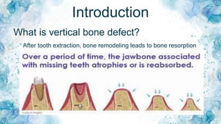
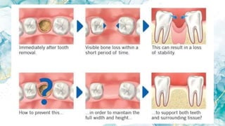
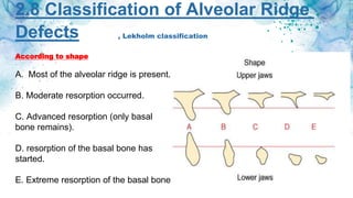
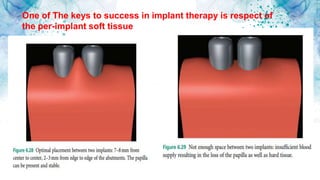
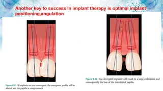
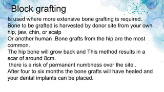
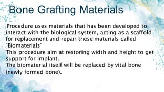
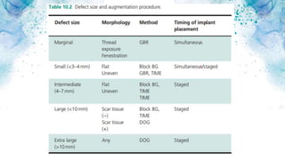
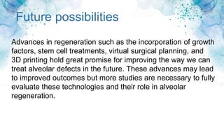
By Ali Hussein
Vertical Bone
Defect In Dental
Implant
Supervised
by
Dr. Hind Sabah
What is vertical bone defect?
After tooth extraction, bone remodeling leads to bone resorption
Introduction
2.8 Classification of Alveolar Ridge
Defects
According to shape
A. Most of the alveolar ridge is present.
B. Moderate resorption occurred.
C. Advanced resorption (only basal
bone remains).
D. resorption of the basal bone has
started.
E. Extreme resorption of the basal bone
, Lekholm classification
According to quality
1-Almost the entire jaw is composed of compact bone.
2- A thick layer of cortical bone surrounds dense
trabecular bone.
3 -A thin layer of cortical bone
surrounds a core of dense trabecular
bone.
4- A thin layer of cortical bone
surrounds a core of low-density
trabecular bone.
Palacci–Ericsson classification
classification of the alveolar
ridge based on the amount of
vertical and horizontal loss of
soft tissue, hard tissue
One of The keys to success in implant therapy is respect of
the per-implant soft tissue
Another key to success in implant therapy is optimal implant
positioning,angulation
Bone grafting techniques
1. Guided bone regeneration (augmentation)
2. Osteoperiosteal flap ridge-split
3. Distraction osteogenesis
4. Block grafting
Guided bone regeneration
In cases where part of the implant is not covered by bone The procedure is
carried out at the same time as implant placement . a small amount of bone
harvested from around the site of the implant. Then bone is placed over the
exposed parts. After placement, the area is covered with a membrane patch,.
The material will heal around the implant .predictible,high success.
Instability of membrane cause delayed healing and formation of fibrous
tissue.
A B C
Osteoperiosteal flap ridge-split
consists of splitting the facial or buccal cortical plate from the lingual or
palatal and further opening the space with osteotomes .This technique
done anywhere in the mouth, but it is recommended for use in the
esthetic zone and posterior mandible. The osteoperiosteal flap ridge-split
technique has had a success rate of 98% to 100% .
greater resistance to resorption and remodeling,access to the endosteal
marrow.
Distraction osteogenesis
can be used to regenerate missing hard and soft tissue..
relies on the body’s ability to generate bone as two segments
of bone are “distracted” apart. The osteotomies are created
and the distraction device is placed.
providing a template to generate new bone as the segments
are distracted apart .
less morbidity, simultaneous bone osteogenesis and soft
tissue regeneration.
Block grafting
Is used where more extensive bone grafting is required.
Bone to be grafted is harvested by donor site from your own
hip, jaw, chin, or scalp
Or another human .Bone grafts from the hip are the most
common.
The hip bone will grow back and This method results in a
scar of around 8cm.
there is a risk of permanent numbness over the site .
After four to six months the bone grafts will have healed and
your dental implants can be placed.
Bone Grafting Materials
Procedure uses materials that has been developed to
interact with the biological system, acting as a scaffold
for replacement and repair these materials called
“Biomaterials”
This procedure aim at restoring width and height to get
support for implant.
The biomaterial itself will be replaced by vital bone
(newly formed bone).
Biomaterials’ ideal properties
• the ideal size of the micropores should be within 180–
600 μ This is of crucial importance as osteoblasts and
stem cells have to proliferate through the pores
• . The biomaterial itself will be replaced by (newly formed
bone).. trabeculae-like structures that leave enough
space for the formation of new vessels by the endothelial
cells that will supply all the nutrients to osseous
In order to be successful, the three
elements must be achieved
Future possibilities
Advances in regeneration such as the incorporation of growth
factors, stem cell treatments, virtual surgical planning, and
3D printing hold great promise for improving the way we can
treat alveolar defects in the future. These advances may lead
to improved outcomes but more studies are necessary to fully
evaluate these technologies and their role in alveolar
regeneration.
Sinus Lift for Vertical Ridge Augmentation in
the Posterior Maxilla
loss of teeth in the posterior maxilla and loss of function resorption. The
majority of the resorption occurs along the buccal portion of the alveolar
ridge. This is due to the thin buccal plate and loosely organized
underlying trabecular bone with low density .
The resorption of the buccal aspect the center of the residual alveolar
crest to be located more palatally ., the maxillary sinus is also resorbing
the residual alveolus from the floor of the sinus as the alveolar ridge
undergoes resorption. This dual resorption often leaves the posterior
maxillary ridge with very thin bone, not enough to support the implant.
Biological basis of the sinus lift
Thank You
And Have a Nice Day

presentation.pptx

  • 1.
    By Ali Hussein VerticalBone Defect In Dental Implant Supervised by Dr. Hind Sabah
  • 2.
    What is verticalbone defect? After tooth extraction, bone remodeling leads to bone resorption Introduction
  • 4.
    2.8 Classification ofAlveolar Ridge Defects According to shape A. Most of the alveolar ridge is present. B. Moderate resorption occurred. C. Advanced resorption (only basal bone remains). D. resorption of the basal bone has started. E. Extreme resorption of the basal bone , Lekholm classification
  • 5.
    According to quality 1-Almostthe entire jaw is composed of compact bone. 2- A thick layer of cortical bone surrounds dense trabecular bone. 3 -A thin layer of cortical bone surrounds a core of dense trabecular bone. 4- A thin layer of cortical bone surrounds a core of low-density trabecular bone.
  • 6.
    Palacci–Ericsson classification classification ofthe alveolar ridge based on the amount of vertical and horizontal loss of soft tissue, hard tissue
  • 7.
    One of Thekeys to success in implant therapy is respect of the per-implant soft tissue
  • 8.
    Another key tosuccess in implant therapy is optimal implant positioning,angulation
  • 9.
    Bone grafting techniques 1.Guided bone regeneration (augmentation) 2. Osteoperiosteal flap ridge-split 3. Distraction osteogenesis 4. Block grafting
  • 10.
    Guided bone regeneration Incases where part of the implant is not covered by bone The procedure is carried out at the same time as implant placement . a small amount of bone harvested from around the site of the implant. Then bone is placed over the exposed parts. After placement, the area is covered with a membrane patch,. The material will heal around the implant .predictible,high success. Instability of membrane cause delayed healing and formation of fibrous tissue. A B C
  • 11.
    Osteoperiosteal flap ridge-split consistsof splitting the facial or buccal cortical plate from the lingual or palatal and further opening the space with osteotomes .This technique done anywhere in the mouth, but it is recommended for use in the esthetic zone and posterior mandible. The osteoperiosteal flap ridge-split technique has had a success rate of 98% to 100% . greater resistance to resorption and remodeling,access to the endosteal marrow.
  • 12.
    Distraction osteogenesis can beused to regenerate missing hard and soft tissue.. relies on the body’s ability to generate bone as two segments of bone are “distracted” apart. The osteotomies are created and the distraction device is placed. providing a template to generate new bone as the segments are distracted apart . less morbidity, simultaneous bone osteogenesis and soft tissue regeneration.
  • 14.
    Block grafting Is usedwhere more extensive bone grafting is required. Bone to be grafted is harvested by donor site from your own hip, jaw, chin, or scalp Or another human .Bone grafts from the hip are the most common. The hip bone will grow back and This method results in a scar of around 8cm. there is a risk of permanent numbness over the site . After four to six months the bone grafts will have healed and your dental implants can be placed.
  • 16.
    Bone Grafting Materials Procedureuses materials that has been developed to interact with the biological system, acting as a scaffold for replacement and repair these materials called “Biomaterials” This procedure aim at restoring width and height to get support for implant. The biomaterial itself will be replaced by vital bone (newly formed bone).
  • 17.
    Biomaterials’ ideal properties •the ideal size of the micropores should be within 180– 600 μ This is of crucial importance as osteoblasts and stem cells have to proliferate through the pores • . The biomaterial itself will be replaced by (newly formed bone).. trabeculae-like structures that leave enough space for the formation of new vessels by the endothelial cells that will supply all the nutrients to osseous
  • 18.
    In order tobe successful, the three elements must be achieved
  • 20.
    Future possibilities Advances inregeneration such as the incorporation of growth factors, stem cell treatments, virtual surgical planning, and 3D printing hold great promise for improving the way we can treat alveolar defects in the future. These advances may lead to improved outcomes but more studies are necessary to fully evaluate these technologies and their role in alveolar regeneration.
  • 21.
    Sinus Lift forVertical Ridge Augmentation in the Posterior Maxilla loss of teeth in the posterior maxilla and loss of function resorption. The majority of the resorption occurs along the buccal portion of the alveolar ridge. This is due to the thin buccal plate and loosely organized underlying trabecular bone with low density . The resorption of the buccal aspect the center of the residual alveolar crest to be located more palatally ., the maxillary sinus is also resorbing the residual alveolus from the floor of the sinus as the alveolar ridge undergoes resorption. This dual resorption often leaves the posterior maxillary ridge with very thin bone, not enough to support the implant. Biological basis of the sinus lift
  • 23.