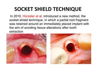
SOCKET SHIELD TECHNIQUE IN
IMPLANT PLACEMENT
PRESENTED BY-ANDREW GNANAMUTTU/CRI
GUIDED BY-DR VIJAY EBENEZER,
DR BALAKRISHNAN RAMALINGAM,
DR BANU SARGUNAR
SREE BALAJI DENTAL COLLEGE AND HOSPITAL
TRADITIONAL BRANEMARK’S
PROTOCOL
HEALING OF EXTRACTION
SOCKET
TISSUE CHANGES AFTER TOOTH
EXTRACTION
 Following an extraction thereis a 25% decreasein the width of the alveolar bone during first
year and an average4mm decrease in height during the first year following multiple
extractions(Carlson 1967)
 Tatum and Misch have observed a 40%-60% decrease in alveolarbone width after the first 2-3
years post extraction.
 Christensen reports an annual resorption rate of at least 0.5% - 1% during the remainder forthe
rest of a patientslife
 Schropp et al (2003) most of the bone gain in the socket occurred in the first 3 months
SOFT TISSUE CHANGES
AFTER EXTRACTION
HARD TISSUE
CHANGES AFER
EXTRACTION
IMMEDIATE IMPLANT PLACEMENT?
Immediate implant placement, defined as the placement of dental implant
immediately into fresh extraction socket site after tooth extraction, has been
considered a predictable and acceptable procedure (Schwartz et al., 2000).
WHY IMMEDIATE IMPLANT
PLACEMENT?
Patients acceptability
Reduces treatment time
Socket as a guide for determination of parallelism and alignment
Surgeon can position the implant more favourably than the original position
Facilitates final restoration and minimizes need for severely angled
abutments
Implants in extraction sites can be placed in the same position as the
extracted teeth
SOCKET SHIELD TECHNIQUE
In 2010, Hürzeler et al. introduced a new method, the
socket shield technique, in which a partial root fragment
was retained around an immediately placed implant with
the aim of avoiding tissue alterations after tooth
extraction
PRINCIPLE OF SOCKET SHIELD
TECHNIQUE
• Preparation of root in such a way that
buccal/facial section remains in-situ with
buccal plate intact
• Tooth root sections periodontal attachment
apparatus remains vital and undamaged to
prevent the expected post extraction socket
remodeling and to support buccal/facial
tissues
INDICATIONS OF SOCKET SHIELD
TECHNIQUE???
• The ideal extraction site for immediate implant placement
Little or no periodontal bone loss on the tooth that is to be
extracted ,such as tooth being extracted due to
1)Endodontic involvement
2)Root fracture,root resorption
3)Periapical pathology
4)Root perforation
5)Unfavorable crown to root ratio
6)Residual deciduous tooth
• A minimum of 4-5mm bone width at the alveolar crest and at least
10mm bone length from the alveolar crest to a safe distance above
closest anatomical structures are recommended(WORTHINGTON
2004)
CONTRAINDICATIONS OF SOCKET
SHIELD TECHNIQUE
Presence of pus
Lack of bone beyond the apex
Close proximity to anatomical vital structures
Clinical conditions preventing primary closure
ADVANTAGES OF SOCKET
SHIELD TECHNIQUE
 Alveolar ridge atrophy after tooth extraction has a negative impact on implant
restoration. The buccal portion of the bone which is more prone to atrophy was
preserved by retaining a part of buccal root segment which shields PDL on
buccofacial aspect of the implant.
 Especially in canine and premolars where the buccal cortical plate is thin and are
more prone to fracture which makes it an unfavourable environment for implant
placement which also requires a secondary grafting procedure. So as to overcome
this difficulty this technique is used.
 No additional material cost, No co-morbidity , Single surgery ,Applicable in sites
with endodontic apical pathology
THIN BUCCAL
CORTICAL PLATE
DISADVANTAGES OF SOCKET SHIELD
TECHNIQUE
• Not yet reliable or predictable
• No long-term data yet
• Technique sensitive
THE RULE OF 5 TRIANGLES
• The presence of a buccal plate
• Primary stability where there is existing apical bone
• Implant design
• Filling of the gap between the buccal plate and the
implant(JUMPING DISTANCE)
• Tissue biotype.
OF THE RULE OF 5 TRIANGLES ,
THE PRESENCE OF BUCCAL
PLATE CAN WELL BE
PRESERVED USING SOCKET
SHIELD TECHNIQUE
PROCEDURE
A 30-year-old malepatient was treated with
a single tooth implantin the maxillarypremolar region. The patient was a
non-smoker and did not present relevant medical conditions. The primary
indication for tooth extraction was an endodontic treatmentfailure.
OCCLUSAL
AND
BUCCAL
VIEW
Without lifting a mucoperiosteal flap or affecting the marginal
gingiva,the tooth was carefully decoronated using a diamond bur.
A 1–2mmthick tooth fragment in the buccal area of the extraction
socket was separated from the rest of the tooth using rotating
tungstencarbide instrumentswith sterilewater irrigation.
Particularattentionwas taken to avoid damage to the bone walls
of the extractionsocket. Following preparation of the implantbed
in the lingualpart of the root, all residualtooth fragments were
completelyremoved. A careful curettage of the extractionsocket
was performed to remove granulationtissue.The buccal fragment
of the root was preserved 1mm coronally to the buccal bone plate
SHIELD WAS
PREPARED
The implant was placed in the lingual part
of the extraction socket, without contact
with the retained root fragment
Radiographic view after
implant placement
Follow-up examinationswere performed
after surgery at follow-up intervals. The
implant site showed uneventful healing.No
socket-shieldexposure was observed.
The final restoration– a screw-retainedceramic-to-
metal implantsingle crown – was delivered four
months after implant placement.It was fixed witha
torque-controldevice using a titaniumscrew.
Functional capabilitieswere checked .
FINAL RESTORATION
CONCLUSION
Tooth loss and subsequent ridge collapse continue to burden restorativeimplant treatment.Especiallyin canine and
premolarswherethe buccal corticalplate is thin and are more prone to fracture which makes it an unfavourable
environment for implantplacement which also requires a secondary grafting procedure. In-lieuof surgical
augmentation to correct a ridge defect, the socket shieldtechnique offers a promising solution.
REFERENCE
1)TANUM, NEHASM,WAKINDERB,ACHYUTS,ANIMESHB,PRATEEKT SOCKETSHIELD
TECHNIQUE.INDIANDENTALJOURNAL2015:7; 31-34
2)HOWARDG,JONATHANDT,MAURICES THE SOCKETSIELD TECHNIQUETO SUPPORT THEBUCCO
FACIALTISSUES AT IMMEDIATEIMPLANT PLACEMENT.INTERNATIONALDENTISTRY-AFRICAN
EDITION2011 :5;6-14
Socket shield technique

Socket shield technique

  • 1.
    SOCKET SHIELD TECHNIQUEIN IMPLANT PLACEMENT PRESENTED BY-ANDREW GNANAMUTTU/CRI GUIDED BY-DR VIJAY EBENEZER, DR BALAKRISHNAN RAMALINGAM, DR BANU SARGUNAR SREE BALAJI DENTAL COLLEGE AND HOSPITAL
  • 2.
  • 3.
  • 4.
    TISSUE CHANGES AFTERTOOTH EXTRACTION  Following an extraction thereis a 25% decreasein the width of the alveolar bone during first year and an average4mm decrease in height during the first year following multiple extractions(Carlson 1967)  Tatum and Misch have observed a 40%-60% decrease in alveolarbone width after the first 2-3 years post extraction.  Christensen reports an annual resorption rate of at least 0.5% - 1% during the remainder forthe rest of a patientslife  Schropp et al (2003) most of the bone gain in the socket occurred in the first 3 months
  • 5.
    SOFT TISSUE CHANGES AFTEREXTRACTION HARD TISSUE CHANGES AFER EXTRACTION
  • 6.
    IMMEDIATE IMPLANT PLACEMENT? Immediateimplant placement, defined as the placement of dental implant immediately into fresh extraction socket site after tooth extraction, has been considered a predictable and acceptable procedure (Schwartz et al., 2000).
  • 7.
    WHY IMMEDIATE IMPLANT PLACEMENT? Patientsacceptability Reduces treatment time Socket as a guide for determination of parallelism and alignment Surgeon can position the implant more favourably than the original position Facilitates final restoration and minimizes need for severely angled abutments Implants in extraction sites can be placed in the same position as the extracted teeth
  • 8.
    SOCKET SHIELD TECHNIQUE In2010, Hürzeler et al. introduced a new method, the socket shield technique, in which a partial root fragment was retained around an immediately placed implant with the aim of avoiding tissue alterations after tooth extraction
  • 9.
    PRINCIPLE OF SOCKETSHIELD TECHNIQUE • Preparation of root in such a way that buccal/facial section remains in-situ with buccal plate intact • Tooth root sections periodontal attachment apparatus remains vital and undamaged to prevent the expected post extraction socket remodeling and to support buccal/facial tissues
  • 10.
    INDICATIONS OF SOCKETSHIELD TECHNIQUE??? • The ideal extraction site for immediate implant placement Little or no periodontal bone loss on the tooth that is to be extracted ,such as tooth being extracted due to 1)Endodontic involvement 2)Root fracture,root resorption 3)Periapical pathology 4)Root perforation 5)Unfavorable crown to root ratio 6)Residual deciduous tooth • A minimum of 4-5mm bone width at the alveolar crest and at least 10mm bone length from the alveolar crest to a safe distance above closest anatomical structures are recommended(WORTHINGTON 2004)
  • 12.
    CONTRAINDICATIONS OF SOCKET SHIELDTECHNIQUE Presence of pus Lack of bone beyond the apex Close proximity to anatomical vital structures Clinical conditions preventing primary closure
  • 13.
    ADVANTAGES OF SOCKET SHIELDTECHNIQUE  Alveolar ridge atrophy after tooth extraction has a negative impact on implant restoration. The buccal portion of the bone which is more prone to atrophy was preserved by retaining a part of buccal root segment which shields PDL on buccofacial aspect of the implant.  Especially in canine and premolars where the buccal cortical plate is thin and are more prone to fracture which makes it an unfavourable environment for implant placement which also requires a secondary grafting procedure. So as to overcome this difficulty this technique is used.  No additional material cost, No co-morbidity , Single surgery ,Applicable in sites with endodontic apical pathology THIN BUCCAL CORTICAL PLATE
  • 14.
    DISADVANTAGES OF SOCKETSHIELD TECHNIQUE • Not yet reliable or predictable • No long-term data yet • Technique sensitive
  • 15.
    THE RULE OF5 TRIANGLES • The presence of a buccal plate • Primary stability where there is existing apical bone • Implant design • Filling of the gap between the buccal plate and the implant(JUMPING DISTANCE) • Tissue biotype. OF THE RULE OF 5 TRIANGLES , THE PRESENCE OF BUCCAL PLATE CAN WELL BE PRESERVED USING SOCKET SHIELD TECHNIQUE
  • 16.
    PROCEDURE A 30-year-old malepatientwas treated with a single tooth implantin the maxillarypremolar region. The patient was a non-smoker and did not present relevant medical conditions. The primary indication for tooth extraction was an endodontic treatmentfailure. OCCLUSAL AND BUCCAL VIEW
  • 17.
    Without lifting amucoperiosteal flap or affecting the marginal gingiva,the tooth was carefully decoronated using a diamond bur. A 1–2mmthick tooth fragment in the buccal area of the extraction socket was separated from the rest of the tooth using rotating tungstencarbide instrumentswith sterilewater irrigation. Particularattentionwas taken to avoid damage to the bone walls of the extractionsocket. Following preparation of the implantbed in the lingualpart of the root, all residualtooth fragments were completelyremoved. A careful curettage of the extractionsocket was performed to remove granulationtissue.The buccal fragment of the root was preserved 1mm coronally to the buccal bone plate SHIELD WAS PREPARED
  • 18.
    The implant wasplaced in the lingual part of the extraction socket, without contact with the retained root fragment Radiographic view after implant placement
  • 19.
    Follow-up examinationswere performed aftersurgery at follow-up intervals. The implant site showed uneventful healing.No socket-shieldexposure was observed. The final restoration– a screw-retainedceramic-to- metal implantsingle crown – was delivered four months after implant placement.It was fixed witha torque-controldevice using a titaniumscrew. Functional capabilitieswere checked .
  • 20.
  • 21.
    CONCLUSION Tooth loss andsubsequent ridge collapse continue to burden restorativeimplant treatment.Especiallyin canine and premolarswherethe buccal corticalplate is thin and are more prone to fracture which makes it an unfavourable environment for implantplacement which also requires a secondary grafting procedure. In-lieuof surgical augmentation to correct a ridge defect, the socket shieldtechnique offers a promising solution. REFERENCE 1)TANUM, NEHASM,WAKINDERB,ACHYUTS,ANIMESHB,PRATEEKT SOCKETSHIELD TECHNIQUE.INDIANDENTALJOURNAL2015:7; 31-34 2)HOWARDG,JONATHANDT,MAURICES THE SOCKETSIELD TECHNIQUETO SUPPORT THEBUCCO FACIALTISSUES AT IMMEDIATEIMPLANT PLACEMENT.INTERNATIONALDENTISTRY-AFRICAN EDITION2011 :5;6-14