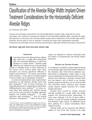
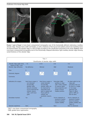
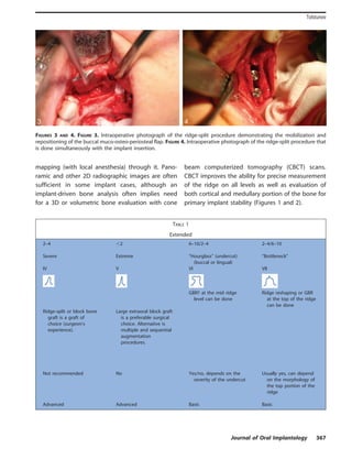
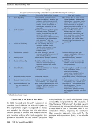
This document discusses the importance of available bone for implant treatment planning and success. It defines available bone as the external architecture and quantity of bone present, and describes how bone is measured in height, width, length, angulation, and crown-height space. Adequate available bone is categorized as Division A, while Division B has barely sufficient bone. Division C bone is deficient in one or more dimensions, and Division D bone is severely atrophic. Treatment options depend on the bone quality and may include osteoplasty, bone augmentation, narrow diameter implants, or subperiosteal implants. Proper evaluation of available bone is critical for determining the appropriate treatment plan.
This document discusses immediate implant placement after tooth extraction. It begins with an introduction that outlines the healing process after extraction and bone resorption over time with traditional protocols. It then covers the advantages and indications of immediate placement, including reducing treatment time and better positioning. Contraindications and classification of extraction sites are presented. The treatment sequence of clinical examination, radiographs, surgical guide fabrication is outlined. Surgical procedures, soft tissue management, post-op care and different treatment protocols like immediate loading are summarized. Clinical trials are briefly discussed showing outcomes of immediate placement. Factors like primary stability, splinting and provisional restoration are highlighted to consider. The conclusion restates the key points about immediate implant placement.
This document discusses occlusal schemes for implants, known as implant protective occlusion (IPO). IPO aims to reduce stress at the implant-bone interface through 14 considerations including eliminating premature contacts, positioning occlusal contacts over implant bodies, reducing cantilever lengths, and decreasing crown heights. The goals of IPO are to reduce force magnification, improve force direction, and increase the implant support area to promote implant longevity and success.
The document discusses various factors that can contribute to dental implant failures, including host factors like poor medical health, smoking, bruxism, and poor oral hygiene; surgical factors like trauma during surgery; and implant selection factors like bone quality. It provides definitions for different types of implant failures and lists criteria for determining implant success. The classifications, predictors, warning signs, and ways to enhance outcomes with implants are also examined.
Zygomatic implants are placed through the alveolar crest and maxillary sinus involving the zygomatic bone for strong anchorage. They provide maximum support and durability compared to conventional implants due to their placement in dense cheek bone. Complications can include zygomatic bone fracture, orbital penetration, or implant head damage during surgery. Post-operative issues may involve screw fracture, implant failure, oroantral communication, soft tissue inflammation, or sinusitis. Zygomatic implants are best for patients with insufficient bone who need a single procedure rather than multiple surgeries, and a fixed prosthesis can be placed in as little as 72 hours.
This document provides an overview of implant supported overdentures, including definitions, history, indications, contraindications, advantages, disadvantages, treatment options, and procedures. Key points discussed include:
- Overdentures are removable prostheses that cover natural tooth roots, implants, or both for support.
- Implant supported overdentures have better outcomes than conventional dentures or overdentures supported only by natural tooth roots.
- Treatment options depend on factors like jaw, bone quality, number of implants, and can involve bar-retained or independent attachments.
- Procedures involve medical evaluation, treatment planning, transitional dentures, surgical placement, attachment connection, and definitive prosthesis fabrication
This document provides an overview of dental implant sinus lift procedures. It begins with brief anatomy of the maxillary sinus and defines a dental implant. It then discusses patient evaluation, including radiographic assessment and anatomical limitations for implantation. Classification systems for the posterior maxilla are presented. The document reviews indications, contraindications, and surgical techniques for sinus lift procedures, including direct and indirect methods. It also discusses graft materials, post-operative instructions, and potential complications.
This document discusses the rehabilitation of the atrophic posterior maxilla using pterygoid implants. It provides background on the challenges of posterior maxillary rehabilitation and outlines treatment options like sinus lifts, short implants and tilted implants. It then focuses on the anatomy of the pterygoid region and classifications for pterygoid implants. The document details the surgical protocol for placing pterygoid implants using guides, angled abutments, impressions and final prosthesis placement. It concludes that pterygoid implants provide an alternative to maxillary reconstruction and avoid cantilevers while allowing for immediate loading.
Split ridge and expansion techniques are effective for the correction of moderately resorbed edentulous ridges in selected cases.
Transverse expansion is based on osseous plasticity obtained by corticotomy. It progressively allows for an adequate transversal intercortical diameter large enough to insert one or several dental implants.
The gap created by sagittal osteotomy expansion undergoes spontaneous ossification, following a mechanism similar to that occurring in fractures.
This document provides an overview of periimplantitis, including its definition, classification systems, epidemiology, etiology, pathogenesis, diagnosis, and treatment. Periimplantitis is defined as an inflammatory process involving both soft and hard tissues around a dental implant, resulting in loss of supporting bone. It is distinguished from peri-implant mucositis, which only involves inflammation of soft tissues. The document discusses various classification systems for periimplantitis and reviews potential etiologic factors such as plaque, biomechanical overload, genetic factors, and iatrogenic causes. Diagnosis involves clinical parameters like bleeding, probing depth, and radiographic bone loss. Treatment aims to eliminate infection and may include nonsurgical and surgical
This seminar deals with implant-related complications that lead to implant failure.this also discus diagnostic criteria and preventive methods for an implant failure.
Vertical ridge augmentation is sometimes required for dental implant placement. The presentation looks at various conventional and newer techniques for ridge augmentation in the oral cavity.
This document provides an overview of guided tissue regeneration (GTR). It begins with definitions of periodontal regeneration and GTR. It then discusses the history and development of GTR from the 1970s onwards. The core concept of GTR is explained, which is based on Melcher's hypothesis that only periodontal ligament cells can regenerate the periodontal attachment apparatus. Indications, contraindications, design criteria and objectives of GTR barriers are covered. The document classifies and compares advantages and disadvantages of absorbable versus non-absorbable membranes. Key factors affecting GTR outcomes are discussed. Surgical techniques and the healing of GTR-treated defects are described. The document concludes with additional considerations like complications and the
This document provides information on various ridge augmentation techniques. It begins with an introduction describing how tooth loss leads to bone resorption and impaired function. It then discusses the history of using autogenous bone grafts for ridge augmentation. The objectives of ridge augmentation are also outlined. Key techniques discussed include ridge preservation, ridge splitting, use of autogenous bone blocks, and distraction osteogenesis. Advantages and disadvantages of different graft sources and incision designs are compared. The document emphasizes the importance of adequate bone volume for successful implant placement and summarizes various methods to augment bone.
This document discusses various suturing techniques used in periodontal flap surgery. It begins with an introduction on the purpose of suturing flaps, which is to maintain the flap in position until desired healing. Resorbable sutures are preferred for patient comfort and elimination of removal appointments. The document then describes different suturing techniques like horizontal mattress suture, continuous independent sling suture, anchor suture, closed anchor suture, and periosteal suture. It provides details on their specific uses and how to perform each technique. A variety of suture materials, both resorbable and non-resorbable, are also listed.
The document discusses treatment planning in periodontics. It begins by defining treatment planning and outlining the short and long-term goals. These include eliminating infection and inflammation and reconstructing a healthy dentition. The treatment plan is the blueprint and involves decisions about emergency treatment, extractions, nonsurgical and surgical therapies, restorations, and maintenance. Phases of treatment are discussed including preliminary, nonsurgical, surgical, and maintenance phases. Factors in deciding whether to extract or preserve a tooth are also outlined.
This document discusses periodontal regeneration and the various factors involved. It begins by defining key terminology related to grafting and regeneration. It then discusses the biology and objectives of periodontal regeneration, including the ideal outcome of new attachment formation and factors that can influence outcomes. The document outlines various techniques for periodontal regeneration including non-graft associated approaches involving removal of epithelium and surgical techniques, as well as graft-associated approaches using various graft materials. Requirements for predictable regeneration and assessment methods are also summarized.
This document discusses patterns of bone destruction in periodontal disease. It covers various causes of bone loss such as the extension of gingival inflammation, trauma from occlusion, and systemic disorders. Factors that determine bone destruction include normal bone variation, exostoses, buttressing bone formation, and food impaction. Common bone destruction patterns include horizontal loss, vertical defects, intrabony defects of one to three walls, furcation involvement, osseous craters, and ledges. Systemic conditions like osteoporosis and Paget's disease can also cause alveolar bone destruction.
This document provides an overview of the history and evolution of dental implants from ancient times to the modern era. It discusses early attempts at implant dentistry dating back thousands of years, including the use of animal teeth, carved ivory, and other materials as implants. The document then outlines several key periods in the more recent history and development of dental implants, including pioneers who advanced implant techniques and materials in the 18th century through the early 20th century. It focuses on the foundational work done in the late 1930s and 1940s that marked the beginning of modern implant dentistry.
A 40-year-old female presented with an inability to eat properly due to a fractured crown. Upon examination, the remaining tooth structure was below the gumline and not accessible. The procedure involves adjusting the gum tissue and bone around the tooth to provide better access and ensure a proper restoration fit. Biologic width and ferrule effect must be considered to prevent inflammation, bone loss, and future issues. Complications can include an unaesthetic appearance, sensitivity, and loss of bone or papilla.
The biological fixation determines the longevity of dental implant treatment. It ensures the long term survival of dental implant. Better the osseointegration,higher will be the survival rate
This document provides an overview of resective osseous surgery techniques. It discusses the anatomical forms of bone, osteoplasty and ostectomy procedures, surgical approaches, and techniques. Osteoplasty involves reshaping bone without removing tooth-supporting bone through techniques like grooving and blending. Ostectomy involves removing tooth-supporting bone to eliminate osseous deformities. Specific techniques like horizontal grooving, scribing, and hand instrumentation are described. Post-operative maintenance and expected osseous changes are also summarized.
1. The document discusses the history, principles, types, and mechanisms of bone grafts. It provides definitions of key terms like graft, flap, osteogenesis, osteoinduction, and osteoconduction.
2. The main types of bone grafts discussed are autogenous grafts, allografts, xenografts, alloplasts, and composite grafts. Autogenous grafts are considered the gold standard due to their osteogenic, osteoinductive and osteoconductive properties but require a second surgical site.
3. Allografts avoid a second surgical site but have reduced osteoinductive potential and risks of disease transmission or immune rejection. Growth factor based grafts and
Ridge preparation for implant placement - part 1Hesham El-Hawary
- criteria of ideal ridge
- implants timing protocol
- implants planning and case selection
- clinical types of bone
- preventive methods to preserve the alveolar ridge
Dr. Ahmed M. Adawy
Professor Emeritus, Dep. Oral & Maxillofacial Surg.
Former Dean, Faculty of Dental Medicine
Al-Azhar University. Numerous etiologies lie behind mandibular defects including pathologic lesions, trauma related, infectious diseases and congenital defects. At present, the methods to restore mandibular defects can be classified into four basic categories:
1.Autogenous bone grafts in the form of nonvascularized free bone transfer, or vascularized tissue transfer, either pedicled or based on microvascular anastomosis
2. Distraction osteogenesis
3. Alloplastic materials (with or without bone)
4. Tissue engineered grafts
The document discusses pre-prosthetic surgery, which aims to modify the oral environment to better support prosthetic appliances. The goals are to provide a broad, flat ridge with height and a firm mucosal covering. Objectives include eliminating disease, conserving structures, and providing support to withstand forces. The document describes various basic surgical procedures like alveoloplasty, tori removal, and soft tissue procedures to reshape ridges and remove excess tissue in preparation for dentures.
Resective osseous surgery involves reshaping the alveolar bone through additive or subtractive techniques to correct deformities caused by periodontal disease. The goal is to reshape the marginal bone to resemble healthy bone. Key steps include vertical grooving, radicular blending, flattening interproximal bone, and gradualizing marginal bone using instruments like chisels and curettes. This technique is best for early to moderate bone loss up to 3mm and can provide reduced pocket depths and stable tissue contours for long-term maintenance when performed with apically positioned flaps.
This study evaluated the healing of mandibular ramus bone block grafts used for alveolar ridge augmentation before implant placement through clinical, histological, and histomorphometric analysis. Bone blocks were harvested from the mandibular ramus in 15 patients and grafted to maxillary defects. After 3-9 months of healing, implants were placed and bone samples were taken. Histological analysis found signs of active remodeling but also substantial amounts of non-vital bone and generally weak neo-vascularization, suggesting that most osteocytes in the grafted bone do not survive and neo-vascularization of non-vital grafted bone is difficult. The outcomes suggest grafted bone undergoes slow remodeling into new vital bone.
This document discusses the use of CBCT scans in evaluating patients for dental implants. It describes how CBCT can be used to assess:
1. Bone quantity and quality, allowing precise measurements of bone height, width, and density.
2. Surrounding anatomical structures like the maxillary sinuses and inferior alveolar canal.
3. Ridge morphology and orientation to plan ideal implant placement.
CBCT provides 3D imaging without superimposition, enabling accurate pre-surgical planning to determine if bone augmentation is needed. This helps ensure implant osseointegration and optimal functional and esthetic outcomes.
This document summarizes a randomized clinical trial comparing the bone quality of free autogenous bone grafts harvested from two mandibular donor sites (chin and retromolar region) for maxillary alveolar ridge augmentation. 14 patients received either chin (n=7) or retromolar (n=7) shell grafts fixed with screws. 6 months post-op, 42 core biopsies were analyzed and found the chin shells had a higher bone area percent (52.53% vs 47.97%) indicating better bone quality and stability compared to retromolar grafts.
This document discusses crown lengthening procedures. It describes different surgical techniques for crown lengthening including external bevel gingivectomy, internal bevel gingivectomy with or without ostectomy, and apically positioned flap techniques. It discusses factors to consider like the amount of bone removal needed, ideal flap design, and management of soft and hard tissues. Proper biologic width dimensions, flap suturing, and esthetic goals are important considerations for achieving good outcomes with crown lengthening surgery.
This document discusses the use of CBCT imaging in dental implant treatment planning and assessment. It provides details on how CBCT can be used to evaluate bone quantity and quality, anatomical structures, ridge morphology, and is recommended for pre-operative planning of sinus lift procedures. CBCT allows accurate measurements and 3D visualization to determine suitable implant size, position, and angulation without superimposition. This helps optimize implant placement and outcomes.
Resective osseous surgery involves removing tooth-supporting alveolar bone to reshape it and eliminate periodontal pockets. It is indicated for inconsistent bone margins, reverse bone architecture, one-wall defects, and other bone irregularities. The surgery involves using burs and chisels to perform vertical grooving of thick bone, blending any bone ledges on tooth roots, flattening interproximal bone in narrow areas, and gradualizing marginal bone. Flaps are closed to cover the new bony margins. Post-operative maintenance like cleaning and antibiotics is needed to allow healing over 2-3 weeks.
The document summarizes research on the healing process that occurs within the alveolar socket after a tooth extraction. It describes the three phases of socket healing: 1) inflammation and blood clot formation, 2) new bone formation through the deposition of woven bone, and 3) bone remodeling where woven bone is replaced with lamellar bone and bone marrow. It notes that socket healing results in dimensional changes to the alveolar ridge over time due to bone modeling and remodeling on the socket walls. Factors like surgical trauma from extraction and lack of a tooth's functional stimulus contribute to reductions in ridge width and height.
The orthodontist can avoid fenestration and dehiscence by determining alveolar morphology through imaging such as CBCT prior to treatment. Heavy forces, ectopic tooth positions, and movement beyond the limits of the alveolar housing can cause bone loss. Fenestrations are more common in the maxilla, especially around molars and canines, while dehiscences occur more in the mandible around incisors. Rapid maxillary expansion can also cause fenestrations and dehiscences. Maintaining an appropriate force level and following the limits of the alveolar bone can help minimize these risks.
The document discusses various techniques for mandibular reconstruction, including non-vascularized bone grafts and vascularized bone flaps. It states that free vascularized bone flaps have become the most popular technique as they heal more rapidly and have less risk of complications compared to non-vascularized grafts. Common types of free flaps used include fibula flaps based on the peroneal artery and iliac crest flaps based on the deep circumflex iliac artery.
This document summarizes a study on using bone grafts and immediate implant placement in areas of high aesthetic value. It describes using block bone grafts taken from the mandible to augment alveolar ridge deficiencies in the maxilla before placing implants. The goals were to assess success rates of implants placed in grafted bone and determine if this technique is suitable for aesthetic areas. The methodology involved using SPI implants and placing immediate provisional restorations on the implants after grafting and implantation to condition the soft tissues during healing. Success was based on radiographic and clinical assessments during outpatient follow-ups.
This document summarizes a study on using bone grafts and immediate implant placement in areas of high aesthetic value. It describes using block bone grafts taken from the mandible to augment alveolar ridge deficiencies in the maxilla before placing implants. The goals were to assess success rates of implants placed in grafted bone and determine if this technique is suitable for aesthetic areas. The protocol involved using SPI implants and placing immediate provisional restorations on the implants after graft integration to condition the soft tissues during healing. The study aims to evaluate if this technique can provide rigid fixation and osseointegration of implants in grafted bone for functional loading.
This document discusses factors affecting the selection of patients for implant retained prostheses. It outlines that a thorough patient evaluation including medical history, dental evaluation through examination and imaging, and informed consent is required. The dental evaluation assesses bone quality and quantity, occlusion, and adjacent teeth. Indications for implants include missing teeth from congenital defects, trauma, or being edentulous. Contraindications include certain medical conditions, smoking, drugs/alcohol, or inadequate bone. Proper patient selection is key for implant success and satisfying treatment outcomes.
This document discusses factors to consider when selecting patients for implant retained prostheses. It outlines general patient factors like medical history and motivation that should be assessed. A thorough dental evaluation including extraoral and intraoral exams, various radiographs, and bone density assessment is important. Patients should provide informed consent and understand expectations, risks, and commitments. Clinical indications for implants include missing teeth due to congenital defects, trauma, or being edentulous. Contraindications include conditions that could compromise bone healing or the patient's ability to maintain implants. A multidisciplinary approach may be needed for complex cases.
It restore alveolar bone to the level existing at the time of surgery or slightly more apical to this level. Aim is to achieve positive bony architecture.
STEPS INCLUDE:
1.VERTICAL GROOVING
2. RADICULAR BLENDING
3. FLATTENING INTERPROXIMAL BONE
4. GRADUALIZING MARGINAL BONE
PREFERRED TREATMENT FOR ONE WALLED PERIODONTAL BONE DEFECTS (HEMISEPTUM)
Similar to Classification of alveolar bone width (20)
Adjunctive role of Orthodontic Therapy in PeriodontologyNavneet Randhawa
This document summarizes the adjunctive role of orthodontic therapy in periodontology. Some key points:
- Orthodontic tooth movement can benefit adult patients by correcting tooth malposition that makes cleaning difficult and increases periodontal disease risk.
- Light, prolonged orthodontic forces can move teeth without damaging tissues if excellent oral hygiene is maintained. However, some tissue necrosis is unavoidable.
- Tooth movement through cortical bone can create dehiscences if the bone is not remodeled quickly enough in front of the tooth.
- Tooth movement into existing infrabony pockets or compromised bone areas does not further periodontal attachment loss if the area is first treated and hygiene is
Osseointegration, definition, history, process of osseointegration, factors influencing osseointegration, methods for evaluation of osseointegration, failure of osseointegration
Definition of periodontal pocket, classification, Histopathology of periodontal pocket, microflora involved, pathogenesis, periodontal pocket as a healing lesion, microtopography of root surface, treatment of periodontal pocket
Smoking and periodontal disease, smoking as a risk factor, incidence of smoking, effects of smoking on periodontium, smoking and gingivitis and smoking and periodontitis, effect of surgical and non surgical therapy on smokers
Cytokines are small soluble proteins that are important mediators of the inflammatory response. They are produced by immune cells like lymphocytes and monocytes and act as signaling molecules between cells. The document defines cytokines and provides classifications of cytokines. It describes the roles of key cytokines like IL-1 and IL-2 in innate immunity and leukocyte recruitment during the early immune response. Cytokines function through binding to specific cell surface receptors and activating intracellular signaling pathways.
Aging is a natural process that affects the entire body in complex ways. It involves the slowing of functions over time due to biological, psychological, and social factors. The elderly population is growing rapidly, with over 20% of the US population expected to be over 65 by 2030. Aging leads to changes in nearly every body system through various proposed mechanisms like the free radical theory of aging. The periodontium is also affected by aging through thinning tissues, decreased function, and increased risk of periodontal disease. Maintaining good oral health is important for the elderly population.
This document discusses the influence of systemic conditions on the periodontium. It begins by introducing periodontitis as a chronic bacterial infection and how host responses can vary between individuals. Systemic disorders can impair the host's immune defenses, creating opportunities for more severe periodontal disease. Several specific systemic factors are then examined in more detail, including hormonal changes, diabetes mellitus, and female sex hormones. The effects of these conditions on the periodontium are explored through their impact on factors like subgingival microbiota, polymorphonuclear leukocyte function, collagen metabolism, and wound healing. Treatment considerations for periodontal disease in systemic disease patients are also briefly addressed.
Gingival enlargement can result from chronic or acute inflammation, drugs, or systemic conditions. Drug-induced enlargement is common with anticonvulsants like phenytoin and presents as a painless, bead-like enlargement of the papillae that progresses to cover tooth crowns. Histologically, there is pronounced hyperplasia of connective tissue and epithelium. While the enlargement is caused by the drug, secondary inflammation from plaque complicates the condition, adding to the size and producing redness. Approximately 50% of patients on phenytoin experience gingival overgrowth.
Supportive periodontal therapy (SPT) involves long-term maintenance programs following active periodontal treatment to maintain periodontal health. SPT involves periodic examination, motivation and instrumentation of sites showing inflammation, treatment of reinfected sites, and polishing. It begins after active treatment and is aimed at preventing recurrence through early detection of disease. The frequency of SPT visits depends on the patient's periodontal risk assessment but generally occurs every 3-4 months. It can be performed by general dentists or specialists depending on the extent of original periodontal destruction. Adjunctive use of antimicrobials may also be included in SPT.
- Trauma from occlusion occurs when occlusal forces exceed the adaptive capacity of the periodontium, causing injury. It can be acute or chronic.
- The magnitude, direction, duration, and frequency of forces impact the periodontium's ability to adapt. Excessive pressure or tension can damage tissues.
- Primary trauma from occlusion is caused by changes in occlusal forces, while secondary trauma occurs when reduced bone support impairs the tissues' resistance to normal forces.
- The periodontium responds to trauma in three stages - injury, repair through new tissue formation, and adaptive remodeling to better withstand forces. Trauma can cause reversible damage if forces are reduced, or lead to irreversible injury if
The document discusses the endodontic-periodontal interrelationship. It begins by introducing how Simring and Goldberg first described this relationship in 1964. It then discusses the classifications of endodontic and periodontal lesions put forth by various studies. The document covers the anatomical considerations between the pulp and periodontium like apical foramina, lateral canals, and dentinal tubules which allow communication between the two tissues. It also discusses the etiological factors involved like bacteria, fungi, and viruses that can lead to endodontic or periodontal diseases.
This document discusses plaque as a biofilm and the microbiology of periodontal diseases. It begins by introducing the complex microbial flora that inhabits the oral cavity. A key point is that while most of these microbes coexist harmlessly with the host, a subset of organisms can lead to periodontal diseases either through overgrowth or new pathogenic properties. The document then examines historical and modern evidence that supports the infectious nature of periodontal diseases. It discusses the unique features of periodontal infections as biofilms outside of the body on tooth surfaces. Finally, it reviews the current understanding of suspected periodontal pathogens and their role in destructive periodontal disease.
The adrenal gland consists of an outer adrenal cortex and inner adrenal medulla. The cortex contains three zones that each secrete different hormone types: the zona glomerulosa secretes mineralocorticoids like aldosterone, the zona fasciculata secretes glucocorticoids like cortisol, and the zona reticularis secretes androgens. The adrenal medulla secretes catecholamines like epinephrine and norepinephrine which modulate stress response. Hormone production is tightly regulated by feedback mechanisms like the HPA axis to maintain homeostasis.
This document provides an overview of cementum, including its definition, physical characteristics, chemical composition, formation, classification, functions, repair capabilities, anomalies, and clinical considerations. Cementum is the mineralized tissue covering tooth roots that anchors periodontal ligament fibers and allows for tooth attachment. It is softer than dentin, continues depositing throughout life, and plays roles in tooth support, compensation, and repair of root surfaces. The document discusses the stages of cementum formation, types based on location/composition, and roles in maintaining tooth structure and occlusion. Pathologies like hypercementosis and cementoma are also summarized.
This study compared different methods for sampling gingival crevicular fluid (GCF) in patients with severe chronic periodontitis. The methods compared were paper strips, paper points, and washing techniques. The goal was to identify a method that could detect both microbiota and immunologic markers like cytokines. The results showed that paper strips detected cytokines like IL-6 and IL-8 more often than paper points. Additionally, washing techniques detected higher levels of proteases from Porphyromonas gingivalis compared to paper strips or points. Therefore, the study concluded that different sampling methods are better suited for detecting certain parameters, and a combination may be needed to analyze both microbiota and inflammatory markers in GCF.
Clinical accuracy outcome of open and closed trayimpressionsNavneet Randhawa
This study was a randomized controlled trial that compared the use of toluidine blue dye to a placebo for oral cancer screening among individuals with high-risk oral habits. Over 7,000 participants were randomized to either receive toluidine blue during their oral exam or a placebo. Examiners found a higher rate of positive initial screens in the toluidine blue group, but after referral to a pathologist, there was no significant difference in detection of oral premalignant lesions or non-premalignant lesions between the groups. No difference was found in oral cancer incidence rates over 5 years of follow-up. The study concluded that toluidine blue did not significantly improve detection of asymptomatic oral premalignant lesions compared to
Michigan HealthTech Market Map 2024. Includes 7 categories: Policy Makers, Academic Innovation Centers, Digital Health Providers, Healthcare Providers, Payers / Insurance, Device Companies, Life Science Companies, Innovation Accelerators. Developed by the Michigan-Israel Business Accelerator
End-tidal carbon dioxide (ETCO2) is the level of carbon dioxide that is released at the end of an exhaled breath. ETCO2 levels reflect the adequacy with which carbon dioxide (CO2) is carried in the blood back to the lungs and exhaled.
Non-invasive methods for ETCO2 measurement include capnometry and capnography. Capnometry provides a numerical value for ETCO2. In contrast, capnography delivers a more comprehensive measurement that is displayed in both graphical (waveform) and numerical form.
Sidestream devices can monitor both intubated and non-intubated patients, while mainstream devices are most often limited to intubated patients.
TEST BANK FOR Health Assessment in Nursing 7th Edition by Weber Chapters 1 - ...rightmanforbloodline
TEST BANK FOR Health Assessment in Nursing 7th Edition by Weber Chapters 1 - 34.
This particular slides consist of- what is hypotension,what are it's causes and it's effect on body, risk factors, symptoms,complications, diagnosis and role of physiotherapy in it.
This slide is very helpful for physiotherapy students and also for other medical and healthcare students.
Here is the summary of hypotension:
Hypotension, or low blood pressure, is when the pressure of blood circulating in the body is lower than normal or expected. It's only a problem if it negatively impacts the body and causes symptoms. Normal blood pressure is usually between 90/60 mmHg and 120/80 mmHg, but pressures below 90/60 are generally considered hypotensive.
The best massage spa Ajman is Chandrima Spa Ajman, which was founded in 2023 and is exclusively for men 24 hours a day. As of right now, our parent firm has been providing massage services to over 50,000+ clients in Ajman for the past 10 years. It has about 8+ branches. This demonstrates that Chandrima Spa Ajman is among the most reasonably priced spas in Ajman and the ideal place to unwind and rejuvenate. We provide a wide range of Spa massage treatments, including Indian, Pakistani, Kerala, Malayali, and body-to-body massages. Numerous massage techniques are available, including deep tissue, Swedish, Thai, Russian, and hot stone massages. Our massage therapists produce genuinely unique treatments that generate a revitalized sense of inner serenely by fusing modern techniques, the cleanest natural substances, and traditional holistic therapists.
Joker Wigs has been a one-stop-shop for hair products for over 26 years. We provide high-quality hair wigs, hair extensions, hair toppers, hair patch, and more for both men and women.
CHAPTER 1 SEMESTER V COMMUNICATION TECHNIQUES FOR CHILDREN.pdfSachin Sharma
Here are some key objectives of communication with children:
Build Trust and Security:
Establish a safe and supportive environment where children feel comfortable expressing themselves.
Encourage Expression:
Enable children to articulate their thoughts, feelings, and experiences.
Promote Emotional Understanding:
Help children identify and understand their own emotions and the emotions of others.
Enhance Listening Skills:
Develop children’s ability to listen attentively and respond appropriately.
Foster Positive Relationships:
Strengthen the bond between children and caregivers, peers, and other adults.
Support Learning and Development:
Aid cognitive and language development through engaging and meaningful conversations.
Teach Social Skills:
Encourage polite, respectful, and empathetic interactions with others.
Resolve Conflicts:
Provide tools and guidance for children to handle disagreements constructively.
Encourage Independence:
Support children in making decisions and solving problems on their own.
Provide Reassurance and Comfort:
Offer comfort and understanding during times of distress or uncertainty.
Reinforce Positive Behavior:
Acknowledge and encourage positive actions and behaviors.
Guide and Educate:
Offer clear instructions and explanations to help children understand expectations and learn new concepts.
By focusing on these objectives, communication with children can be both effective and nurturing, supporting their overall growth and well-being.
VEDANTA AIR AMBULANCE SERVICES IN REWA AT A COST-EFFECTIVE PRICE.pdfVedanta A
Air Ambulance Services In Rewa works in close coordination with ground-based emergency services, including local Emergency Medical Services, fire departments, and law enforcement agencies.
More@: https://tinyurl.com/2shrryhx
More@: https://tinyurl.com/5n8h3wp8
As Mumbai's premier kidney transplant and donation center, L H Hiranandani Hospital Powai is not just a medical facility; it's a beacon of hope where cutting-edge science meets compassionate care, transforming lives and redefining the standards of kidney health in India.
The facial nerve, also known as cranial nerve VII, is one of the 12 cranial nerves originating from the brain. It's a mixed nerve, meaning it contains both sensory and motor fibres, and it plays a crucial role in controlling various facial muscles, as well as conveying sensory information from the taste buds on the anterior two-thirds of the tongue.
practical guide of obesityfinal hyper1د حاتم البيطار القمة اكاديمي 0120238902...
Classification of alveolar bone width
1. ClassificationoftheAlveolarRidgeWidth:Implant-Driven
Treatment Considerations for the Horizontally Deficient
Alveolar Ridges
Len Tolstunov, DDS, DMD1
Among many techniques advocated for the horizontally deficient alveolar ridges, ridge-split has many
advantages. Here, treatment management strategies of the horizontally collapsed ridges, especially the ridge-
split approach, are discussed and a clinically relevant implant-driven classification of the alveolar ridge width is
proposed, with the goal to assist an operator in choosing the proper bone augmentation technique.
Comparison and advantages of two commonly used techniques, ridge-split and block bone graft, are presented.
Key Words: ridge-split, block bone graft, alveolar ridge
INTRODUCTION
I
t has been shown that although bone collapse
after tooth loss is usually three dimensional
(3D), the horizontal deficiency or width loss
develops to a larger extent.1,2
Alveolar width
deficiency can represent loss of buccal (labial)
cortical or medullary bone, or both. Deficiency of
the buccal cortex (cortical plate) after tooth
extraction can present significant difficulty in
implant reconstruction.3,4
The buccal cortical plate
with a thickness ,2 mm next to an implant appears
to have a higher risk of subsequent resorption.5
A variety of implant-driven bone augmentation
techniques for the deficient alveolar bone have
been proposed.6–8
Four of these techniques are
frequently performed: (1) guided bone regeneration
(GBR)/particulate bone grafting;9,10
(2) onlay (ve-
neer) block bone grafting with intraoral sources,
such as chin, ramus, posterior mandible, zygomatic
buttress, and maxillary tuberosity;11–13
(3) ridge-
split/bone graft procedure;14–16
and (4) alveolar
distraction osteogenesis.17–19
Most of these tech-
niques are designed to improve horizontal bone
loss before or simultaneously with dental implant
placement.
DIAGNOSIS AND TREATMENT PLANNING
It is important to establish a proper diagnosis based
on the alveolar ridge assessment before initiation of
the treatment plan. Initial clinical evaluation sup-
plemented by radiographic images helps in most
cases to distinguish two-dimensional (2D) versus 3D
alveolar bone deficiency. Although minimal bone
loss and patient’s lack of desire to go through
grafting surgical procedure(s) can be circumvented
with restorative means, extensive bone atrophy
usually requires surgical correction for a proper
implant placement.
Alveolar bone should be initially assessed
clinically (visually) for a rough width and height
analysis and interarch-occlusal relationships. In
some cases, although 7–8 mm of bone width is
present, it could be lingually (palatally) positioned
and therefore might require an additional buccal
bone grafting for a proper restoratively driven
implant insertion.
Alveolar width can be measured with different
calipers on top of the thin mucosa or by ridge
1
Private practice, Oral and Maxillofacial Surgery, San Francisco,
Calif, and Departments of Oral and Maxillofacial Surgery,
University of the Pacific, Arthur A. Dugoni School of Dentistry
and University of California San Francisco, San Francisco, Calif.
Corresponding author, e-mail: tolstunov@yahoo.com
DOI: 10.1563/AAID-JOI-D-14-00023
Journal of Oral Implantology 365
CLINICAL
2. FIGURES 1 AND 2. FIGURE 1. Cone beam computerized tomography scan of the horizontally deficient edentulous maxillary
alveolar ridge. Alveolar bone width and height, as well as thickness of the buccal and palatal cortical and medullary bone
are demonstrated. This alveolar ridge is a class III ridge according to the classification presented in the article. FIGURE 2. Axial
cone beam computerized tomography scan of the horizontally collapsed edentulous right maxillary alveolar ridge showing
varied thickness of the alveolar ridge.
TABLE 1
Classification of alveolar ridge width
Alveolar ridge width (mm),
based on CBCT* scan
.10 8–10 6–8 4–6
Alveolar ridge deficiency No deficiency Minimal Mild Moderate
Class 0 I II III
Schematic diagram
Comments
Indications for surgery Hard tissue surgery is
not indicated.
Occasionally,
alveolar width
(buccal convexity)
can be improved for
esthetic reasons
with a soft tissue
graft.
Hard tissue surgery is
rarely indicated.
Occasionally,
alveolar width can
be improved by
particulate bone
graft or palatal soft
tissue graft for
esthetic and
prosthetic reasons.
Particulate (GBR)
grafting or ridge-
split is often needed
to improve labial
bone projection and
proper occlusal
implant position.
An ideal width for the
ridge-split
procedure that can
be done in a single-
or two-stage
approach (see
Figure 3). Block
graft or GBR can
also be done.
Immediate insertion Yes Yes Yes/no, depends on
presence of apical
bone for primary
implant stability
Yes/no, depends on
presence of apical
bone for primary
implant stability (see
Figure 4)
Operator experience Basic Basic Basic Basic to advanced
*CBCT, cone beam computerized tomography.
ÀGBR, guided bone regeneration.
366 Vol. XL/Special Issue/2014
Classification of the Alveolar Ridge Width
3. mapping (with local anesthesia) through it. Pano-
ramic and other 2D radiographic images are often
sufficient in some implant cases, although an
implant-driven bone analysis often implies need
for a 3D or volumetric bone evaluation with cone
beam computerized tomography (CBCT) scans.
CBCT improves the ability for precise measurement
of the ridge on all levels as well as evaluation of
both cortical and medullary portion of the bone for
primary implant stability (Figures 1 and 2).
TABLE 1
Extended
2–4 ,2 6–10/2–4 2–4/6–10
Severe Extreme ‘‘Hourglass’’ (undercut)
(buccal or lingual)
‘‘Bottleneck’’
IV V VI VII
GBRÀ at the mid ridge
level can be done
Ridge reshaping or GBR
at the top of the ridge
can be done
Ridge-split or block bone
graft is a graft of
choice (surgeon’s
experience).
Large extraoral block graft
is a preferable surgical
choice. Alternative is
multiple and sequential
augmentation
procedures.
Not recommended No Yes/no, depends on the
severity of the undercut
Usually yes, can depend
on the morphology of
the top portion of the
ridge
Advanced Advanced Basic Basic
FIGURES 3 AND 4. FIGURE 3. Intraoperative photograph of the ridge-split procedure demonstrating the mobilization and
repositioning of the buccal muco-osteo-periosteal flap. FIGURE 4. Intraoperative photograph of the ridge-split procedure that
is done simultaneously with the implant insertion.
Journal of Oral Implantology 367
Tolstunov
4. CLASSIFICATION OF THE ALVEOLAR RIDGE WIDTH
In 1988, Cawood and Howell20
suggested an
anatomic classification of the edentulous jaws for
the preprosthetic surgery. It proposed six classes
and detailed the changes that the edentulous
alveolar process in anterior and posterior maxilla
and mandible undergo after teeth extraction (the
pattern of resorption). In 1989, Jensen21
proposed
an implant-driven site classification by bone quality
and quantity and proximity to vital structures. In
2002, Wang and Al-Shammari22
described a practi-
cal (therapeutically oriented) classification of alveo-
lar ridge defects, that is, horizontal, vertical, and
combination defects, proposing the edentulous
ridge expansion approach (ridge-split) for the
horizontal and combination defects of the alveolar
ridge.
TABLE 2
Ten-point comparison of ridge-split and monocortical block bone graft techniques
Comparison Monocortical Block Grafting (Intraoral) Ridge-split Procedure
1 Type of grafting Onlay: external, ‘‘cortex to cortex’’;
donor cortical graft is added to the
collapsed recipient buccal cortical
bone, resulting in the grafted bone
that has cortical environment on
one side and periosteum on the
other side
Inlay: internal (like an ‘‘open book’’);
cortical envelope is preserved and
expanded and a particulate grafting
is done ‘‘from within,’’ resulting in a
bilateral proximity of the grafted
bone to both cortices (similar to a 4-
wall defect of extraction socket)
2 Graft resorption Free (devascularized) graft; the grafted
bone may contain a substantial
amount of nonvital bone that did
not survive detachment,
devascularization, and transportation;
an increased risk of postoperative
graft resorption27
; slow and
incomplete neovascularization rate28
Vascular bone flap (muco-osteo-
periosteal flap) (see Figure 3),
vascularization is preserved at all
times; ‘‘cancellous bone grafts are
more rapidly and completely
revascularize than cortical grafts’’29
Decreased risk of postoperative graft
resorption16
3 Donor site morbidity Yes: pain, swelling, IAN* injury
(posterior mandible, ramus),
‘‘wooden teeth sensation’’ (chin),
sinus perforation (zygomatic
buttress), others
No
4 Recipient site morbidity Soft tissue dehiscence and graft
exposure, loose fixation screws and
graft mobility; graft loss
Soft tissue dehiscence and graft
exposure, buccal plate malfracture;
inadequate split
5 Wound closure Primary wound closure is mandatory Closure by secondary intention is
preferred
6 Buccal soft tissue flap Buccal flap is lifted and often
stretched; tension-free primary
closure is important, but can be
challenging
Buccal flap is not compromised; it is
not lifted and left attached to the
buccal periosteum
7 Wound healing By plasmatic imbibition from the host
(recipient) tissue
Internal ‘‘coagulum’’ is easily converted
in the woven bone due to
protection and excellent
vascularization from both cortices
throughout the whole process
8 Immediate implant insertion Traditionally not done Can be done in some cases (see Figure
4)
9 Delayed implant insertion Implants are placed into the cortical
bone interface 4 to 6 months later
Implants are placed into the cancellous
bone interface 4 to 6 months later
10 Environmental factors and
long-term stability of a
graft
More subject to a postoperative injury
(‘‘external’’ grafting); less long-term
stability and more long-term
resorption28
Less subject to a postoperative injury
during mastication; it is more
protected (‘‘internal’’ or
interpositional grafting); less long-
term resorption and more long-term
stability30,31
*IAN, inferior alveolar nerve.
368 Vol. XL/Special Issue/2014
Classification of the Alveolar Ridge Width
5. Here, a clinically relevant implant-driven classi-
fication of the alveolar ridge width based on
precise measurement of the alveolar width with
computerized tomography/CBCT scans is recom-
mended; it is presented in the Table 1. The
classification attempts to match the specific ridge
(its width and topography) with the appropriate
surgical technique (GBR, ridge-split, or block graft)
that can be used in the particular case of
horizontal bone atrophy. Although each opera-
tor’s experience ultimately determines the chosen
surgical technique, it is important to compare
benefits and drawbacks of different surgical
procedures for certain ridges to improve the
selection process.
COMPARISON OF THE RIDGE-SPLIT AND BLOCK BONE GRAFTING
TECHNIQUES
A literature review showed few similarities and
many differences between autogenous intraoral
monocortical (veneer) block graft and ridge-split/
bone graft techniques. Both procedures require a
skilled surgical practitioner equipped with knowl-
edge of regional anatomy and vascularization and
prepared for risks and complications of the proce-
dure. Both the ridge-split and block grafting
techniques are used mainly for a 2D horizontal
alveolar ridge augmentation (alveolar bone widen-
ing; some height gain can also be achieved with
both techniques).
Autogenous block bone grafting demonstrates
high osteogenic potential and effective in severe
anterior alveolar atrophy in maxilla and mandi-
ble.23–25
Two main disadvantages of monocortical
block grafts are donor site morbidity and late-term
graft resorption.26
The monocortical block bone
resorption has been reported to have up to 5%
early bone loss and up to 40% late bone loss of the
entire graft volume due to remodeling and
inadequate consolidation.27
Table 2 shows differences (10-point comparison)
between the ridge-split procedure and autogenous
intraoral monocortical block bone grafting. Factors
that are presented include donor- and recipient site
morbidity, type of wound closure, buccal flap
integrity and vascularity, specifics of wound healing,
type of bone interface, and possibility of an
immediate implant placement.
CONCLUSION
Knowledge of 3D bone anatomy with CBCT scan
helps to establish a proper ridge diagnosis before
initiation of implant treatment. The recommended
ridge width classification for the horizontally
deficient alveolar ridges is designed to be a clinically
relevant implant-driven anatomic guide for choos-
ing an appropriate surgical modality for the specific
collapsed alveolar ridge. Operator experience and
surgical comfort ultimately determines the choice of
the technique. The ridge-split approach tends to
have many advantages, including lack of donor site
morbidity and a graft stability over time.
ABBREVIATIONS
CBCT: cone beam computerized tomography
GBR: guided bone regeneration
REFERENCES
1. Schropp L, Wenzel A, Kostopoulos L, Karring T. Bone
healing and soft tissue contour changes following single tooth
extraction: a clinical and radiographic 12-month prosthetic study.
Int J Periodont Restor Dent. 2003;23:313–323.
2. Botticelli D, Berglundh T, Lindhe J. Hard tissue alterations
following immediate implant placement in extraction sites. J Clin
Periodontol. 2004;31:820–828.
3. Cardaropoli G, Arau´jo M, Lindhe J. Dynamics of bone tissue
formation in tooth extraction sites. An experimental study in dogs.
J Clin Periodontol. 2003;30:809–818.
4. Arau´jo M, Lindhe J. Dimensional ridge alterations following
tooth extraction. An experimental study in the dog. J Clin
Periodontol. 2005;32:212–218.
5. Qahash M, Susin C, Polimeni G, Hall J, Wikesjo¨ UM. Bone
healing dynamics at buccal peri-implant sites. Clin Oral Implants
Res. 2008;19:166–172.
6. Aghaloo TL, Moy PK. Which hard tissue augmentation
techniques are the most successful in furnishing bony support for
implant placement? Int J Oral Maxillofac Implants. 2007;22(suppl):
49–70.
7. McAllister BS, Haghighat K. Bone augmentation tech-
niques. J Periodontol. 2007;78:377–396.
8. Chiapasco M, Zaniboni M, Boisco M. Augmentation
procedures for the rehabilitation of deficient edentulous ridges
with oral implants. Clin Oral Implants Res. 2006;1(suppl 2):136–159.
9. Buser D, Bra¨gger U, Lang NP, Nyman S. Regeneration and
enlargement of jaw bone using guided tissue regeneration. Clin
Oral Implants Res. 1990;1:22–32.
10. Annibali S, Bignozzi I, Sammartino G, La Monaca G, Cristalli
MP. Horizontal and vertical ridge augmentation in localized alveolar
deficient sites: a retrospective case series. Implant Dent. 2012;21:
175–185.
11. Bedrossian E, Tawfilis A, Alijanian A. Veneer grafting: a
technique for augmentation of the resorbed alveolus prior to
implant placement. A clinical report. Int J Oral Maxillofac Implants.
2000;15:853–858.
Journal of Oral Implantology 369
Tolstunov
6. 12. Pikos MA. Mandibular block autografts for alveolar ridge
augmentation. Atlas Oral Maxillofac Clin North Am. 2005;13:91–107.
13. Tolstunov L. Maxillary tuberosity block bone graft:
innovative technique and case report. J Oral Maxillofac Surg.
2009;67:1723–1729.
14. Simion M, Baldoni M, Zaffe D. Jawbone enlargement using
immediate implant placement associated with a split-crest
technique and guided tissue regeneration. Int J Periodontics
Restorative Dent. 1992;12:462–473.
15. Scipioni A, Bruschi GB, Calesini G. The edentulous ridge
expansion technique: a five-year study. Int J Periodontics Restor
Dent. 1994;14:451–459.
16. Jensen OT, Cullum DR, Baer D. Marginal bone stability
using 3 different flap approaches for alveolar split expansion for
dental implants: a 1-year clinical study. J Oral Maxillofac Surg. 2009;
67:1921–1930.
17. McCarthy JG. The role of distraction osteogenesis in the
reconstruction of the mandible in unilateral craniofacial microso-
mia. Clin Plast Surg. 1994;21:625–631.
18. Chin M, Toth BA. Distraction osteogenesis in maxillofacial
surgery using internal devises: review of five cases. J Oral Maxillofac
Surg. 1996;54:45–53.
19. Laster Z, Reem Y, Nagler R. Horizontal alveolar ridge
distraction in an edentulous patient. J Oral Maxillofac Surg. 2011;69:
502–506.
20. Cawood JI, Howell RA. A classification of the edentulous
jaws. Int J Oral Maxillofac Surg. 1988;17:232–236.
21. Jensen O. Site classification for the osseointegrated
implant. J Prosthet Dent. 1989;61:228–234.
22. Wang HL, Al-Shammari K. HVC ridge deficiency classifica-
tion: a therapeutic oriented classification. Int J Periodontics Restor
Dent. 2002;22:335–343.
23. Barone A, Covani U. Maxillary alveolar ridge reconstruction
with nonvascularized autogenous block bone: clinical results. J Oral
Maxillofac Surg. 2007;65:2039–2046.
24. Cordaro L, Amade´ DS, Cordaro M. Clinical results of alveolar
ridge augmentation with mandibular block bone graft in partially
edentulous patients prior to implant placement. Clin Oral Implants
Res. 2002;13:103–111.
25. Adeyemo WL, Reuther T, Bloch W, et al. Influence of host
periosteum and recipient bed perforation on the healing of onlay
mandibular bone graft: an experimental pilot study in the sheep.
Oral Maxillofac Surg. 2008;12:19–28.
26. Casap N, Brand M, Mogyros R, et al. Island osteoperiosteal
flaps with interpositional bone grafting in rabbit tibia: preliminary
study for development of new bone augmentation technique. J
Oral Maxillofac Surg. 2011;69:3045–3051.
27. Romero-Olid Mde N, Vallencillo-Capilla M. A pilot study in
the development of indices for predicting the clinical outcomes of
oral bone grafts. Int J Oral Maxillofac Implants. 2005;20:595–604.
28. Acocella A, Bertolai R, Calafranceschi M, Sacco R. Clinical,
histological and histomorphometric evaluation of the healing of
mandibular ramus bone block grafts for the alveolar ridge
augmentation before implant placement. J Craniomaxillofac Surg.
2010;38:222–230.
29. Oppenheimer AJ, Tong L, Buchman SR. Craniofacial bone
grafting: Wolff’s law revisited. Craniomaxillofac Trauma Reconstr.
2008;1:49–61.
30. Gonzalez-Garcia R, Monje F, Moreno C. Alveolar split
osteotomy for the treatment of the severe narrow ridge maxillary
atrophy: a modified technique. Int J Oral Maxillofac Surg. 2011;40:
57–64.
31. de Wijs FL, Cune MS. Immediate labial contour restoration
for improved esthetics: a radiographic study on bone splitting in
anterior single-tooth replacement. Int J Oral Maxillofac Implants.
1997;12:686–696.
370 Vol. XL/Special Issue/2014
Classification of the Alveolar Ridge Width