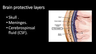
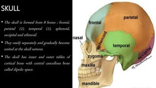
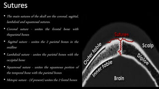
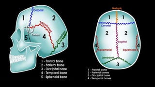
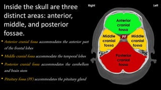
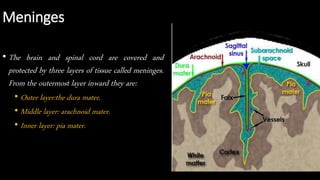
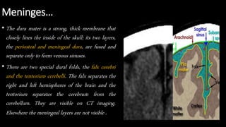
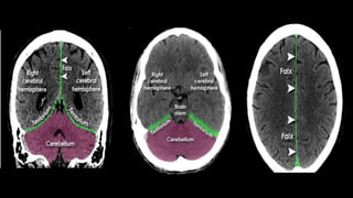
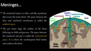
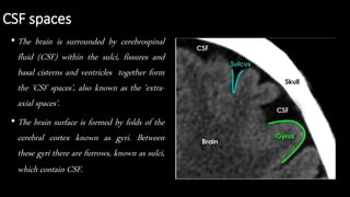
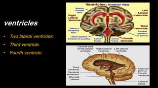
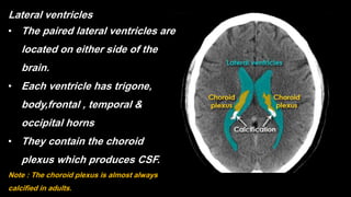
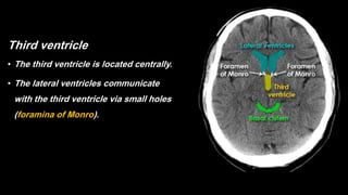
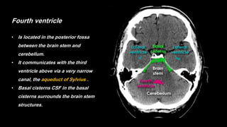
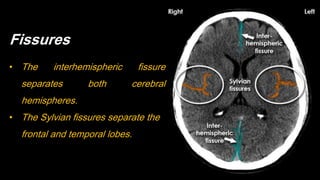
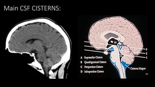
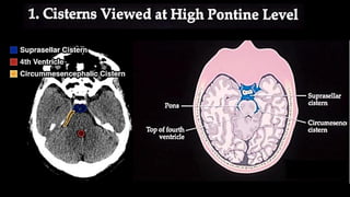
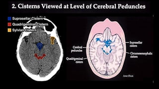
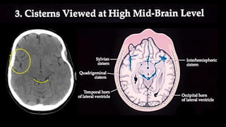
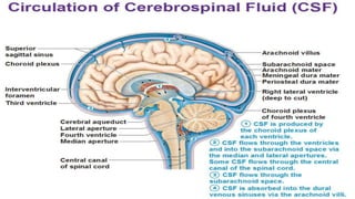
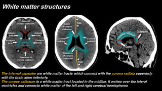
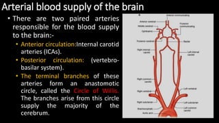
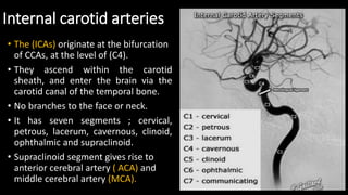
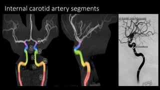
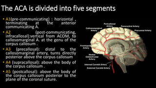
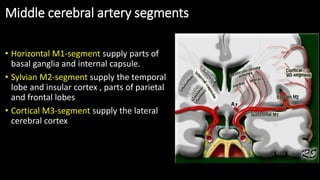
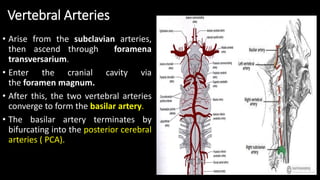
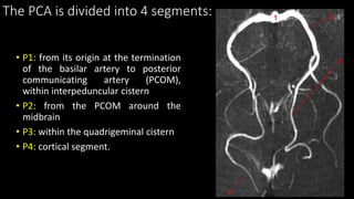
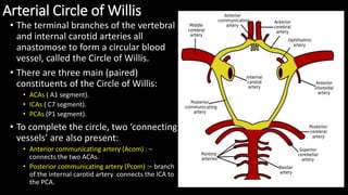
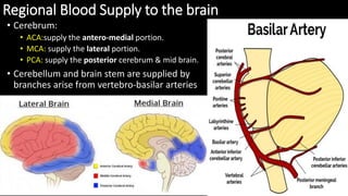
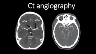
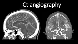
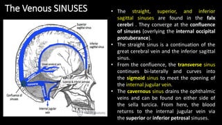
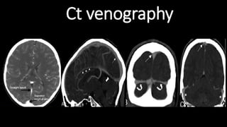
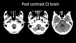
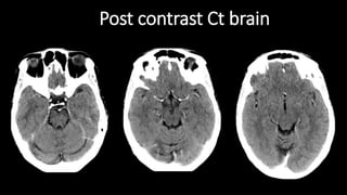
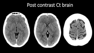
CROSS
SECTIONAL
ANATOMY
OF THE
BRAIN
Dr.Mohamed Kamel,MD.
Lecturer of diagnostic radiology
Faculty of medicine-Menoufia university
Brain protective layers
• Skull .
• Meninges.
• Cerebrospinsal
fluid (CSF).
SKULL
• The skull is formed from 8 bones ; frontal,
parietal (2), temporal (2), sphenoid,
occipital and ethmoid.
• They ossify separately and gradually become
united at the skull sutures.
• The skull has inner and outer tables of
cortical bone with central cancellous bone
called dipolic space.
Sutures
• The main sutures of the skull are the coronal, sagittal,
lambdoid and squamosal sutures.
• Coronal suture - unites the frontal bone with
theparietal bones
• Sagittal suture - unites the 2 parietal bones in the
midline
• Lambdoid suture - unites the parietal bones with the
occipital bone
• Squamosal suture - unites the squamous portion of
the temporal bone with the parietal bones
• Metopic suture - (if present) unites the 2 fontal bones
Inside the skull are three
distinct areas: anterior,
middle, and posterior
fossae.
• Anterior cranial fossa accommodates the anterior part
of the frontal lobes
• Middle cranial fossa accommodate the temporal lobes
• Posterior cranial fossa accommodates the cerebellum
and brain stem
• Pituitary fossa (PF) accommodates the pituitary gland
Meninges
• The brain and spinal cord are covered and
protected by three layers of tissue called meninges.
From the outermost layer inward they are:
• Outer layer:the dura mater.
• Middle layer: arachnoid mater.
• Inner layer: pia mater.
Meninges…
• The dura mater is a strong, thick membrane that
closely lines the inside of the skull; its two layers,
the periosteal and meningeal dura, are fused and
separate only to form venous sinuses.
• There are two special dural folds, the falx cerebri
and the tentorium cerebelli. The falx separates the
right and left hemispheres of the brain and the
tentorium separates the cerebrum from the
cerebellum. They are visible on CT imaging.
Elsewhere the meningeal layers are not visible .
Meninges…
• The arachnoid mater is a thin, web-like membrane
that covers the entire brain. The space between the
dura and arachnoid membranes is called the
subdural space.
• The pia mater hugs the surface of the brain
following its folds and grooves. The space between
the arachnoid and pia is called the subarachnoid
space. It is where the cerebrospinal fluid bathes
and cushions the brain.
CSF spaces
• The brain is surrounded by cerebrospinal
fluid (CSF) within the sulci, fissures and
basal cisterns and ventricles together form
the 'CSF spaces', also known as the 'extra-
axial spaces'.
• The brain surface is formed by folds of the
cerebral cortex known as gyri. Between
these gyri there are furrows, known as sulci,
which contain CSF.
ventricles
• Two lateral ventricles.
• Third ventricle.
• Fourth ventricle.
Lateral ventricles
• The paired lateral ventricles are
located on either side of the
brain.
• Each ventricle has trigone,
body,frontal , temporal &
occipital horns
• They contain the choroid
plexus which produces CSF.
Note : The choroid plexus is almost always
calcified in adults.
Third ventricle
• The third ventricle is located centrally.
• The lateral ventricles communicate
with the third ventricle via small holes
(foramina of Monro).
Fourth ventricle
• Is located in the posterior fossa
between the brain stem and
cerebellum.
• It communicates with the third
ventricle above via a very narrow
canal, the aqueduct of Sylvius .
• Basal cisterns CSF in the basal
cisterns surrounds the brain stem
structures.
Fissures
• The interhemispheric fissure
separates both cerebral
hemispheres.
• The Sylvian fissures separate the
frontal and temporal lobes.
Main CSF CISTERNS:
Brain.
•The brain is composed of the:
•Cerebrum.
•Cerebellum.
•Brain stem.
Brain.
• The cerebrum is the largest part of the brain
and is composed of right and left
hemispheres. It performs higher functions like
interpreting, touch, vision and hearing, as well
as speech, reasoning, emotions, learning, and
fine control of movement.
• The cerebral hemispheres have distinct
fissures, which divide the brain into lobes.
Each hemisphere has 4 lobes: frontal,
temporal, parietal, and occipital.
Brain lobes - CT brain (superior slice) On both
sides the frontal lobes are separated from the
parietal lobes by the central sulcus
Note: The large frontal lobes as compared to
parietal and occipital lobes
Brain lobes - CT brain (inferior slice) the frontal
lobes at anterior cranial fossae .The temporal
lobes at middle cranial fossae .The cerebellum
and brain stem at the posterior fossa
Brain...
• The cerebellum is located under the cerebrum.
• it’s two hemishperes with midline structure
named vermis.
• Its function is to coordinate muscle
movements, maintain posture, and balance.
• Note the gyri in cerebellum is named foli
Brain...
• The brainstem is the lower extension of the brain,
located in front of the cerebellum and connected to
the spinal cord.
• It consists of three structures; the midbrain, pons
and medulla oblongata.
• It performs many automatic functions such as
breathing, heart rate, body temperature, wake and
sleep cycles, digestion, sneezing, coughing, vomiting,
and swallowing. Ten of the twelve cranial nerves
originate in the brainstem.
Destruction of these regions of the brain will cause "brain death." Without these key functions, humans cannot survive.
• The brain consists of grey and white matter
structures which are differentiated on CT by
differences in density.
• White matter has a high content of myelinated
axons.
• Grey matter contains relatively few axons and a
higher number of cell bodies.
• As myelin is a fatty substance it is of relatively low
density compared to the cellular grey matter. White
matter, therefore, appears blacker than grey
matter.
Grey / white matter
Grey matter structures
Important grey matter structures visible on CT :
• Cortex, insula.
• Basal ganglia( lentiform nucleus + caudate nucleus ).
• Thalamus .
White matter structures
The internal capsules are white matter tracts which connect with the corona radiata superiorly
with the brain stem inferiorly.
The corpus callosum is a white matter tract located in the midline. It arches over the lateral
ventricles and connects white matter of the left and right cerebral hemispheres.
AXIAL SECTIONS
OF CT HEAD
POSTERIOR FOSSA CUTS
-ABOVE THE FORAMEN MAGNUM
LEVEL
-LEVEL OF THE FOURTH VENTRICLE
-ABOVE THE FOURTH VENTRICULAR
LEVEL
-TENTORIAL
SUPRATENTORIAL CUTS
-THIRD VENTRICULAR LEVEL
-LATERAL VENTRICULAR LEVEL
-ABOVE THE VENTRICULAR LEVEL
Arterial blood supply of the brain
• There are two paired arteries
responsible for the blood supply
to the brain:-
• Anterior circulation:Internal carotid
arteries (ICAs).
• Posterior circulation: (vertebro-
basilar system).
• The terminal branches of these
arteries form an anastomotic
circle, called the Circle of Willis.
The branches arise from this circle
supply the majority of the
cerebrum.
Internal carotid arteries
• The (ICAs) originate at the bifurcation
of CCAs, at the level of (C4).
• They ascend within the carotid
sheath, and enter the brain via the
carotid canal of the temporal bone.
• No branches to the face or neck.
• It has seven segments ; cervical,
petrous, lacerum, cavernous, clinoid,
ophthalmic and supraclinoid.
• Supraclinoid segment gives rise to
anterior cerebral artery ( ACA) and
middle cerebral artery (MCA).
Internal carotid artery segments
The ACA is divided into five segments
• A1(pre-communicating) : horizontal ,
terminating at the anterior
communicating A.
• A2 (post-communicating,
infracallosal):vertical from ACOM, to
callosomarginal A. at the genu of the
corpus callosum .
• A3 (precallosal): distal to the
callosomarginal artery, turns directly
posterior above the corpus callosum
• A4 (supracallosal): above the body of
the corpus callosum .
• A5 (postcallosal): above the body of
the corpus callosum posterior to the
plane of the coronal suture.
Middle cerebral artery segments
• Horizontal M1-segment supply parts of
basal ganglia and internal capsule.
• Sylvian M2-segment supply the temporal
lobe and insular cortex , parts of parietal
and frontal lobes
• Cortical M3-segment supply the lateral
cerebral cortex
Vertebral Arteries
• Arise from the subclavian arteries,
then ascend through foramena
transversarium.
• Enter the cranial cavity via
the foramen magnum.
• After this, the two vertebral arteries
converge to form the basilar artery.
• The basilar artery terminates by
bifurcating into the posterior cerebral
arteries ( PCA).
The PCA is divided into 4 segments:
• P1: from its origin at the termination
of the basilar artery to posterior
communicating artery (PCOM),
within interpeduncular cistern
• P2: from the PCOM around the
midbrain
• P3: within the quadrigeminal cistern
• P4: cortical segment.
Arterial Circle of Willis
• The terminal branches of the vertebral
and internal carotid arteries all
anastomose to form a circular blood
vessel, called the Circle of Willis.
• There are three main (paired)
constituents of the Circle of Willis:
• ACAs ( A1 segment).
• ICAs ( C7 segment).
• PCAs (P1 segment).
• To complete the circle, two ‘connecting
vessels’ are also present:
• Anterior communicating artery (Acom) : –
connects the two ACAs.
• Posterior communicating artery (Pcom) :– branch
of the internal carotid artery connects the ICA to
the PCA.
Regional Blood Supply to the brain
• Cerebrum:
• ACA:supply the antero-medial portion.
• MCA: supply the lateral portion.
• PCA: supply the posterior cerebrum & mid brain.
• Cerebellum and brain stem are supplied by
branches arise from vertebro-basilar arteries
Ct angiography
Ct angiography
The Venous Drainage of the Central Nervous System
• The dural venous sinuses lie between
the periosteal and meningeal layers
of the dura mater. They are best
thought of as collecting pools of
blood, which drain the brain.
• Unlike most veins of the body, the
dural venous sinuses do not
have valves.
• All the dural venous sinuses
ultimately drain into the internal
jugular vein.
• The straight, superior, and inferior
sagittal sinuses are found in the falx
cerebri . They converge at the confluence
of sinuses (overlying the internal occipital
protuberance).
• The straight sinus is a continuation of the
great cerebral vein and the inferior sagittal
sinus.
• From the confluence, the transverse sinus
continues bi-laterally and curves into
the sigmoid sinus to meet the opening of
the internal jugular vein.
• The cavernous sinus drains the ophthalmic
veins and can be found on either side of
the sella turcica. From here, the blood
returns to the internal jugular vein via
the superior or inferior petrosal sinuses.
The Venous SINUSES
Ct venography
Post contrast Ct brain
brain.pdf

brain.pdf

  • 1.
    CROSS SECTIONAL ANATOMY OF THE BRAIN Dr.Mohamed Kamel,MD. Lecturerof diagnostic radiology Faculty of medicine-Menoufia university
  • 2.
    Brain protective layers •Skull . • Meninges. • Cerebrospinsal fluid (CSF).
  • 3.
    SKULL • The skullis formed from 8 bones ; frontal, parietal (2), temporal (2), sphenoid, occipital and ethmoid. • They ossify separately and gradually become united at the skull sutures. • The skull has inner and outer tables of cortical bone with central cancellous bone called dipolic space.
  • 4.
    Sutures • The mainsutures of the skull are the coronal, sagittal, lambdoid and squamosal sutures. • Coronal suture - unites the frontal bone with theparietal bones • Sagittal suture - unites the 2 parietal bones in the midline • Lambdoid suture - unites the parietal bones with the occipital bone • Squamosal suture - unites the squamous portion of the temporal bone with the parietal bones • Metopic suture - (if present) unites the 2 fontal bones
  • 6.
    Inside the skullare three distinct areas: anterior, middle, and posterior fossae. • Anterior cranial fossa accommodates the anterior part of the frontal lobes • Middle cranial fossa accommodate the temporal lobes • Posterior cranial fossa accommodates the cerebellum and brain stem • Pituitary fossa (PF) accommodates the pituitary gland
  • 7.
    Meninges • The brainand spinal cord are covered and protected by three layers of tissue called meninges. From the outermost layer inward they are: • Outer layer:the dura mater. • Middle layer: arachnoid mater. • Inner layer: pia mater.
  • 8.
    Meninges… • The duramater is a strong, thick membrane that closely lines the inside of the skull; its two layers, the periosteal and meningeal dura, are fused and separate only to form venous sinuses. • There are two special dural folds, the falx cerebri and the tentorium cerebelli. The falx separates the right and left hemispheres of the brain and the tentorium separates the cerebrum from the cerebellum. They are visible on CT imaging. Elsewhere the meningeal layers are not visible .
  • 10.
    Meninges… • The arachnoidmater is a thin, web-like membrane that covers the entire brain. The space between the dura and arachnoid membranes is called the subdural space. • The pia mater hugs the surface of the brain following its folds and grooves. The space between the arachnoid and pia is called the subarachnoid space. It is where the cerebrospinal fluid bathes and cushions the brain.
  • 11.
    CSF spaces • Thebrain is surrounded by cerebrospinal fluid (CSF) within the sulci, fissures and basal cisterns and ventricles together form the 'CSF spaces', also known as the 'extra- axial spaces'. • The brain surface is formed by folds of the cerebral cortex known as gyri. Between these gyri there are furrows, known as sulci, which contain CSF.
  • 12.
    ventricles • Two lateralventricles. • Third ventricle. • Fourth ventricle.
  • 13.
    Lateral ventricles • Thepaired lateral ventricles are located on either side of the brain. • Each ventricle has trigone, body,frontal , temporal & occipital horns • They contain the choroid plexus which produces CSF. Note : The choroid plexus is almost always calcified in adults.
  • 14.
    Third ventricle • Thethird ventricle is located centrally. • The lateral ventricles communicate with the third ventricle via small holes (foramina of Monro).
  • 15.
    Fourth ventricle • Islocated in the posterior fossa between the brain stem and cerebellum. • It communicates with the third ventricle above via a very narrow canal, the aqueduct of Sylvius . • Basal cisterns CSF in the basal cisterns surrounds the brain stem structures.
  • 16.
    Fissures • The interhemisphericfissure separates both cerebral hemispheres. • The Sylvian fissures separate the frontal and temporal lobes.
  • 17.
  • 22.
    Brain. •The brain iscomposed of the: •Cerebrum. •Cerebellum. •Brain stem.
  • 23.
    Brain. • The cerebrumis the largest part of the brain and is composed of right and left hemispheres. It performs higher functions like interpreting, touch, vision and hearing, as well as speech, reasoning, emotions, learning, and fine control of movement. • The cerebral hemispheres have distinct fissures, which divide the brain into lobes. Each hemisphere has 4 lobes: frontal, temporal, parietal, and occipital.
  • 24.
    Brain lobes -CT brain (superior slice) On both sides the frontal lobes are separated from the parietal lobes by the central sulcus Note: The large frontal lobes as compared to parietal and occipital lobes Brain lobes - CT brain (inferior slice) the frontal lobes at anterior cranial fossae .The temporal lobes at middle cranial fossae .The cerebellum and brain stem at the posterior fossa
  • 25.
    Brain... • The cerebellumis located under the cerebrum. • it’s two hemishperes with midline structure named vermis. • Its function is to coordinate muscle movements, maintain posture, and balance. • Note the gyri in cerebellum is named foli
  • 26.
    Brain... • The brainstemis the lower extension of the brain, located in front of the cerebellum and connected to the spinal cord. • It consists of three structures; the midbrain, pons and medulla oblongata. • It performs many automatic functions such as breathing, heart rate, body temperature, wake and sleep cycles, digestion, sneezing, coughing, vomiting, and swallowing. Ten of the twelve cranial nerves originate in the brainstem. Destruction of these regions of the brain will cause "brain death." Without these key functions, humans cannot survive.
  • 27.
    • The brainconsists of grey and white matter structures which are differentiated on CT by differences in density. • White matter has a high content of myelinated axons. • Grey matter contains relatively few axons and a higher number of cell bodies. • As myelin is a fatty substance it is of relatively low density compared to the cellular grey matter. White matter, therefore, appears blacker than grey matter. Grey / white matter
  • 28.
    Grey matter structures Importantgrey matter structures visible on CT : • Cortex, insula. • Basal ganglia( lentiform nucleus + caudate nucleus ). • Thalamus .
  • 29.
    White matter structures Theinternal capsules are white matter tracts which connect with the corona radiata superiorly with the brain stem inferiorly. The corpus callosum is a white matter tract located in the midline. It arches over the lateral ventricles and connects white matter of the left and right cerebral hemispheres.
  • 31.
    AXIAL SECTIONS OF CTHEAD POSTERIOR FOSSA CUTS -ABOVE THE FORAMEN MAGNUM LEVEL -LEVEL OF THE FOURTH VENTRICLE -ABOVE THE FOURTH VENTRICULAR LEVEL -TENTORIAL SUPRATENTORIAL CUTS -THIRD VENTRICULAR LEVEL -LATERAL VENTRICULAR LEVEL -ABOVE THE VENTRICULAR LEVEL
  • 40.
    Arterial blood supplyof the brain • There are two paired arteries responsible for the blood supply to the brain:- • Anterior circulation:Internal carotid arteries (ICAs). • Posterior circulation: (vertebro- basilar system). • The terminal branches of these arteries form an anastomotic circle, called the Circle of Willis. The branches arise from this circle supply the majority of the cerebrum.
  • 41.
    Internal carotid arteries •The (ICAs) originate at the bifurcation of CCAs, at the level of (C4). • They ascend within the carotid sheath, and enter the brain via the carotid canal of the temporal bone. • No branches to the face or neck. • It has seven segments ; cervical, petrous, lacerum, cavernous, clinoid, ophthalmic and supraclinoid. • Supraclinoid segment gives rise to anterior cerebral artery ( ACA) and middle cerebral artery (MCA).
  • 42.
  • 43.
    The ACA isdivided into five segments • A1(pre-communicating) : horizontal , terminating at the anterior communicating A. • A2 (post-communicating, infracallosal):vertical from ACOM, to callosomarginal A. at the genu of the corpus callosum . • A3 (precallosal): distal to the callosomarginal artery, turns directly posterior above the corpus callosum • A4 (supracallosal): above the body of the corpus callosum . • A5 (postcallosal): above the body of the corpus callosum posterior to the plane of the coronal suture.
  • 44.
    Middle cerebral arterysegments • Horizontal M1-segment supply parts of basal ganglia and internal capsule. • Sylvian M2-segment supply the temporal lobe and insular cortex , parts of parietal and frontal lobes • Cortical M3-segment supply the lateral cerebral cortex
  • 45.
    Vertebral Arteries • Arisefrom the subclavian arteries, then ascend through foramena transversarium. • Enter the cranial cavity via the foramen magnum. • After this, the two vertebral arteries converge to form the basilar artery. • The basilar artery terminates by bifurcating into the posterior cerebral arteries ( PCA).
  • 46.
    The PCA isdivided into 4 segments: • P1: from its origin at the termination of the basilar artery to posterior communicating artery (PCOM), within interpeduncular cistern • P2: from the PCOM around the midbrain • P3: within the quadrigeminal cistern • P4: cortical segment.
  • 47.
    Arterial Circle ofWillis • The terminal branches of the vertebral and internal carotid arteries all anastomose to form a circular blood vessel, called the Circle of Willis. • There are three main (paired) constituents of the Circle of Willis: • ACAs ( A1 segment). • ICAs ( C7 segment). • PCAs (P1 segment). • To complete the circle, two ‘connecting vessels’ are also present: • Anterior communicating artery (Acom) : – connects the two ACAs. • Posterior communicating artery (Pcom) :– branch of the internal carotid artery connects the ICA to the PCA.
  • 48.
    Regional Blood Supplyto the brain • Cerebrum: • ACA:supply the antero-medial portion. • MCA: supply the lateral portion. • PCA: supply the posterior cerebrum & mid brain. • Cerebellum and brain stem are supplied by branches arise from vertebro-basilar arteries
  • 49.
  • 50.
  • 51.
    The Venous Drainageof the Central Nervous System • The dural venous sinuses lie between the periosteal and meningeal layers of the dura mater. They are best thought of as collecting pools of blood, which drain the brain. • Unlike most veins of the body, the dural venous sinuses do not have valves. • All the dural venous sinuses ultimately drain into the internal jugular vein.
  • 52.
    • The straight,superior, and inferior sagittal sinuses are found in the falx cerebri . They converge at the confluence of sinuses (overlying the internal occipital protuberance). • The straight sinus is a continuation of the great cerebral vein and the inferior sagittal sinus. • From the confluence, the transverse sinus continues bi-laterally and curves into the sigmoid sinus to meet the opening of the internal jugular vein. • The cavernous sinus drains the ophthalmic veins and can be found on either side of the sella turcica. From here, the blood returns to the internal jugular vein via the superior or inferior petrosal sinuses. The Venous SINUSES
  • 53.
  • 54.
  • 55.
  • 56.