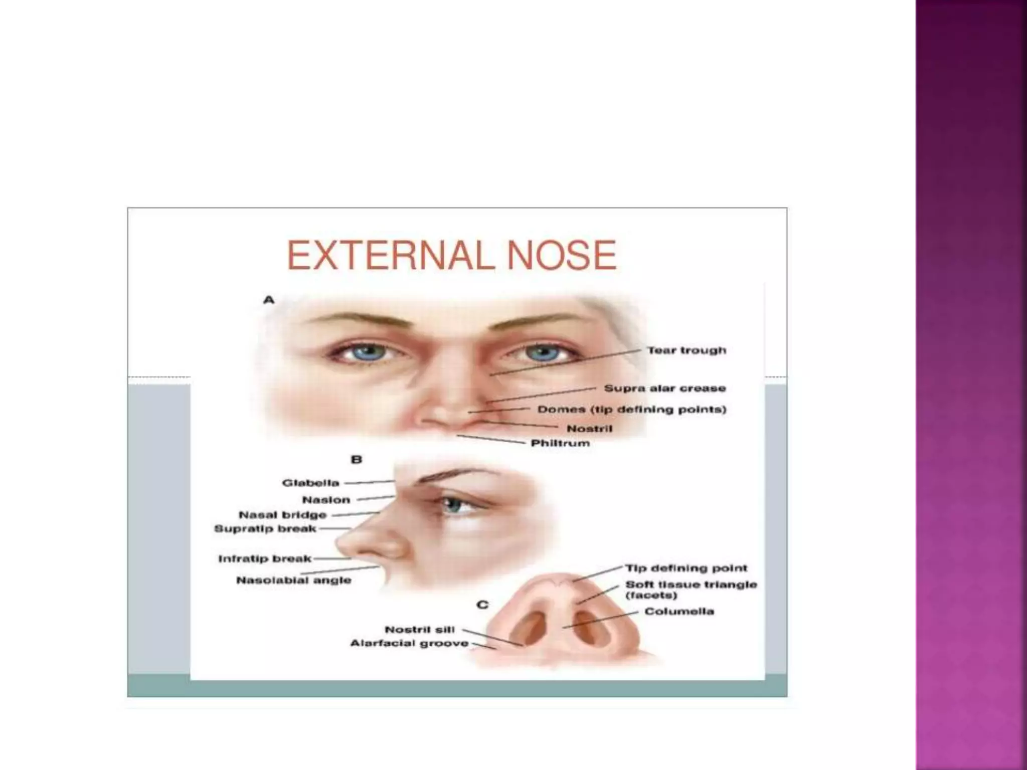
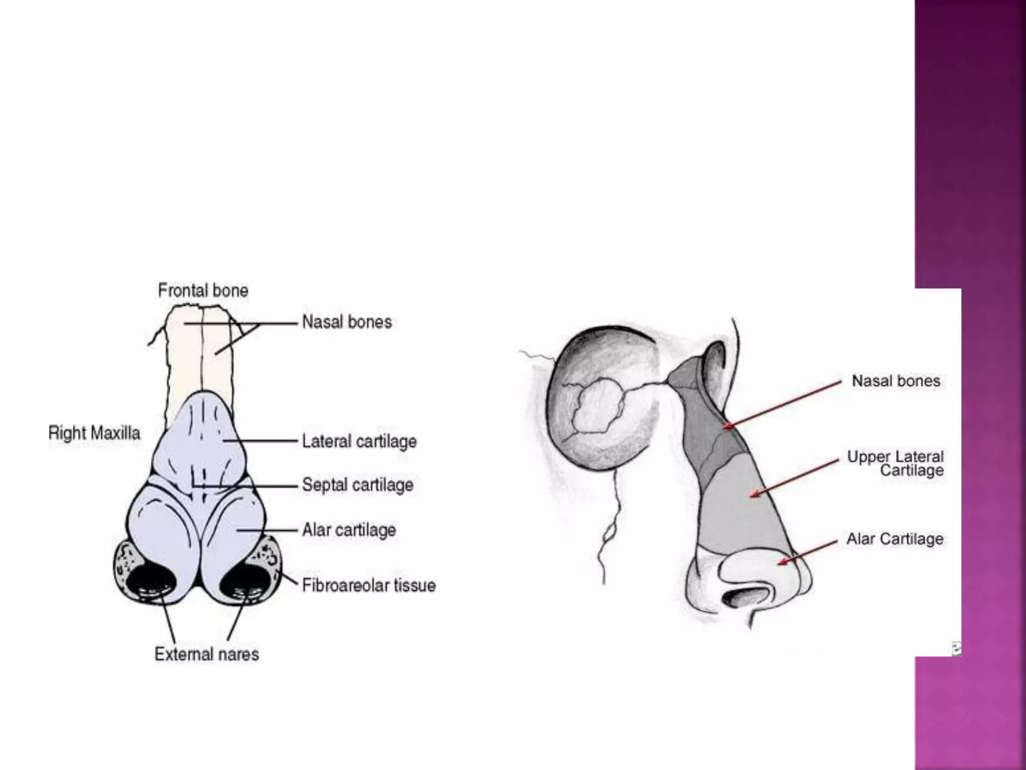
 External Nose
 Internal Nose
 Blood Supply
 Nerve Supply
 Lymphatic Drainage
 PNS
 Embryology
 Triangular pyramidal structure
 Bony (upper 1/3rd) and Cartilaginous (lower 2/3rd
)
 Root of nose – upper angle of nose where it is
continous with forehead.
 Base of nose – triangular, directed downwards
 Tip of nose – free angle of nose below
 Dorsum of nose – lower cartilaginous part
connected to tip
 Bridge of nose – upper bony part of dorsum
connected to root. It is a junction between 2
nasal bones
 Glabella – point between root of nose and
forehead
 Upper 1/3rd
 Paired nasal bones
 Paired frontal process of maxilla
 Nasal process of frontal bones
 Nasal bones articulate
 Sup – nasal process of frontal bone
 Inf – upper lateral cartilage
 Lat – frontal process of maxilla
 Med – opp nasal bone
 Nasion – upper point where they meet
 Rhinion – lower point where they meet
 Paired upper lateral cartilages
 Paired lower lateral cartilages (alar)
 Sesamoid cartilage (lesser alar)
 Septal cartilage (ant sup part)
 Hyaline cartilage
 Bone and cartilage connected to each other
by periosteum and perichondrium which is
continous
 Upper lateral cartilages
 Triangular in shape
 Form dorsum of nose
 Middle 1/3rd
 Lower lateral cartilages (alar)
 Maintain projection and shape of tip
 Lower 1/3rd
 Parts
 Slender medial crus – 2 in midline form columella
 Wider lateral crus – form ala of nose each side
 Projection between two – form tip of nose
 Sesamoid cartilage (lesser alar)
 Minor
 2 or more in number
 Present between upper and lower nasal cartilage
 External Nasal Skin
 Continous with skin of vestibule and columella
 Thin and freely mobile – nasal bones and upper
lateral cartilage
 Thick and adherent – alar cartilage, contains
sebaceous glands – hypertrophy (rhinophyma)
 Facial muscles bring about movement of nose
 Procerus
 Nasalis (transverse and alar)
 Levator labii superioris alaeque nasi
 Depressor septi
 Ant and post dilator nares
 Arise from fascia and inserted into skin
 Facial N
 Right and left by nasal septum
 Communicates with external (ant nares),
nasopharynx (choanae), sinuses (ostium)
 Each nasal cavity 5-7 cm length, 5 cm height,
1.5 cm transversely at floor, 1-2 mm at roof
 Parts
 Skin lined – Vestibule
 Mucosa lined – cavity proper
 Nasal cavity proper – medial wall, lateral
wall, roof, floor
 Ant inf part of nasal cavity, entrance
 Lined by skin
 Contains hairs (vibrissae), hair follicles,
sebaceous glands, sweat glands
 Limen nasi – sup margin of lower lateral
cartilage, separates vestibule from nasal
mucosa. Upper limit of nasal vestibule
 Part of dangerous area of face – triangular
area extending from nasion to angle of
mouth (includes external nose and upper lip)
– without valve venous drainage – lead to CST
 Narrowest part of nasal cavity
 Triangular shaped
 Area of high resistance, mild narrowing
causes nasal obstruction
 Regulates air flow
 Medially – cartilaginous septum, caudally –
floor, laterally – ant end of inf turbinate,
lower border of upper lateral cartilage
 Columella – between two nasal vestibules,
formed by medial crusa of alar cart, forms
caudal end of nasal septum
 Framework – Floor, Roof, Medial wall and
lateral wall
 Floor
 Made up of hard palate
 Palatine process of maxilla (ant 3/4th)
 Horizontal process of palatine bones (post
1/4th)
 5cm long, 1.25 cm wide
 Highest part of nasal cavity
 Ant sloping part – nasal bones
 Post sloping part – body of sphenoid
 Middle horizontal part – cribriform plate of
ethmoid
 Part of olfactory area
 Olfactory area
 Bounded lat by sup turbinate, med by septum
and sup by cribriform plate
 Dangerous area as olfactory nerve fibres from
this area pass through cribriform plate to ant
cranial fossa and carry infection, ass with CSF
rhinorrhoea
 Bony and cartilaginous framework lined by
mucoperiosteum and mucoperichondrium
 Columellar septum – covered by skin, medial crus
 Membranous septum – b/w columella and septum
proper, no bone or cartilage, double layer of skin
 Septum proper
 Cartilaginous part – large quadrangular cartilage
with contributions from upper and lower lateral
cartilage
 Septal cartilage provides support to tip and
dorsum of nose
 Bony part
 Perpendicular plate of ethmoid – post superior
part
 Vomer – post inferior part
 Maxillary crest – inf most part
 Palatine crest
 Small contributions from nasal spine of frontal
bone, rostrum of sphenoid, ant nasal spine of
maxilla
 Jacobson’s organ – concerned with smell in
lower animals, vestigeal in adult humans,
defined in foetus as blind tubular pouch 2-6 mm
long
 Irregular
 Atrium – lies b/w vestibule and concha in the
lateral wall
 Covered by respiratory epithelium
 Formed by various bones
 Main area for drainage of sinus secretions
 Formed by medial wall of maxilla, middle and
sup concha (ethmoid bone), inf concha, medial
pterygoid process of sphenoid, nasal bones,
perpendicular plate of palatine bones
 Has scroll/shelf shaped ridges/ bony horizontal
elevations called turbinates (sup/middle/inf)
and beneath and lateral to them depressions
called meatus (sup/middle/inf)
 Superior turbinate
 Smallest
 Part of ethmoid bone
 The area above it and its medial part lined
by olfactory epithelium
 Ostium of sphenoid sinus lies medial to it
 Supreme turbinate
 60% of cases, U/L or B/L
 Small ridge above sup turbinate
 Middle turbinate
 Part of ethmoid bone
 Formed by middle concha
 Has basal lamina – joins lamina papyracea
 Ascending limb – joins cribriform plate
 Bony lamella (basal or ground) – attached to lateral wall
 Concha bullosa – pneumatized middle turbinate by
ethmoidal air cells
 Paradoxically curved – narrowed middle meatus
 Post end – sphenopalatine foramen (gap b/w fusion of
sphenoid, palatine and ethmoidal bones)

 Inferior turbinate
 Largest
 Separate bone
 Composed of inf concha
 Submucosa – contains rich cavernous venous plexus
 Superior meatus
 Smallest, lies beneath sup concha
 Opening for post ethmoid air cells (1-5)
 Sphenoethmoidal recess
 Triangular area above sup turbinate
 Sphenoidal sinus opens here
 Middle meatus
 Lies below middle concha
 Major structure jere forms osteo meatal complex
 Frontal, maxillary, ant ethmoidal sinus drain
here
 Inferior meatus
 Largest
 Present along the whole length of lateral wall
 NLD opens in the ant part, under genu of inf
turbinate, opening guarded by a valve – hasner’s
valve (plica lacrimalis)
 Skin
 Stratified squamous epithelium covers vestibule
 Olfactory epithelium
 Upper 1/3rd of lateral wall till sup concha, roof, nasal
septum
 Mucous membrane – pale (yellowish) in colour
 Respiratory epithelium
 Lower 2/3rd of nasal cavity
 Thickest over concha, thick over septum
 Thin over meatus and floor
 Pink in colour, pseudostratified ciliated columnar
epithelium
 Arterial supply
 ICA and ECA
 ECA – maxillary artery (sphenopalatine, greater
palatine), facial artery (sup labial)
 ICA – ophthalmic artery (ant and post ethmoidal)
 Lateral wall
 Main artery – sphenopalatine artery – all meatus
and turbinates
 Roof and sup part – ant ethmoidal artery
 Alae nasi – sup labial artery
 Medial wall (septum)
 Ant sup part – ant ethmoidal artery
 Ant inf part – sphenopalatine artery – main
artery to septum, also called artery of epistaxis
 Keisselbach’s plexus (little’s area)
 Anastomotic plexus b/w sphenopalatine, greater
palatine, sup labial and ant ethmoidal artery
 Frequent site for bleeding (90%, ant epistaxis)
 Site for origin of bleeding polyposis
(haemangioma of nose)
 Ant part – ant facial vein and ophthalmic vein
 Middle part – pterygoid venous plexus
 Post part – pharyngeal plexus of veins
 Cavernous plexus – beneath the middle meatus formed of
sphenopalatine vein, ophthalmic vein, sup saggital sinus
 Retrocolumellar vein – just behind the columella,
common site for venous bleeding
 Woodruff’s plexus – site for venous bleed inf to post end
of inferior turbinate
 Arterial bleed post to middle turbinate
 Site for post epistaxis, maxillary sinus ostia dividing line
 Erectile tissue
 Subepithelial venous plexus
 Seen mainly in inf turbinate, post part of
middle turbinate, septum
 Controlled by ANS – maintains congestion and
decongestion of turbinates
 Trigeminal nerve – sensory supply
 Ophthalmic nerve – ant ethmoidal nerve – ant
and sup part of nasal cavity – blocked by placing
pledgets high up
 Maxillary nerve – sphenopalatine branch – post
2/3rd of nasal cavity – blocked by placing
pledgets post extension of middle meatus
 Infra orbital nerve – vestibule of nose
 Facial nerve – motor supply
 Olfactory nerves – 20 in number - smell
 Secretomotor supply – vidian nerve (nerve of
pterygoid canal)
 Formed by
 Deep petrosal nerve – sympathetic plexus
around ICA
 Greater sup petrosal nerve – parasympathetic
supply to nasal glands and controls nasal
secretions
 Submental lymph nodes (anterior part and
external nose)
 Retropharyngeal ln (post part)
 Upper deep cervical ln (post part)
 Preauricular ln

Anatomy of nose and pns

  • 2.
     External Nose Internal Nose  Blood Supply  Nerve Supply  Lymphatic Drainage  PNS  Embryology
  • 3.
     Triangular pyramidalstructure  Bony (upper 1/3rd) and Cartilaginous (lower 2/3rd )  Root of nose – upper angle of nose where it is continous with forehead.  Base of nose – triangular, directed downwards  Tip of nose – free angle of nose below  Dorsum of nose – lower cartilaginous part connected to tip  Bridge of nose – upper bony part of dorsum connected to root. It is a junction between 2 nasal bones  Glabella – point between root of nose and forehead
  • 5.
     Upper 1/3rd Paired nasal bones  Paired frontal process of maxilla  Nasal process of frontal bones  Nasal bones articulate  Sup – nasal process of frontal bone  Inf – upper lateral cartilage  Lat – frontal process of maxilla  Med – opp nasal bone  Nasion – upper point where they meet  Rhinion – lower point where they meet
  • 6.
     Paired upperlateral cartilages  Paired lower lateral cartilages (alar)  Sesamoid cartilage (lesser alar)  Septal cartilage (ant sup part)  Hyaline cartilage  Bone and cartilage connected to each other by periosteum and perichondrium which is continous
  • 8.
     Upper lateralcartilages  Triangular in shape  Form dorsum of nose  Middle 1/3rd  Lower lateral cartilages (alar)  Maintain projection and shape of tip  Lower 1/3rd  Parts  Slender medial crus – 2 in midline form columella  Wider lateral crus – form ala of nose each side  Projection between two – form tip of nose
  • 9.
     Sesamoid cartilage(lesser alar)  Minor  2 or more in number  Present between upper and lower nasal cartilage  External Nasal Skin  Continous with skin of vestibule and columella  Thin and freely mobile – nasal bones and upper lateral cartilage  Thick and adherent – alar cartilage, contains sebaceous glands – hypertrophy (rhinophyma)
  • 10.
     Facial musclesbring about movement of nose  Procerus  Nasalis (transverse and alar)  Levator labii superioris alaeque nasi  Depressor septi  Ant and post dilator nares  Arise from fascia and inserted into skin  Facial N
  • 12.
     Right andleft by nasal septum  Communicates with external (ant nares), nasopharynx (choanae), sinuses (ostium)  Each nasal cavity 5-7 cm length, 5 cm height, 1.5 cm transversely at floor, 1-2 mm at roof  Parts  Skin lined – Vestibule  Mucosa lined – cavity proper  Nasal cavity proper – medial wall, lateral wall, roof, floor
  • 13.
     Ant infpart of nasal cavity, entrance  Lined by skin  Contains hairs (vibrissae), hair follicles, sebaceous glands, sweat glands  Limen nasi – sup margin of lower lateral cartilage, separates vestibule from nasal mucosa. Upper limit of nasal vestibule  Part of dangerous area of face – triangular area extending from nasion to angle of mouth (includes external nose and upper lip) – without valve venous drainage – lead to CST
  • 14.
     Narrowest partof nasal cavity  Triangular shaped  Area of high resistance, mild narrowing causes nasal obstruction  Regulates air flow  Medially – cartilaginous septum, caudally – floor, laterally – ant end of inf turbinate, lower border of upper lateral cartilage  Columella – between two nasal vestibules, formed by medial crusa of alar cart, forms caudal end of nasal septum
  • 15.
     Framework –Floor, Roof, Medial wall and lateral wall  Floor  Made up of hard palate  Palatine process of maxilla (ant 3/4th)  Horizontal process of palatine bones (post 1/4th)  5cm long, 1.25 cm wide
  • 16.
     Highest partof nasal cavity  Ant sloping part – nasal bones  Post sloping part – body of sphenoid  Middle horizontal part – cribriform plate of ethmoid  Part of olfactory area  Olfactory area  Bounded lat by sup turbinate, med by septum and sup by cribriform plate  Dangerous area as olfactory nerve fibres from this area pass through cribriform plate to ant cranial fossa and carry infection, ass with CSF rhinorrhoea
  • 17.
     Bony andcartilaginous framework lined by mucoperiosteum and mucoperichondrium  Columellar septum – covered by skin, medial crus  Membranous septum – b/w columella and septum proper, no bone or cartilage, double layer of skin  Septum proper  Cartilaginous part – large quadrangular cartilage with contributions from upper and lower lateral cartilage  Septal cartilage provides support to tip and dorsum of nose
  • 18.
     Bony part Perpendicular plate of ethmoid – post superior part  Vomer – post inferior part  Maxillary crest – inf most part  Palatine crest  Small contributions from nasal spine of frontal bone, rostrum of sphenoid, ant nasal spine of maxilla  Jacobson’s organ – concerned with smell in lower animals, vestigeal in adult humans, defined in foetus as blind tubular pouch 2-6 mm long
  • 20.
     Irregular  Atrium– lies b/w vestibule and concha in the lateral wall  Covered by respiratory epithelium  Formed by various bones  Main area for drainage of sinus secretions  Formed by medial wall of maxilla, middle and sup concha (ethmoid bone), inf concha, medial pterygoid process of sphenoid, nasal bones, perpendicular plate of palatine bones  Has scroll/shelf shaped ridges/ bony horizontal elevations called turbinates (sup/middle/inf) and beneath and lateral to them depressions called meatus (sup/middle/inf)
  • 21.
     Superior turbinate Smallest  Part of ethmoid bone  The area above it and its medial part lined by olfactory epithelium  Ostium of sphenoid sinus lies medial to it  Supreme turbinate  60% of cases, U/L or B/L  Small ridge above sup turbinate
  • 22.
     Middle turbinate Part of ethmoid bone  Formed by middle concha  Has basal lamina – joins lamina papyracea  Ascending limb – joins cribriform plate  Bony lamella (basal or ground) – attached to lateral wall  Concha bullosa – pneumatized middle turbinate by ethmoidal air cells  Paradoxically curved – narrowed middle meatus  Post end – sphenopalatine foramen (gap b/w fusion of sphenoid, palatine and ethmoidal bones) 
  • 23.
     Inferior turbinate Largest  Separate bone  Composed of inf concha  Submucosa – contains rich cavernous venous plexus  Superior meatus  Smallest, lies beneath sup concha  Opening for post ethmoid air cells (1-5)  Sphenoethmoidal recess  Triangular area above sup turbinate  Sphenoidal sinus opens here
  • 24.
     Middle meatus Lies below middle concha  Major structure jere forms osteo meatal complex  Frontal, maxillary, ant ethmoidal sinus drain here  Inferior meatus  Largest  Present along the whole length of lateral wall  NLD opens in the ant part, under genu of inf turbinate, opening guarded by a valve – hasner’s valve (plica lacrimalis)
  • 26.
     Skin  Stratifiedsquamous epithelium covers vestibule  Olfactory epithelium  Upper 1/3rd of lateral wall till sup concha, roof, nasal septum  Mucous membrane – pale (yellowish) in colour  Respiratory epithelium  Lower 2/3rd of nasal cavity  Thickest over concha, thick over septum  Thin over meatus and floor  Pink in colour, pseudostratified ciliated columnar epithelium
  • 27.
     Arterial supply ICA and ECA  ECA – maxillary artery (sphenopalatine, greater palatine), facial artery (sup labial)  ICA – ophthalmic artery (ant and post ethmoidal)  Lateral wall  Main artery – sphenopalatine artery – all meatus and turbinates  Roof and sup part – ant ethmoidal artery  Alae nasi – sup labial artery
  • 28.
     Medial wall(septum)  Ant sup part – ant ethmoidal artery  Ant inf part – sphenopalatine artery – main artery to septum, also called artery of epistaxis  Keisselbach’s plexus (little’s area)  Anastomotic plexus b/w sphenopalatine, greater palatine, sup labial and ant ethmoidal artery  Frequent site for bleeding (90%, ant epistaxis)  Site for origin of bleeding polyposis (haemangioma of nose)
  • 30.
     Ant part– ant facial vein and ophthalmic vein  Middle part – pterygoid venous plexus  Post part – pharyngeal plexus of veins  Cavernous plexus – beneath the middle meatus formed of sphenopalatine vein, ophthalmic vein, sup saggital sinus  Retrocolumellar vein – just behind the columella, common site for venous bleeding  Woodruff’s plexus – site for venous bleed inf to post end of inferior turbinate  Arterial bleed post to middle turbinate  Site for post epistaxis, maxillary sinus ostia dividing line
  • 32.
     Erectile tissue Subepithelial venous plexus  Seen mainly in inf turbinate, post part of middle turbinate, septum  Controlled by ANS – maintains congestion and decongestion of turbinates
  • 33.
     Trigeminal nerve– sensory supply  Ophthalmic nerve – ant ethmoidal nerve – ant and sup part of nasal cavity – blocked by placing pledgets high up  Maxillary nerve – sphenopalatine branch – post 2/3rd of nasal cavity – blocked by placing pledgets post extension of middle meatus  Infra orbital nerve – vestibule of nose  Facial nerve – motor supply  Olfactory nerves – 20 in number - smell
  • 35.
     Secretomotor supply– vidian nerve (nerve of pterygoid canal)  Formed by  Deep petrosal nerve – sympathetic plexus around ICA  Greater sup petrosal nerve – parasympathetic supply to nasal glands and controls nasal secretions
  • 36.
     Submental lymphnodes (anterior part and external nose)  Retropharyngeal ln (post part)  Upper deep cervical ln (post part)  Preauricular ln