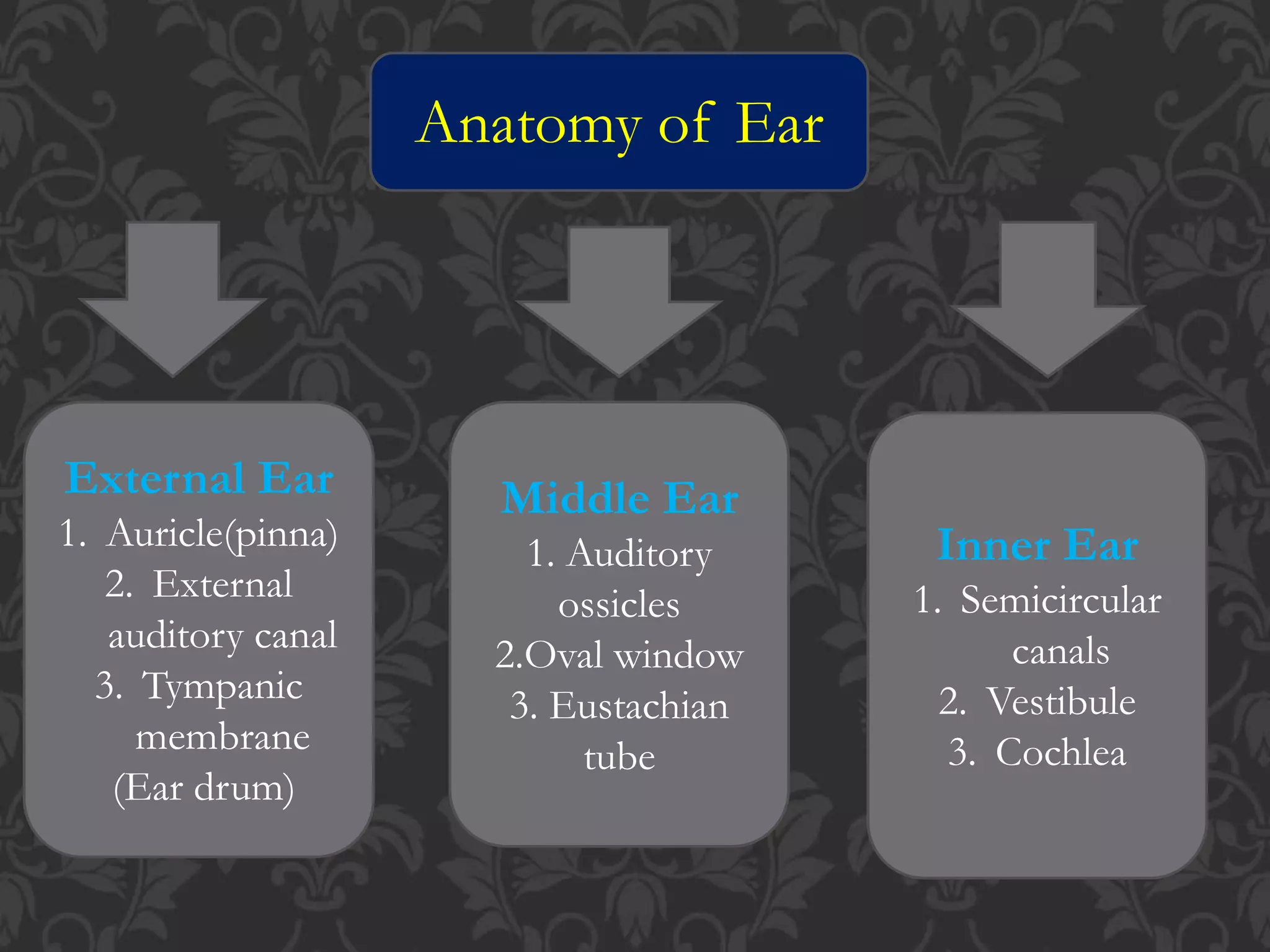
EAR
COVERED BY :
DEEPAK KUMAR
ANATOMY OF EAR
 Ear is divided into three main regions
1. External Ear
2. Middle Ear
3. Inner Ear
Anatomy of Ear
External Ear
1. Auricle(pinna)
2. External
auditory canal
3. Tympanic
membrane
(Ear drum)
Middle Ear
1. Auditory
ossicles
2.Oval window
3. Eustachian
tube
Inner Ear
1. Semicircular
canals
2. Vestibule
3. Cochlea
A).External Ear (Outer Ear)
1.Auricle /pinna
> it is flap of elastic cartilage
> it is covered by skin
> Rim of auricle is called helix
> Inferior portion is called lobule
> Plays important role in
localization of sound
> it opens External auditory canal
2. External auditory canal External auditory meatus
Curved tube ,extends pinna to ear drum
2.5 cm long
Near the external opening the external auditory
canal contains a few hair and ceruminous glands.
Ceruminous glands secrete the cerumen (ear wax )
Combination of hairs and cerumen helps prevent
dust and foreign objects from entering the ear .
3.Tympanic membrane /Ear drum
External auditory canal ends at the
Tympanic membrane also called as ear
drum .
It is a thin, semitransparent partition
b/w external auditory canal and middle
ear .
It is covered by epidermis
Lined by simple cuboidal epithelium
B). Middle Ear
> Middle ear is small, air filled cavity in
temporal bone.
 It is lined by epithelium.
 It separated from external ear by ear drum.
 It separated from inner ear by oval
window .
 The structures of middle ear are :
1. Auditory Ossicles
2. Oval window
3. Eustachian tube
1). Auditory Ossicles
> These are the smallest three bones of the body
Connected by synovial joints
a. The
Malleus Or
The Hammer
b. The Incus
Or
The Anvil
C. The Stapes
Or
The Stirrup
a)Malleus :
Word malleus is latin for hammer .
It is first bone of the middle ear.
The handle of malleus is attached with
internal surface of eardrum .
Head of malleus is attached with body
of incus.
The primary function of the malleus is
the transmission of sound waves or
vibration from the eardrum to the incus
b) Incus (Anvil)
It is second bone
Located in b/w the malleus and the
stapes .
The incus transmits vibrations from
the malleus to the stapes.
c) Stapes
Stapes is the third and final bone of the
middle ear
It is the smallest and lightest bone of the
human body .
The stapes connects to the incus on the
outward side and to the oval window .
The primary function of the stapes is
transmitting sound waves from the incus to
the membrane of the inner ear .
The base or footplate of stapes is fits into
oval window
2) Oval window
It is a membrane – covered opening that
leads from the middle ear to the vastibule of
the inner ear
The oval window is the intersection of the
middle ear with the inner ear and is directly
by the stapes .
By the time vibrations reach the oval
window, they have been amplified over 10
time from what they were when they
contacted the tympanic membrane
w
3) Eustachian Tube
The middle ear is an air – filled space
It consists of bone and hyaline cartilage
This runs from the middle ear to the naso –
pharynx behind the nose .
It is normally closed at pharyngeal end
During swallowing ,chewing and yawing it
opens
It helps maintaning equal air pressure on the
two sides of ear drum .
If pressure disturbed hinders clear normal
hearing .
(3) Inner Ear
It is also called as labyrinth
Two main division of labyrinth
a. Outer Bony labyrinth
b. Inner membranous labyrinth
a. Outer Bony labyrinth :- Bony labyrinth is
a series of cavities in the temporal bone
it is divided into three regions ,
* Semicircular canals
* Vestibule
* Cochlea
* Semicircular Canals
The are named :
. Anterior Semicircular Canals
. Posterior Semicircular Canals
. Lateral Semicircular Canals
Anterior and Posterior are vertically oriented
Lateral is horizontally oriented
Contains criste ,site of hair cells ,maintain static
equilibrium.
Ampula
> One end of each canal is swollen enlargement
is called ampula
* Vestibule
It is the central part ,Lies b/w cochlea in
front and semicircular canal behind .
It contains utricles and sacule which are part
of membranous labyrinth .
The walls of both saccule and utricle contain
small thickened region is called macula
Contains receptor for static equilibrium
Maintains poster and balance
Maculae contains two type of cells
. Hair cells
. Supporting cells
 Bony spiral canal
Divided into three channels
. Cochlear duct
. Scala vestibuli
. Scala tampani
Snail shaped
* Cochlea
b . Inner membranous labyrinth
It is series of sacs and tubes inside of bony
labyrinth
Membranous labyrinth is lined with epithelium
It contains endolymph
The level of potassium ions are high in
endolymph
Potassium ions generates the of auditory
signals
Membranous labyrinth consists of two sacs
. Utricle
. Saccule
Physiology of Hearing
1.Auricle /pinna directs sound waves into
external auditory canal.
2.When sound waves strike eardrum that cause
eardrum to vibrate.
3.The central area of ear drum is connected
to malleus which start to vibration is
transmitted from malleus to incus then to
stapes.
4.As a stapes moves back and forth it pushes
the membrane of oval window in and out .
5. The movements of oval window sets up
fluid pressure waves in the perilymph .
6. So the vibrations are transmitted to “ organ
of corti” through perilymph and endolymph .
7. From the organ of corti , the impulses
(produced by vibrations ) are carried to brain
through 8th cranial nerve to auditory centers
of brain which is present in temporal lobe .
Thank you

EAR ppt..pptx

  • 1.
  • 2.
    ANATOMY OF EAR Ear is divided into three main regions 1. External Ear 2. Middle Ear 3. Inner Ear
  • 3.
    Anatomy of Ear ExternalEar 1. Auricle(pinna) 2. External auditory canal 3. Tympanic membrane (Ear drum) Middle Ear 1. Auditory ossicles 2.Oval window 3. Eustachian tube Inner Ear 1. Semicircular canals 2. Vestibule 3. Cochlea
  • 5.
    A).External Ear (OuterEar) 1.Auricle /pinna > it is flap of elastic cartilage > it is covered by skin > Rim of auricle is called helix > Inferior portion is called lobule > Plays important role in localization of sound > it opens External auditory canal
  • 7.
    2. External auditorycanal External auditory meatus Curved tube ,extends pinna to ear drum 2.5 cm long Near the external opening the external auditory canal contains a few hair and ceruminous glands. Ceruminous glands secrete the cerumen (ear wax ) Combination of hairs and cerumen helps prevent dust and foreign objects from entering the ear .
  • 9.
    3.Tympanic membrane /Eardrum External auditory canal ends at the Tympanic membrane also called as ear drum . It is a thin, semitransparent partition b/w external auditory canal and middle ear . It is covered by epidermis Lined by simple cuboidal epithelium
  • 10.
    B). Middle Ear >Middle ear is small, air filled cavity in temporal bone.  It is lined by epithelium.  It separated from external ear by ear drum.  It separated from inner ear by oval window .  The structures of middle ear are : 1. Auditory Ossicles 2. Oval window 3. Eustachian tube
  • 11.
    1). Auditory Ossicles >These are the smallest three bones of the body Connected by synovial joints a. The Malleus Or The Hammer b. The Incus Or The Anvil C. The Stapes Or The Stirrup
  • 12.
    a)Malleus : Word malleusis latin for hammer . It is first bone of the middle ear. The handle of malleus is attached with internal surface of eardrum . Head of malleus is attached with body of incus. The primary function of the malleus is the transmission of sound waves or vibration from the eardrum to the incus
  • 13.
    b) Incus (Anvil) Itis second bone Located in b/w the malleus and the stapes . The incus transmits vibrations from the malleus to the stapes.
  • 14.
    c) Stapes Stapes isthe third and final bone of the middle ear It is the smallest and lightest bone of the human body . The stapes connects to the incus on the outward side and to the oval window . The primary function of the stapes is transmitting sound waves from the incus to the membrane of the inner ear . The base or footplate of stapes is fits into oval window
  • 15.
    2) Oval window Itis a membrane – covered opening that leads from the middle ear to the vastibule of the inner ear The oval window is the intersection of the middle ear with the inner ear and is directly by the stapes . By the time vibrations reach the oval window, they have been amplified over 10 time from what they were when they contacted the tympanic membrane
  • 16.
  • 17.
    3) Eustachian Tube Themiddle ear is an air – filled space It consists of bone and hyaline cartilage This runs from the middle ear to the naso – pharynx behind the nose . It is normally closed at pharyngeal end During swallowing ,chewing and yawing it opens It helps maintaning equal air pressure on the two sides of ear drum . If pressure disturbed hinders clear normal hearing .
  • 19.
    (3) Inner Ear Itis also called as labyrinth Two main division of labyrinth a. Outer Bony labyrinth b. Inner membranous labyrinth a. Outer Bony labyrinth :- Bony labyrinth is a series of cavities in the temporal bone it is divided into three regions , * Semicircular canals * Vestibule * Cochlea
  • 21.
    * Semicircular Canals Theare named : . Anterior Semicircular Canals . Posterior Semicircular Canals . Lateral Semicircular Canals Anterior and Posterior are vertically oriented Lateral is horizontally oriented Contains criste ,site of hair cells ,maintain static equilibrium. Ampula > One end of each canal is swollen enlargement is called ampula
  • 22.
    * Vestibule It isthe central part ,Lies b/w cochlea in front and semicircular canal behind . It contains utricles and sacule which are part of membranous labyrinth . The walls of both saccule and utricle contain small thickened region is called macula Contains receptor for static equilibrium Maintains poster and balance Maculae contains two type of cells . Hair cells . Supporting cells
  • 23.
     Bony spiralcanal Divided into three channels . Cochlear duct . Scala vestibuli . Scala tampani Snail shaped * Cochlea
  • 25.
    b . Innermembranous labyrinth It is series of sacs and tubes inside of bony labyrinth Membranous labyrinth is lined with epithelium It contains endolymph The level of potassium ions are high in endolymph Potassium ions generates the of auditory signals Membranous labyrinth consists of two sacs . Utricle . Saccule
  • 26.
    Physiology of Hearing 1.Auricle/pinna directs sound waves into external auditory canal. 2.When sound waves strike eardrum that cause eardrum to vibrate. 3.The central area of ear drum is connected to malleus which start to vibration is transmitted from malleus to incus then to stapes. 4.As a stapes moves back and forth it pushes the membrane of oval window in and out .
  • 27.
    5. The movementsof oval window sets up fluid pressure waves in the perilymph . 6. So the vibrations are transmitted to “ organ of corti” through perilymph and endolymph . 7. From the organ of corti , the impulses (produced by vibrations ) are carried to brain through 8th cranial nerve to auditory centers of brain which is present in temporal lobe .
  • 28.