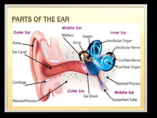
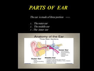
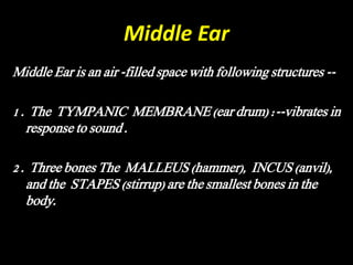
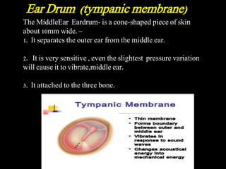
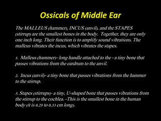
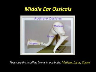
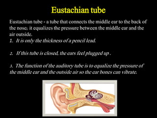
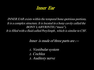
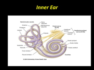
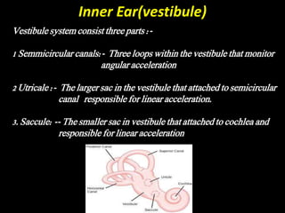
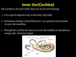
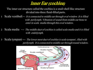
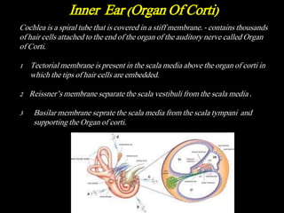
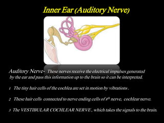
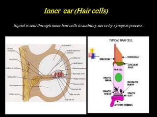
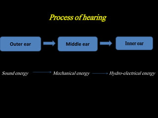
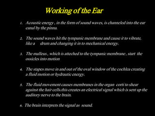
3. PARTS OF EAR
The ear is made of three portion -----
1 . The outer ear
2 . The middle ear
3 . The inner ear
4. The outer ear
• 1. The outer ear is an acoustical chamber that picks up sounds
from the environment and resonates at particular frequencies .
• 2. The curved formation on the outside ( the pinna) helps funnel
sound down the ear canal to the eardrum. The pinna is the
cartilage of the ear.
• 3 . THE OUTER EAR Pinna (auricle)- receives sound waves
that travel though the auditory canal or ear canal. Auditory
canal (ear canal)- acts as a funnel with an approximate length of
2.5cm and leads to the ear drum.
• 4 . It also protects the eardrum from shock and intrusion by
external objects.
5. Middle Ear
Middle Ear is an air -filled space with following structures --
1 . The TYMPANIC MEMBRANE (ear drum) :--vibrates in
response to sound .
2 . Three bones The MALLEUS (hammer), INCUS (anvil),
and the STAPES (stirrup) are the smallest bones in the
body.
6. The MiddleEar Eardrum- is a cone-shaped piece of skin
about 10mm wide. –
1. It separates the outer ear from the middle ear.
2. It is very sensitive , even the slightest pressure variation
will cause it to vibrate,middle ear.
3. It attached to the three bone.
Ear Drum (tympanic membrane)
7. Ossicals of Middle Ear
The MALLEUS (hammer), INCUS (anvil), and the STAPES
(stirrup) are the smallest bones in the body. Together, they are only
one inch long. Their function is to amplify sound vibrations. The
malleus vibrates the incus, which vibrates the stapes.
1. Malleus (hammer)- long handle attached to the - a tiny bone that
passes vibrations from the eardrum to the anvil.
2. Incus (anvil)- a tiny bone that passes vibrations from the hammer
to the stirrup.
3. Stapes (stirrups)- a tiny, U-shaped bone that passes vibrations from
the stirrup to the cochlea. -This is the smallest bone in the human
body (it is 0.25 to 0.33 cm long).
9. Eustachian tube - a tube that connects the middle ear to the back of
the nose; it equalizes the pressure between the middle ear and the
air outside.
1. It is only the thickness of a pencil lead.
2. If this tube is closed, the ears feel plugged up .
3. The function of the auditory tube is to equalize the pressure of
the middle ear and the outside air so the ear bones can vibrate.
Eustachian tube
10. INNER EAR exists within the temporal bone (petrious portion).
It is a complex structure. It is located in a bony cavity called the
BONY LABYRINTH (“maze”).
It is filled with a fluid called Perylimph , which is similar to CSF.
Inner is made of three parts are :--
1. Vestibular system
2. Cochlea
3. Auditory nerve
Inner Ear
12. Inner Ear(vestibule)
Vestibule system consist three parts :-
1 Semmicircular canals:- Three loops within the vestibule that monitor
angular acceleration
2 Utricale :- The larger sac in the vestibule that attached to semicircular
canal responsible for linear acceleration.
3. Saccule: -- The smaller sac in vestibule that attached to cochlea and
responsible for linear acceleration
13. Inner Ear(Cochlea)
The cochlea is the part of the inner ear involved in hearing.
1. It is a spiral-shaped cavity in the bony labyrinth,
2. In humans making 2 turns(full) and a 3/4(3 quarters) turn around
its axis, the modiolus.
3. Through the cochlea the inner converts the middle ear mechinacal
energy into electrical signal.
14. The inner ear structure called the cochlea is a snail-shell like structure
divided into three fluid-filled parts.
1 Scala vestibuli -- It is connected to middle ear through oval window .It is filled
with perilymph. Vibration of sound from middle ear bone to
enter in scala media through this oval window.
2 Scala media --- The middle duct of cochlea is called scala media and it is filled
with endolymph.
3 Scala tympani --- The lower most duct of cochlea is scala tympani , filed with
perilymph. It is connected to middle ear through round window.
Inner Ear (cochlea)
15. Cochlea is a spiral tube that is covered in a stiff membrane. - contains thousands
of hair cells attached to the end of the organ of the auditory nerve called Organ
of Corti.
1 Tectorial membrane is present in the scala media above the organ of corti in
which the tips of hair cells are embedded.
2 Reissner’s membrane separate the scala vestibuli from the scala media .
3 Basilar membrane seprate the scala media from the scala tympani and
supporting the Organ of corti.
Inner Ear (Organ Of Corti)
16. Inner Ear (Auditory Nerve)
Auditory Nerve- These nerves receive the electrical impulses generated
by the ear and pass this information up to the brain so it can be interpreted.
1 The tiny hair cells of the cochlea are set in motion by vibrations .
2 These hair cells connected to nerve ending cells of 8th nerve, cochlear nerve.
3 The VESTIBULAR COCHLEAR NERVE , which takes the signals to the brain.
17. Signal is sent through inner hair cells to auditory nerve by synapsis process
Inner ear (Hair cells)
18. Outer ear Middle ear Inner ear
Sound energy Mechanical energy Hydro-electrical energy
Process of hearing
19. 1. Acoustic energy , in the form of sound waves, is channeled into the ear
canal by the pinna.
2. The sound waves hit the tympanic membrane and cause it to vibrate,
like a drum and changing it in to mechanical energy.
3. The malleus , which is atteched to the tympanic membrane , start the
ossicles into motion
4. The stapes move in and out of the oval window of the cochlea creating
a fluid motion or hydrautic energy.
5. The fluid movement causes membranes in the organ corti to shear
against the hair cells.this creates an electrical signal which is sent up the
auditory nerve to the brain.
6. The brain interprets the signal as sound.
Working of the Ear