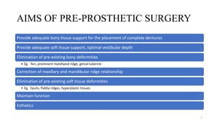
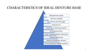
PRE-PROSTHETIC SURGERY
DR ASWITHA G
1ST YEAR PG
DEPT OF PROSTHODONTICS
1
CONTENTS
• Introduction
• Definition
• Objectives
• Aims of pre-prosthetic surgery
• Pre-prosthetic surgical procedures
• Conclusion
• References
2
INTRODUCTION
• Prosthetics is the replacement of missing teeth and contiguous oral and
maxillofacial tissues with artificial substitute
• Now, there remains significant number of patients, who can never be made to use
dentures effectively, because of
- Bone atrophy
- Soft tissue hypertrophy
- Localized soft and hard tissue problems
• In these patients, pre-prosthetic surgery offers significant contribution by
removing hindrance for prosthesis stability and retention
3
DEFINITION
• According to GPT 9,
“Pre-prosthetic surgery is defined as surgical procedures designed to
facilitate fabrication of a prosthesis or to improve the prognosis of prosthodontic
care”
• Pre-prosthetic surgery is carried out to redesign soft/hard tissues, by eliminating
hindrances to receive comfortable and stable prosthesis
4
AIMS OF PRE-PROSTHETIC SURGERY
Provide adequate bony tissue support for the placement of complete dentures
Provide adequate soft tissue support, optimal vestibular depth
Elimination of pre-existing bony deformities
• Eg. Tori, prominent mylohyoid ridge, genial tubercle
Correction of maxillary and mandibular ridge relationship
Elimination of pre-existing soft tissue deformities
• Eg. Epulis, flabby ridges, hyperplastic tissues
Maintain function
Esthetics
5
CHARACTERSTICS OF IDEAL DENTURE BASE
Adequate bone support
Soft tissue coverage
No undercuts and sharp edges
Adequate sulci
No soft tissue folds/ hypertrophies
No neoplastic lesions
Proper maxillomandibular relationships
Adequate palatal vault
Absence of peripheral scar bands
No muscle fibers to mobilize prosthesis
6
PATIENT EVALUATION AND TREATMENT
PLANNING
• Pre-prosthetic surgical treatment must begin with a proper case history and
physical examination
• Special attention should be given to systemic diseases that may be responsible for
the severe degree of bone resorption
• Esthetic and functional goals of the patient must be assessed carefully
• Long term maintenance of the underlying tissues as well as prosthetic appliances
should be kept in mind
7
PRE-PROSTHETIC SURGICAL PROCEDURES
Can be classified as,
• Basic procedures : Can be carried out under local anesthesia on a day care
basis
• Advanced surgery procedure : Require hospitalization and general anesthesia
Procedures are carried out for the following,
• Alveolar ridge correction
• Alveolar ridge extension
• Alveolar ridge augmentation
8
I. ALVEOLAR RIDGE CORRECTION
9
• Alveolectomy
• Alveoloplasty
• Elimination of unfavourable undercuts
• - Reduction of genial tubercle
• - Reduction of mylohyoid ridge
• Excision of tori
• Maxillary tuberosity reduction and exostosis removal
BONY
SURGERIES
• Removal of redundant crestal soft tissues
• Frenectomy
• Excision of epulis fissurata
• Excision palatal papillary hyperplasia
SOFT
TISSUE
SURGERIES
10
BONY SURGERIES
11
ALVEOLECTOMY
• Surgical removal or trimming of the alveolar process is termed as ‘alveolectomy’
• Procedure :
After extraction whenever there is presence of sharp margins at interdental,
interseptal or labiobuccal alveolar crest, they should be trimmed with bone rounger
or round bur and smoothened with bone file
12
ALVEOLOPLASTY
• Alveoloplasty defined as surgical recontouring of the alveolar process
• This procedure is done with the purpose to take care of bony projections, sharp
crestal bone or undercuts
• Types
Simple alveoloplasty
Interseptal alveoloplasty
• Dean’s alveoloplasty
• Obwegeser’s modification
Post-extraction alveoloplasty
13
SIMPLE ALVEOLOPLASTY
PROCEDURE
• Bony areas requiring recontouring should be
exposed using an envelop type of flap
• A mucoperiosteal incision along the crest of the
ridge with adequate A-P extension is given
• Adequate visualization and access to the alveolar
ridge obtained
• Vertical incisions given if necessary
• Excessive flap reflection may result in devitalized
areas of bone which may resorb rapidly after
surgery
• Recontouring can be accomplished with rongeur,
bone file, bone bur in hand piece
14
• Copious saline irrigation should be done throughout the recontouring procedure
to avoid overheating and bone necrosis
• After this the edges of the flap are trimmed and then sutured with continuous or
non-continuous sutures
15
DEAN’S INTERSEPTAL ALVEOLOPLASTY
• Only done in maxillary anterior region to reduce gross maxillary
over jet
• Mostly done immediately after extraction of anterior teeth
• This technique is best used in an area where the ridge is of
relative regular contour and adequate height but presents an
undercut to the depth of the labial vestibule
Advantages
Labial prominence is reduced without reducing the height of the
ridge
The periosteal attachment to the bone can be maintained hereby
reducing bone resorption
Muscle attachment are left undisturbed
Disadvantages
Decrease in ridge thickness
16
Procedure
• After anterior teeth extraction, interseptal bone
is cut with the bur from canine to canine region
• With the same bur vertical cuts are made only
in the labial cortex at distal end of the canine
extraction socket bilaterally without perforation
of labial mucosa
• Now labial cortex is fractured with periosteal
elevator and compressed into palatal direction
in approximation with palatal plate
• After removing any sharp margin, suturing is
done
17
OBWEGESER’S MODIFICATION OF DEAN’S
ALVEOLOPLASTY
• In this both the labial and palatal cortices are repositioned
• This is done when the anterior over jet is too gross that cannot reduced by labial plate repositioning
Procedure
Procedure is same to Dean’s alveoloplasty
but the only addition is that, here palatal
plate is fractured too at its base and
repositioned with labial plate in palatal
direction
18
ELIMINATION OF UNFAVORABLE UNDERCUTS
• Unfavorable undercuts are developed due to severe atrophy of the mandible which hinders in
proper denture construction
• These undercuts are mostly present on lingual aspect of mandible like genial tubercle
prominences, sharp mylohyoid ridge prominence
• Most of the times, patient wearing old dentures comes with the complaint of ulceration or
inflammation on these lingual prominence
• So surgical reduction should be carried out to relieve these undercuts
19
REDUCTION OF GENIAL TUBERCLES
Procedure
In this, lingual flap is reflected in anterior region of mandible and genial tubercle are reduced with
the bur
Then genioglossus muscle is sutured below at geniohyoid tubercle and flap is closed
20
REDUCTION OF MYLOHYOID RIDGE
Procedure
Linear incision is made over the crest of the ridge in the posterior aspect of the mandible
Full thickness mucoperiosteal flap is elevated to expose the muscles
Bone file is used to remove the sharp prominence of the mylohyoid ridge
Mylohyoid muscle is sutured below and flap is closed
21
EXCISION OF TORI – PALATAL TORUS
• Palatal tori are usually present on the midline of the hard palate. Most palatal tori are less than 2
cm in diameter, but their size can change throughout life
INDICATIONS
Small tori can be relieved during denture
construction but large tori should be surgically
removed
 An extremely large torus filling the palatal
vault
 A torus that extend beyond the posterior
dam area
 Traumatized mucosa over the torus
 Deep bony undercuts interfering with
denture insertion and stability
 Interferences with function (speech,
deglutition)
22
Procedure
In this, midline incision is given in palate and
flap is reflected with Y-shaped releasing
incisions
Torus is removed by making multiple cuts of
it and flap is sutured
A palatal splint is given to prevent hematoma
formation
23
EXCISION OF TORI – MANDIBULAR TORUS
• Torus mandibularis is an exostosis found on the lingual surface of the mandible opposite the canine
and premolars region
• They too interfere with denture retention because of the loss of marginal seal in premolar region
• Indication
Tori causing lingual undercuts and
interfering with lingual flange
extension of the planned prosthesis
When the mucosal covering is ulcerated
Large tori interfering with speech and
deglutition
24
PROCEDURE
• Bilateral lingual and inferior alveolar anesthesia is given
• Incision extending from 1 to 1.5 cm beyond each tori is
given
• Always leave behind a band of tissue attached to the
midline between the anterior extent of the two incisions
• When the torus has a small pedunculated base, a mallet
and an osteotome is used to cleave the tori from the
medial aspect of the mandible
• The direction of the initial bur is parallel to the medial
aspect of the mandible to prevent fracture of the lingual or
inferior cortex
• A bone file is used to smoothen the lingual cortex
• Palpation is done to check for proper contour and presence
of any undercuts
• Continuous suturing is done and gauze packs are placed
and retained for the next 12 hrs
25
MAXILLARY TUBEROSITY REDUCTION AND
EXOSTOSIS REMOVAL
• The main reason for tubercle overgrowth is extraction of
opposing mandibular 3rd molar and subsequent supra-
eruption of maxillary 3rd molar, where remains as bony
overgrowth after maxillary 3rd molar extraction
• Maxillary tubercle interfere with denture construction
because it decreases inter-arch space
26
PROCEDURE
 Crestal incision from tuberosity to premolar area
should be given and hyperplastic soft tissue or
bony overgrowth should be removed with the help
of chisel, mallet or burs
 After desired contour is achieved, the excess soft
tissue is trimmed and flap is sutured followed by
splint over it
27
SOFT TISSUE SURGERIES
28
REDUNDANT CRESTAL SOFT TISSUE REMOVAL
• The presence of the fibrous, hyperplastic tissue gives rise to flabby ridge form
• These flabby ridges results in unstable base for dentures
• In maxilla – enlarged tuberosity
• In mandible – enlarged retromolar pad
• Surgical removal is done by particular requirements of the tissue growth
29
FRENECTOMY
• Many times, there is high frenum attached near to the
crest of the ridge which may be too broad which
interfere in getting proper peripheral seal in dentures
• Lingual frenum may be too short and attached till the
tip of the tongue which interfere with normal tongue
movements and causes speech problem to the patient,
so surgical correction is advocated in these cases
30
LABIAL FRENECTOMY
CROSS-DIAMOND EXCISION
• This is done when there is lot of tissue is
available. Here base of the frenum at the
alveolar crest is grasped with haemostat and
incision is taken above and below the
haemostat
• The surgical defect is created by excision of
fibrous band. The closure is done by
interrupted sutures and small defect at
alveolar crest is left to granule
SEMILUNAR INCISIONS
• These incisions are used for broad premolar
and molar region freni
Z-PLASTY
• This procedure is used when the frenum is
broad and vestibule is short
V-Y INCISIONS
• These incisions are used for lengthening
localized area
31
LINGUAL FRENECTOMY
• Here lingual frenum is reduced by giving cross-diamond incision
• After incision, sub-mucosal dissection is done on either side and vertical suturing is given
32
EXCISION OF EPULIS FISSURATUM
• These are benign, pedunculated lesions present as excessive or redundant tissue of the vestibule,
frequently associated with over extended denture border
• These lesions are removed by sharp excision, electrocauterization, cryosurgery or laser excision
33
EXCISION OF PALATAL PAPILLARY HYPERPLASIA
• This happens because of chronic denture irritation, because of ill-fitting dentures
• There can be superimposed candida infection
• Denture should be relieved in this region and anti-fungal agent should be applied
• Supra-periosteal excision with a electrocautery can be done
34
II. ALVEOLAR RIDGE EXTENSION
35
• Whenever there is an inadequate vestibular depth present (due to mandibular atrophy and high
muscle or soft tissue attachment) so to increase retention and stability of denture, deepening of
vestibule is considered
• Sufficient amount of bone should be present ( Minimum 15mm bone height) for alveolar ridge
extension / vestibuloplasty procedure
36
VESTIBULOPLASTY
LABIAL VESTIBULOPLASTY
For mandibular
ridge
- Kazanjian technique
(1924)
- Godwin’s
modification (1947)
- Clark’s technique
- Obwegeser’s
modification (1959)
For maxillary ridge
- Maxillary pocket inlay
vestibuloplasty
LINGUAL VESTIBULOPLASTY
1. Trauner’s
technique
2. Caldwell’s
technique
3. Mental nerve
repositioning
37
LABIAL VESTIBULOPLASTY (MANDIBULAR RIDGE)
KAZANJIAN TECHNIQUE
Mucosal flap from inner aspect of the lower lip is used to increase the vestibular depth in anterior
mandibular labial vestibule (premolar to premolar region)
Periosteum of the bone is left intact
Drawback – Scaring of mucosa with subsequent decreased flexibility of lower lip
38
GODWIN’S MODIFICATION
In this procedure, flap is reflected from the inner aspect of the lip till the alveolar crest and
periosteum is reflected from crest of the ridge till the desired depth of the vestibule
This periosteum is now sutured to the lip mucosal margin and then lip flap is sutured at the
required vestibular depth
Stent or splint is used for adaptation
Advantage – Less scaring of lip mucosa
39
CLARK’S TECHNIQUE
Flap is reflected from alveolar crest till vermillion
border of the lip
Supra-periosteal dissection is done till desired
vestibular depth is obtained
This flap is held in position with sutures passed
through the chin area extraorally and tied around the
rubber catheter
Here alveolar bone is covered by periosteum which
heals quickly by granulation
40
OBWEGESER’S MODIFICATION
 The modification is that the alveolar bone with
periosteal attachment is covered with the split
thickness skin graft or mucosal graft
41
LABIAL VESTIBULOPLASTY (MAXILLARY RIDGE)
MAXILLARY POCKET – INLAY VESTIBULOPLASTY
• Incision is made in the vestibule from molar to molar region and supra-periosteal dissection is
carried out
• Now a split thickness graft is placed on the extended flange of prefabricated denture and this
denture is positioned in extended vestibular depth which is fixed with circumzygomatic wiring
• Wound margins are sutured to the graft
• New denture will be constructed after 6 weeks
Advantages
Better retention of the dentures
Helps to restore deficiency in the region of nasolabial fold with improved contour
42
LINGUAL VESTIBULOPLASTY
TRAUNER’S TECHNIQUE
• This procedure is used to increase the depth of floor of the mouth in mylohyoid region
• Incision is given over lingual side of the alveolar ridge bilaterally in posterior region (2nd molar
region)
• Supra-periosteal dissection is done to identify mylohyoid muscle, which is separated from its
attachment and sutured to the new desired vestibular depth
• Skin graft is placed and sutured with the prefabricated stent over it
43
CALDWELL’S TECHNIQUE
• Entire lingual mucoperiosteal flap is reflected from molar to molar region
• Mylohyoid and genioglossus muscle attachments are dissected and sutured below to the desired
depth of the vestibule
• Rubber tubing is placed in the lingual vestibule and the flap is held in place at desired vestibular
depth which is sutured with the sutures passing extra-orally at inferior border of the mandible
44
MENTAL NERVE TRANSPOSITION
• Many times patient with severe atrophic ridge,
complains of pain after wearing complete dentures
• This is because that the position of mental nerve is
superior because of severe mandibular atrophy so this is
the reason for pain on compression
Procedure
The flap is reflected on buccal aspect in the mental nerve
region
The nerve is held with the hook lightly and a bony
groove is cut below mental foramen only in buccal
cortex
Then nerve is positioned in that groove secured in place
with gelfoam and flap is sutured
45
III. ALVEOLAR RIDGE AUGMENTATION
46
• This procedure is done when alveolar bone has been completely disappeared to the point where in
maxilla, a flat surface is present between vestibule and palate and in mandible, mental nerve is
positioned almost at the crest
• Here alveolar bone height is less than 15mm
• So, vestibuloplasty is out of consideration in this case until the replacement of necessary supportive
bone is done
47
AIM
• Restoration of optimum height, width, ridge form, vestibular depth and optimum denture bearing
area
• Protection of neurovascular bundle
• Establishment of proper interarch relationship
• Improvement of retention and stability of denture
• Improve the patient comfort for wearing the denture
48
MATERIALS USED FOR RIDGE AUGMENTATION
1. Autogenous bone graft – iliac crest, rib grafts
2. Allogenic bone grafts – freeze dried cadaver bone
3. Alloplastic material – hydroxyapatite
4. Metal mesh with autogenous cancellous bone
5. Metal mesh with hydroxyapatite
49
RIDGE AUGMENTATION PROCEDURE
1. Superior border augmentation
2. Inferior border augmentation
3. Interpositional or sandwich bone grafts
4. Onlay grafting
5. Visor osteotomy
6. Modified visor osteotomy
50
SUPERIOR BORDER GRAFTING
• First described by Davis in 1970
• Remaining bone <10 mm
• Here 2 autogenous bone grafts of 15cm each are used
• One rib is scored to the cortex followed by giving shape of the mandible and attached at the
superior border of the mandible by circum-mandibular wiring
• The other rib graft is made into cortico-cancellous particles and moulded around the first rib graft
• Surgical flap is then closed
DISADVANTAGES
ⅹ Donor site morbidity
ⅹ Surgical site is necessary
ⅹ Continued resorption at the graft sites
51
INFERIOR BORDER GRAFTING
• This procedure is indicated when alveolar ridge height is less than 5 to 8 mm and is at risk of
pathological fracture
• In this procedure, a cadaver mandible used for grafting which is filled with cancellous graft
material for revascularization
• This mandible is then fixed to the inferior border with vicryl sutures, by circum-mandibular
fixation and neck flap is closed
• Osteointegrated implants can be placed after 4-6 months
52
ADVANTAGES
• Does not obliterate the
vestibule
• Interim denture can be worn
• No change in vertical dimension
• Graft is not subjected to direct
masticatory forces
DISADVANTAGES
• It will not correct the
abnormalities of denture
bearing areas
• it will not protect a highly
placed mental nerve
• Resorption of the graft
• Presence of scar
53
INTERPOSITIONAL BONE GRAFT (SANDWICH
TECHNIQUE)
• In this procedure, a horizontal osteotomy is
performed by splitting of the maxilla or mandible and
bone is grafted in the gap
• In mandible, this procedure is mainly used in anterior
mandible
• Prosthetic appliance is given after 3-5 months
ADVANTAGES
Less resorption than onlay grafting
More predictable long term results
Decreased incidence of nerve paresthesia than the
visor osteotomy
Can be used in conjuction with implants
54
ONLAY GRAFTING
• This procedure helps in increasing width of the ridge
• Here graft material is placed on the buccal cortex either is putty form by mixing with saline/ blood
or in the form of blocks or split thickness rib/ iliac crest graft
ADVANTAGES
Improves width and to some extent height too
Can be used in anterior and posterior region
55
VISOR OSTEOTOMY
• Originated by Harle and modified by Peterson and Slade
• It is used where insufficient vertical mandibular bone height
is present for the horizontal osteotomy technique but
adequate bone width (approx. 10 mm) is present
• The mandible is split vertically and the lingual section is
elevated to increase the mandibular height
• Cancellous bone or particulate bone and marrow is placed to
correct the contours and fill in the gaps on the facial side of
the elevated segment
• Transosteal wires hold the segments in place for a period of
3-4 months before vestibuloplasties are performed
• DISADVANTAGE- unavoidable nerve trauma and the
resultant parasthesia
56
MODIFIED VISOR OSTEOTOMY
• Procedure of choice for mandibular ridge augmentation include
the combination of osteotomy techniques (horizontal or
vertical)with interpositional bone grafting
• These procedures involve the movement of a pedicle of bone
along with its blood supply
• Theoretically, the viability of the bone will be greater and the
resorption decreased because the blood supply to the bone is
maintained
PROCEDURE
Vertical osteotomy cut is made in the posterior region to divide
the segments buccolingually
A horizontal osteotomy is performed in the anterior mandible to
divide the anterior segment superiorly and inferiorly and bone
grafting was done into the osteotomised gap
Two osteotomised segments are fixed with wires
57
ADVANTAGES
Increased bone height which is relatively stable
Shortened post-operative period (3 months)
Rate of resorption is less when compared to onlay grafts
DISADVANTAGES
ⅹ Parasthesia
ⅹ Nerve trauma
ⅹ Mandibular fracture
ⅹ Flap dehiscence
58
PROBLEMS ENCOUNTERED WITH AUGMENTATION
PROCEDURE
1. Inadequate soft tissue cover
2. Rejection of the autografts (failure of union with the host bone)
3. Dehiscence of overlying mucosa
4. Migration of the graft material
5. Resorption of the graft
59
CONCLUSION
• Accurate diagnosis of the problem areas during denture construction and determination of the
necessity of surgery is accomplished by careful evaluation of the information systemically obtained
from the patient
• Proper knowledge of the available surgical procedures helps in achieving the best results
60
REFERENCES
• BOUCHER’S – Prosthodontic treatment for edentulous patients 11th edition
• CHARLES HEARTWELL & ARTHUR O RAHN – Syllabus of complete dentures 4th edition
• JOHN J SHARRY – Complete denture prosthodontics 2nd edition
• SHELDON WINKLER – Essentials of complete dentures 2nd edition
• ZARB BOLENDER - Prosthodontic treatment for edentulous patients 12th edition
• PETERSON – Principles of oral and maxillofacial surgery
61

PRE-PROSTHETIC SURGERY.pptx

  • 1.
    PRE-PROSTHETIC SURGERY DR ASWITHAG 1ST YEAR PG DEPT OF PROSTHODONTICS 1
  • 2.
    CONTENTS • Introduction • Definition •Objectives • Aims of pre-prosthetic surgery • Pre-prosthetic surgical procedures • Conclusion • References 2
  • 3.
    INTRODUCTION • Prosthetics isthe replacement of missing teeth and contiguous oral and maxillofacial tissues with artificial substitute • Now, there remains significant number of patients, who can never be made to use dentures effectively, because of - Bone atrophy - Soft tissue hypertrophy - Localized soft and hard tissue problems • In these patients, pre-prosthetic surgery offers significant contribution by removing hindrance for prosthesis stability and retention 3
  • 4.
    DEFINITION • According toGPT 9, “Pre-prosthetic surgery is defined as surgical procedures designed to facilitate fabrication of a prosthesis or to improve the prognosis of prosthodontic care” • Pre-prosthetic surgery is carried out to redesign soft/hard tissues, by eliminating hindrances to receive comfortable and stable prosthesis 4
  • 5.
    AIMS OF PRE-PROSTHETICSURGERY Provide adequate bony tissue support for the placement of complete dentures Provide adequate soft tissue support, optimal vestibular depth Elimination of pre-existing bony deformities • Eg. Tori, prominent mylohyoid ridge, genial tubercle Correction of maxillary and mandibular ridge relationship Elimination of pre-existing soft tissue deformities • Eg. Epulis, flabby ridges, hyperplastic tissues Maintain function Esthetics 5
  • 6.
    CHARACTERSTICS OF IDEALDENTURE BASE Adequate bone support Soft tissue coverage No undercuts and sharp edges Adequate sulci No soft tissue folds/ hypertrophies No neoplastic lesions Proper maxillomandibular relationships Adequate palatal vault Absence of peripheral scar bands No muscle fibers to mobilize prosthesis 6
  • 7.
    PATIENT EVALUATION ANDTREATMENT PLANNING • Pre-prosthetic surgical treatment must begin with a proper case history and physical examination • Special attention should be given to systemic diseases that may be responsible for the severe degree of bone resorption • Esthetic and functional goals of the patient must be assessed carefully • Long term maintenance of the underlying tissues as well as prosthetic appliances should be kept in mind 7
  • 8.
    PRE-PROSTHETIC SURGICAL PROCEDURES Canbe classified as, • Basic procedures : Can be carried out under local anesthesia on a day care basis • Advanced surgery procedure : Require hospitalization and general anesthesia Procedures are carried out for the following, • Alveolar ridge correction • Alveolar ridge extension • Alveolar ridge augmentation 8
  • 9.
    I. ALVEOLAR RIDGECORRECTION 9
  • 10.
    • Alveolectomy • Alveoloplasty •Elimination of unfavourable undercuts • - Reduction of genial tubercle • - Reduction of mylohyoid ridge • Excision of tori • Maxillary tuberosity reduction and exostosis removal BONY SURGERIES • Removal of redundant crestal soft tissues • Frenectomy • Excision of epulis fissurata • Excision palatal papillary hyperplasia SOFT TISSUE SURGERIES 10
  • 11.
  • 12.
    ALVEOLECTOMY • Surgical removalor trimming of the alveolar process is termed as ‘alveolectomy’ • Procedure : After extraction whenever there is presence of sharp margins at interdental, interseptal or labiobuccal alveolar crest, they should be trimmed with bone rounger or round bur and smoothened with bone file 12
  • 13.
    ALVEOLOPLASTY • Alveoloplasty definedas surgical recontouring of the alveolar process • This procedure is done with the purpose to take care of bony projections, sharp crestal bone or undercuts • Types Simple alveoloplasty Interseptal alveoloplasty • Dean’s alveoloplasty • Obwegeser’s modification Post-extraction alveoloplasty 13
  • 14.
    SIMPLE ALVEOLOPLASTY PROCEDURE • Bonyareas requiring recontouring should be exposed using an envelop type of flap • A mucoperiosteal incision along the crest of the ridge with adequate A-P extension is given • Adequate visualization and access to the alveolar ridge obtained • Vertical incisions given if necessary • Excessive flap reflection may result in devitalized areas of bone which may resorb rapidly after surgery • Recontouring can be accomplished with rongeur, bone file, bone bur in hand piece 14
  • 15.
    • Copious salineirrigation should be done throughout the recontouring procedure to avoid overheating and bone necrosis • After this the edges of the flap are trimmed and then sutured with continuous or non-continuous sutures 15
  • 16.
    DEAN’S INTERSEPTAL ALVEOLOPLASTY •Only done in maxillary anterior region to reduce gross maxillary over jet • Mostly done immediately after extraction of anterior teeth • This technique is best used in an area where the ridge is of relative regular contour and adequate height but presents an undercut to the depth of the labial vestibule Advantages Labial prominence is reduced without reducing the height of the ridge The periosteal attachment to the bone can be maintained hereby reducing bone resorption Muscle attachment are left undisturbed Disadvantages Decrease in ridge thickness 16
  • 17.
    Procedure • After anteriorteeth extraction, interseptal bone is cut with the bur from canine to canine region • With the same bur vertical cuts are made only in the labial cortex at distal end of the canine extraction socket bilaterally without perforation of labial mucosa • Now labial cortex is fractured with periosteal elevator and compressed into palatal direction in approximation with palatal plate • After removing any sharp margin, suturing is done 17
  • 18.
    OBWEGESER’S MODIFICATION OFDEAN’S ALVEOLOPLASTY • In this both the labial and palatal cortices are repositioned • This is done when the anterior over jet is too gross that cannot reduced by labial plate repositioning Procedure Procedure is same to Dean’s alveoloplasty but the only addition is that, here palatal plate is fractured too at its base and repositioned with labial plate in palatal direction 18
  • 19.
    ELIMINATION OF UNFAVORABLEUNDERCUTS • Unfavorable undercuts are developed due to severe atrophy of the mandible which hinders in proper denture construction • These undercuts are mostly present on lingual aspect of mandible like genial tubercle prominences, sharp mylohyoid ridge prominence • Most of the times, patient wearing old dentures comes with the complaint of ulceration or inflammation on these lingual prominence • So surgical reduction should be carried out to relieve these undercuts 19
  • 20.
    REDUCTION OF GENIALTUBERCLES Procedure In this, lingual flap is reflected in anterior region of mandible and genial tubercle are reduced with the bur Then genioglossus muscle is sutured below at geniohyoid tubercle and flap is closed 20
  • 21.
    REDUCTION OF MYLOHYOIDRIDGE Procedure Linear incision is made over the crest of the ridge in the posterior aspect of the mandible Full thickness mucoperiosteal flap is elevated to expose the muscles Bone file is used to remove the sharp prominence of the mylohyoid ridge Mylohyoid muscle is sutured below and flap is closed 21
  • 22.
    EXCISION OF TORI– PALATAL TORUS • Palatal tori are usually present on the midline of the hard palate. Most palatal tori are less than 2 cm in diameter, but their size can change throughout life INDICATIONS Small tori can be relieved during denture construction but large tori should be surgically removed  An extremely large torus filling the palatal vault  A torus that extend beyond the posterior dam area  Traumatized mucosa over the torus  Deep bony undercuts interfering with denture insertion and stability  Interferences with function (speech, deglutition) 22
  • 23.
    Procedure In this, midlineincision is given in palate and flap is reflected with Y-shaped releasing incisions Torus is removed by making multiple cuts of it and flap is sutured A palatal splint is given to prevent hematoma formation 23
  • 24.
    EXCISION OF TORI– MANDIBULAR TORUS • Torus mandibularis is an exostosis found on the lingual surface of the mandible opposite the canine and premolars region • They too interfere with denture retention because of the loss of marginal seal in premolar region • Indication Tori causing lingual undercuts and interfering with lingual flange extension of the planned prosthesis When the mucosal covering is ulcerated Large tori interfering with speech and deglutition 24
  • 25.
    PROCEDURE • Bilateral lingualand inferior alveolar anesthesia is given • Incision extending from 1 to 1.5 cm beyond each tori is given • Always leave behind a band of tissue attached to the midline between the anterior extent of the two incisions • When the torus has a small pedunculated base, a mallet and an osteotome is used to cleave the tori from the medial aspect of the mandible • The direction of the initial bur is parallel to the medial aspect of the mandible to prevent fracture of the lingual or inferior cortex • A bone file is used to smoothen the lingual cortex • Palpation is done to check for proper contour and presence of any undercuts • Continuous suturing is done and gauze packs are placed and retained for the next 12 hrs 25
  • 26.
    MAXILLARY TUBEROSITY REDUCTIONAND EXOSTOSIS REMOVAL • The main reason for tubercle overgrowth is extraction of opposing mandibular 3rd molar and subsequent supra- eruption of maxillary 3rd molar, where remains as bony overgrowth after maxillary 3rd molar extraction • Maxillary tubercle interfere with denture construction because it decreases inter-arch space 26
  • 27.
    PROCEDURE  Crestal incisionfrom tuberosity to premolar area should be given and hyperplastic soft tissue or bony overgrowth should be removed with the help of chisel, mallet or burs  After desired contour is achieved, the excess soft tissue is trimmed and flap is sutured followed by splint over it 27
  • 28.
  • 29.
    REDUNDANT CRESTAL SOFTTISSUE REMOVAL • The presence of the fibrous, hyperplastic tissue gives rise to flabby ridge form • These flabby ridges results in unstable base for dentures • In maxilla – enlarged tuberosity • In mandible – enlarged retromolar pad • Surgical removal is done by particular requirements of the tissue growth 29
  • 30.
    FRENECTOMY • Many times,there is high frenum attached near to the crest of the ridge which may be too broad which interfere in getting proper peripheral seal in dentures • Lingual frenum may be too short and attached till the tip of the tongue which interfere with normal tongue movements and causes speech problem to the patient, so surgical correction is advocated in these cases 30
  • 31.
    LABIAL FRENECTOMY CROSS-DIAMOND EXCISION •This is done when there is lot of tissue is available. Here base of the frenum at the alveolar crest is grasped with haemostat and incision is taken above and below the haemostat • The surgical defect is created by excision of fibrous band. The closure is done by interrupted sutures and small defect at alveolar crest is left to granule SEMILUNAR INCISIONS • These incisions are used for broad premolar and molar region freni Z-PLASTY • This procedure is used when the frenum is broad and vestibule is short V-Y INCISIONS • These incisions are used for lengthening localized area 31
  • 32.
    LINGUAL FRENECTOMY • Herelingual frenum is reduced by giving cross-diamond incision • After incision, sub-mucosal dissection is done on either side and vertical suturing is given 32
  • 33.
    EXCISION OF EPULISFISSURATUM • These are benign, pedunculated lesions present as excessive or redundant tissue of the vestibule, frequently associated with over extended denture border • These lesions are removed by sharp excision, electrocauterization, cryosurgery or laser excision 33
  • 34.
    EXCISION OF PALATALPAPILLARY HYPERPLASIA • This happens because of chronic denture irritation, because of ill-fitting dentures • There can be superimposed candida infection • Denture should be relieved in this region and anti-fungal agent should be applied • Supra-periosteal excision with a electrocautery can be done 34
  • 35.
    II. ALVEOLAR RIDGEEXTENSION 35
  • 36.
    • Whenever thereis an inadequate vestibular depth present (due to mandibular atrophy and high muscle or soft tissue attachment) so to increase retention and stability of denture, deepening of vestibule is considered • Sufficient amount of bone should be present ( Minimum 15mm bone height) for alveolar ridge extension / vestibuloplasty procedure 36
  • 37.
    VESTIBULOPLASTY LABIAL VESTIBULOPLASTY For mandibular ridge -Kazanjian technique (1924) - Godwin’s modification (1947) - Clark’s technique - Obwegeser’s modification (1959) For maxillary ridge - Maxillary pocket inlay vestibuloplasty LINGUAL VESTIBULOPLASTY 1. Trauner’s technique 2. Caldwell’s technique 3. Mental nerve repositioning 37
  • 38.
    LABIAL VESTIBULOPLASTY (MANDIBULARRIDGE) KAZANJIAN TECHNIQUE Mucosal flap from inner aspect of the lower lip is used to increase the vestibular depth in anterior mandibular labial vestibule (premolar to premolar region) Periosteum of the bone is left intact Drawback – Scaring of mucosa with subsequent decreased flexibility of lower lip 38
  • 39.
    GODWIN’S MODIFICATION In thisprocedure, flap is reflected from the inner aspect of the lip till the alveolar crest and periosteum is reflected from crest of the ridge till the desired depth of the vestibule This periosteum is now sutured to the lip mucosal margin and then lip flap is sutured at the required vestibular depth Stent or splint is used for adaptation Advantage – Less scaring of lip mucosa 39
  • 40.
    CLARK’S TECHNIQUE Flap isreflected from alveolar crest till vermillion border of the lip Supra-periosteal dissection is done till desired vestibular depth is obtained This flap is held in position with sutures passed through the chin area extraorally and tied around the rubber catheter Here alveolar bone is covered by periosteum which heals quickly by granulation 40
  • 41.
    OBWEGESER’S MODIFICATION  Themodification is that the alveolar bone with periosteal attachment is covered with the split thickness skin graft or mucosal graft 41
  • 42.
    LABIAL VESTIBULOPLASTY (MAXILLARYRIDGE) MAXILLARY POCKET – INLAY VESTIBULOPLASTY • Incision is made in the vestibule from molar to molar region and supra-periosteal dissection is carried out • Now a split thickness graft is placed on the extended flange of prefabricated denture and this denture is positioned in extended vestibular depth which is fixed with circumzygomatic wiring • Wound margins are sutured to the graft • New denture will be constructed after 6 weeks Advantages Better retention of the dentures Helps to restore deficiency in the region of nasolabial fold with improved contour 42
  • 43.
    LINGUAL VESTIBULOPLASTY TRAUNER’S TECHNIQUE •This procedure is used to increase the depth of floor of the mouth in mylohyoid region • Incision is given over lingual side of the alveolar ridge bilaterally in posterior region (2nd molar region) • Supra-periosteal dissection is done to identify mylohyoid muscle, which is separated from its attachment and sutured to the new desired vestibular depth • Skin graft is placed and sutured with the prefabricated stent over it 43
  • 44.
    CALDWELL’S TECHNIQUE • Entirelingual mucoperiosteal flap is reflected from molar to molar region • Mylohyoid and genioglossus muscle attachments are dissected and sutured below to the desired depth of the vestibule • Rubber tubing is placed in the lingual vestibule and the flap is held in place at desired vestibular depth which is sutured with the sutures passing extra-orally at inferior border of the mandible 44
  • 45.
    MENTAL NERVE TRANSPOSITION •Many times patient with severe atrophic ridge, complains of pain after wearing complete dentures • This is because that the position of mental nerve is superior because of severe mandibular atrophy so this is the reason for pain on compression Procedure The flap is reflected on buccal aspect in the mental nerve region The nerve is held with the hook lightly and a bony groove is cut below mental foramen only in buccal cortex Then nerve is positioned in that groove secured in place with gelfoam and flap is sutured 45
  • 46.
    III. ALVEOLAR RIDGEAUGMENTATION 46
  • 47.
    • This procedureis done when alveolar bone has been completely disappeared to the point where in maxilla, a flat surface is present between vestibule and palate and in mandible, mental nerve is positioned almost at the crest • Here alveolar bone height is less than 15mm • So, vestibuloplasty is out of consideration in this case until the replacement of necessary supportive bone is done 47
  • 48.
    AIM • Restoration ofoptimum height, width, ridge form, vestibular depth and optimum denture bearing area • Protection of neurovascular bundle • Establishment of proper interarch relationship • Improvement of retention and stability of denture • Improve the patient comfort for wearing the denture 48
  • 49.
    MATERIALS USED FORRIDGE AUGMENTATION 1. Autogenous bone graft – iliac crest, rib grafts 2. Allogenic bone grafts – freeze dried cadaver bone 3. Alloplastic material – hydroxyapatite 4. Metal mesh with autogenous cancellous bone 5. Metal mesh with hydroxyapatite 49
  • 50.
    RIDGE AUGMENTATION PROCEDURE 1.Superior border augmentation 2. Inferior border augmentation 3. Interpositional or sandwich bone grafts 4. Onlay grafting 5. Visor osteotomy 6. Modified visor osteotomy 50
  • 51.
    SUPERIOR BORDER GRAFTING •First described by Davis in 1970 • Remaining bone <10 mm • Here 2 autogenous bone grafts of 15cm each are used • One rib is scored to the cortex followed by giving shape of the mandible and attached at the superior border of the mandible by circum-mandibular wiring • The other rib graft is made into cortico-cancellous particles and moulded around the first rib graft • Surgical flap is then closed DISADVANTAGES ⅹ Donor site morbidity ⅹ Surgical site is necessary ⅹ Continued resorption at the graft sites 51
  • 52.
    INFERIOR BORDER GRAFTING •This procedure is indicated when alveolar ridge height is less than 5 to 8 mm and is at risk of pathological fracture • In this procedure, a cadaver mandible used for grafting which is filled with cancellous graft material for revascularization • This mandible is then fixed to the inferior border with vicryl sutures, by circum-mandibular fixation and neck flap is closed • Osteointegrated implants can be placed after 4-6 months 52
  • 53.
    ADVANTAGES • Does notobliterate the vestibule • Interim denture can be worn • No change in vertical dimension • Graft is not subjected to direct masticatory forces DISADVANTAGES • It will not correct the abnormalities of denture bearing areas • it will not protect a highly placed mental nerve • Resorption of the graft • Presence of scar 53
  • 54.
    INTERPOSITIONAL BONE GRAFT(SANDWICH TECHNIQUE) • In this procedure, a horizontal osteotomy is performed by splitting of the maxilla or mandible and bone is grafted in the gap • In mandible, this procedure is mainly used in anterior mandible • Prosthetic appliance is given after 3-5 months ADVANTAGES Less resorption than onlay grafting More predictable long term results Decreased incidence of nerve paresthesia than the visor osteotomy Can be used in conjuction with implants 54
  • 55.
    ONLAY GRAFTING • Thisprocedure helps in increasing width of the ridge • Here graft material is placed on the buccal cortex either is putty form by mixing with saline/ blood or in the form of blocks or split thickness rib/ iliac crest graft ADVANTAGES Improves width and to some extent height too Can be used in anterior and posterior region 55
  • 56.
    VISOR OSTEOTOMY • Originatedby Harle and modified by Peterson and Slade • It is used where insufficient vertical mandibular bone height is present for the horizontal osteotomy technique but adequate bone width (approx. 10 mm) is present • The mandible is split vertically and the lingual section is elevated to increase the mandibular height • Cancellous bone or particulate bone and marrow is placed to correct the contours and fill in the gaps on the facial side of the elevated segment • Transosteal wires hold the segments in place for a period of 3-4 months before vestibuloplasties are performed • DISADVANTAGE- unavoidable nerve trauma and the resultant parasthesia 56
  • 57.
    MODIFIED VISOR OSTEOTOMY •Procedure of choice for mandibular ridge augmentation include the combination of osteotomy techniques (horizontal or vertical)with interpositional bone grafting • These procedures involve the movement of a pedicle of bone along with its blood supply • Theoretically, the viability of the bone will be greater and the resorption decreased because the blood supply to the bone is maintained PROCEDURE Vertical osteotomy cut is made in the posterior region to divide the segments buccolingually A horizontal osteotomy is performed in the anterior mandible to divide the anterior segment superiorly and inferiorly and bone grafting was done into the osteotomised gap Two osteotomised segments are fixed with wires 57
  • 58.
    ADVANTAGES Increased bone heightwhich is relatively stable Shortened post-operative period (3 months) Rate of resorption is less when compared to onlay grafts DISADVANTAGES ⅹ Parasthesia ⅹ Nerve trauma ⅹ Mandibular fracture ⅹ Flap dehiscence 58
  • 59.
    PROBLEMS ENCOUNTERED WITHAUGMENTATION PROCEDURE 1. Inadequate soft tissue cover 2. Rejection of the autografts (failure of union with the host bone) 3. Dehiscence of overlying mucosa 4. Migration of the graft material 5. Resorption of the graft 59
  • 60.
    CONCLUSION • Accurate diagnosisof the problem areas during denture construction and determination of the necessity of surgery is accomplished by careful evaluation of the information systemically obtained from the patient • Proper knowledge of the available surgical procedures helps in achieving the best results 60
  • 61.
    REFERENCES • BOUCHER’S –Prosthodontic treatment for edentulous patients 11th edition • CHARLES HEARTWELL & ARTHUR O RAHN – Syllabus of complete dentures 4th edition • JOHN J SHARRY – Complete denture prosthodontics 2nd edition • SHELDON WINKLER – Essentials of complete dentures 2nd edition • ZARB BOLENDER - Prosthodontic treatment for edentulous patients 12th edition • PETERSON – Principles of oral and maxillofacial surgery 61