
Recommended
More Related Content
What's hot
What's hot (20)
Similar to Preprosthetic surgery.pdf
Similar to Preprosthetic surgery.pdf (20)
Recently uploaded
Recently uploaded (20)
Preprosthetic surgery.pdf
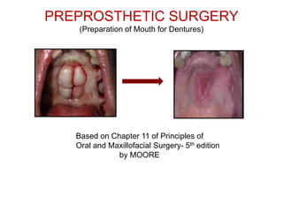
- 1. PREPROSTHETIC SURGERY (Preparation of Mouth for Dentures) Based on Chapter 11 of Principles of Oral and Maxillofacial Surgery- 5th edition by MOORE
- 2. Specific Learning outcomes List the objectives of preprosthetic surgical procedures and describe the preoperative patient selection(C1) Give the classification of preprosthetic surgical procedures (C2) Apply the knowledge to plan the treatment for patients requiring alveolar ridge correction procedures (C3) Explain various alveolar ridge extension and alveolar ridge augmentation procedures (C2)
- 3. LECTURE OUTLINE Aims & Objectives of Pre Prosthetic Surgery Bone Loss- Causes, Patterns Preservation of Alveolar Ridge Preprosthetic Surgical Techniques Surgery involving bony irregularities Surgery involving soft tissue irregularities
- 4. The prosthetic replacement of lost teeth frequently involves surgical preparation of the remaining oral tissues. Aims To leave a satisfactory base for subsequent placement of prosthetic appliances. To provide a better anatomic environment and to create proper supporting structures for denture construction. Ideally treatment for these procedures should be planned jointly (by a prosthodontist and oral surgeon)
- 6. Objectives • Provide adequate bony tissue support for the placement of the prosthesis. • Provide adequate soft tissue support. Optimum vestibular depth. • Elimination of pre-existing bony and soft tissue deformities. • Relocation of frenal/muscle attachments.
- 7. CAUSES OF BONE LOSS Causes of bone loss : 1.Metabolicfactors:osteoporosis,osteomalacia. 2. Aging. 3. Trauma. 4. Periodontal disease. 5. Disuse atrophy. 6. Long term denture use.
- 8. PATTERNS OF BONE LOSS • Tallgren in 1972 stated that most of the bone loss occurs in the first year of denture wearing (ten times). • Four times more bone loss in the mandible. • Usual resorption of the maxilla is on the buccal and inferior portion of the alveolar ridge. • Anterior maxilla less horizontal bone loss.
- 9. • In the posterior maxilla there is inward drift of the posterior crest. • The width of the maxilla is reduced. • Decrease in the palatal vault. • Mandible resorbs downwards and outwards. • Edentulous bone loss is upto 1mm per year with greatest loss within 18 months after extraction.
- 10. Bone height loss can be up to 1.5 mm in 3 months and decrease in the width of alveolar ridge can be as much as 50% within12 months.
- 11. MERCIER 1995. RESORPTIVE PATTERN OF THE EDENTULOUS RIDGE. • TYPE 1 : minor ridge modelling. • TYPE 2 : sharp atrophic residual ridge. • TYPE 3 : basal bone ridge. • TYPE 4 : basal bone resorption.
- 12. Preservation of alveolar ridge at time of extraction Preservation of alveolar ridge for prosthesis starts – when the first permanent tooth is extracted itself. Careful planning to be done during removal of buried teeth and lesions. Difficult extractions (multirooted teeth)---best removed— by transalveolar extraction----to avoid alveolar fracture.
- 13. Preservation of alveolar ridge at time of extraction– contd Access to deeply buried roots----made through lateral aspect of alveolus---leaving the ridge intact. Bone cutting to be limited to one side----leaving palatal or lingual plate with its mucoperiosteum untouched. More conservative approach is important where periodontal disease has already caused more bone loss.
- 14. Preservation of alveolar bone in edentulous patients Resorption-----makes alveolar ridge more weak Symptomless root fragments in thin mandibles Buried teeth unlikely to come to surface during their life time Use of Blunt burs Failure to provide irrigation SHOULD BE AVOIDED Those teeth lying superficially Those associated with cysts and granulomas NEED NOT BE EXTRACTED need to be EXTRACTED
- 15. IDEAL DENTURE BASE AREA • Adequate bone support. • Adequate firm soft tissue coverage. • No bony or soft tissue undercuts or prominences. • No sharp ridges. • No high muscle or frenal attachments.
- 16. Classification of Surgical Techniques • 1.Alveolar ridge correction. • 2.Alveolar ridge extension. • 3.Alveolar ridge augmentation.
- 17. SURGERY INVOLVING BONE IRREGULARITIES Alveoloplasty Tori Removal Maxillary Tuberosity reduction Mylohyoid Ridge Reduction Genial Tubercle Reduction Alveolar Ridge Correction Procedures
- 18. SURGERY INVOLVING SOFT TISSUE IRREGULARITIES Maxillary Tuberosity Reduction (soft tissue) Frenectomy Denture irritation hyperplasia Flabby ridge
- 19. ALVEOLOPLASTY Minor surgical procedure done to smoothen irregular ridges, bony spicules and to remove undercuts Horizontal incision is made on the alveolus with (vertical release incision if needed) under LA Flap is reflected to expose the alveolar crest Primary Secondary
- 20. Bone ---- trimmed with large rosehead bur or rongeur smoothen----with bone file Operator then replaces the flap and runs his finger over the ridge to check—smoothness Followed by irrigation with saline and suturing
- 21. REMOVAL OF PALATAL TORI Torus is exostosis/overgrowth of cortical/cortico-cancellous Bone. Palatal tori should be removed if they cause denture instability or repeated fracture or where there is soft tissue trauma.
- 22. REMOVAL OF PALATAL TORI
- 23. Steps involved A) Reflection of mucoperiosteal flaps---Y shaped incision B) Sectioning of torus with fissure bur C) Small osteotome used to remove sections of torus D) Large bone bur used to produce final desired contour E,F) Removal of excess soft tissue and closure
- 24. REMOVAL OF LINGUAL TORI - Steps involved Step1 Incision Step 2 Exposure of torus Step 3 Trough created between mandible and torus using fissure bur
- 25. Step 4 Removal of torus en mass is done with osteotome Step 5 Burs are used to get final desired contour Step 6 Soft tissue closure
- 26. MAXILLARY TUBEROSITY REDUCTION Technique Step 1 Administration of LA Incision extended along the crest of alveolar ridge distally to superior extent of tuberosity area Step 2 Elevation of mucoperiosteal flap
- 27. Step 3 Elimination of bony excess using rongeur (avoid perforation to floor of sinus) and suturing. Cross sectional view of posterior tuberosity area showing vertical reduction of bone and reapposition of mucoperiosteal flap
- 28. MAXILLARY TUBEROSITY REDUCTION----contd If perforated and sinus floor NOT violated, no specific treatment required Initial denture impressions can be made 4 weeks after surgery In case of sinus infections-----antibiotics (penicillin) ,sinus decongestants (pseudoephedrine) should be given 7-10 days post operatively. Patient has to be cautioned against creating excessive sinus pressure, such as nose blowing or sucking with a straw for 10-14 days.
- 29. MYLOHYOID RIDGE REDUCTION Is needed when ridge is sharp and denture pressure can cause significant pain in that area. Technique Administration of LA ----inferior alveolar, buccal, lingual Incision, exposure and removal of sharp bone with rongeur in mylohyoid ridge area Bone file used to complete recontouring of mylohyoid area
- 30. GENIAL TUBERCLE REDUCTION They can become prominent in floor of mouth due to alveolar recession. Occasionally the upper part of prominent genial tubercle may require excision to facilitate denture wearing.
- 31. SURGERY INVOLVING SOFT TISSUE IRREGULARITIES Maxillary Tuberosity Reduction (soft tissue) Frenectomy Denture irritation hyperplasia Flabby ridge
- 32. SURGERY INVOLVING SOFT TISSUES FRENECTOMY Musculo -fibrous band attached to alveolus Labial, Buccal, Lingual frenum are most prominent When they become prominent they lift the denture and break peripheral seal
- 33. LABIAL FRENECTOMY Diamond shaped incision is made round the margins deep enough. Extent of frenum is noted by drawing the upper lip forward.
- 35. LINGUAL FRENECTOMY An abnormal lingual frenal attachment usually consists of mucosa, dense fibrous connective tissue, and occasionally superior fibers of genioglossus muscle. This attachment binds the tip of the tongue to the posterior surface of the mandibular alveolar ridge. Such attachments can affect speech and after teeth loss they interfere with denture stability.
- 36. Step1 Retraction suture placed in tip of tongue Diamond shaped incision Step 2 Undermining of lateral borders of wound margin Step 3 Soft tissue closure
- 37. MAXILLARY TUBEROSITY REDUCTION (SOFT TISSUE) The primary objective for soft tissue tuberosity is to provide adequate interarch space for proper denture construction Amount of soft tissue reduction can be determined by presurgical OPG or the thickness can be measured with a sharp probe after administration of LA Technique Step 1 Elliptical incision over the tuberosity area requiring reduction Step 2 Soft tissue are excised with initial incision
- 38. Step 3 Undermining of buccal and palatal flaps to provide adequate soft tissue contour and tension –free closure After tissue removal Step 4 Soft tissue closure
- 39. DENTURE IRRITATION HYPERPLASIA Fibroepithelial overgrowth in response to chronic trauma Because of overextended denture flange Presents as one or series of folds like leaves of a book Management includes removal of irritation (denture)--- by leaving out. Review after one month and if still hyperplasia persist ---do surgical excision.
- 40. Surgical correction of localized area of fibrous hyperplasia Step 1 Simple excision Step 2 Closure of wound margins
- 41. Surgical correction of large areas of fibrous hyperplasia Large multi leaved hyperplasia require excision Raw area present covered with a split thickness skin graft Cryosurgery /Co2 laser are other alternatives available.
- 42. Surgical correction of large areas of fibrous hyperplasia----contd Surgical splint is placed with soft tissue conditioner for 5-7 days Oral hygiene should be maintained in this period with saline rinses Secondary epithelization usually takes place and denture impressions can be made after 4 weeks.
- 43. Alveolar Ridge Extension Procedures (Vestibuloplasty or Sulcus deepening procedures) Whenever there is an inadequate vestibular depth present, (due to mandibular atrophy and high muscle and soft tissue attachments) to increase the retention and stability of the denture, deepening of the vestibule is considered.
- 45. Alveolar Ridge Augmentation Procedures When there is extreme alveolar ridge resorption , the ridge augmentation procedures are advised. Superior Border Inferior Border Visor Osteotomy Sandwich Grafting
- 46. THANK YOU