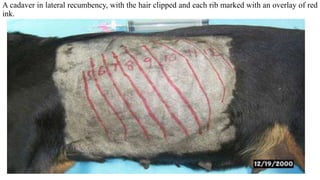
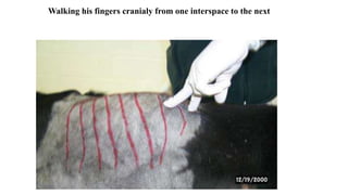
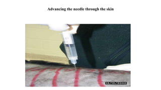
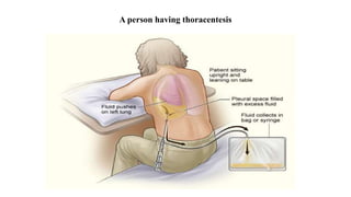
Thoracentesis is a procedure to remove fluid or air from the pleural space for diagnostic or therapeutic purposes. It requires inserting a needle between the ribs to drain fluid. The document outlines the materials, steps, and potential complications of performing thoracentesis on dogs, cats, and horses. Diagnostic thoracentesis is indicated for animals showing respiratory distress and reduced breath sounds to identify the cause of pleural effusion. Analysis of drained pleural fluid can help determine the underlying disease process.
Exfoliative vaginal cytology help to determine the stage of estrus, is very simple method, cost-effective and comparatively accurate test for predicting the she dogs fit for breeding.
one of the most commonly used techniques of the lung drainage is the postural drainage its non invasive and easy technique ans very useful in hospital as well as home settings.
procedure of thoracenthesis ( removal of water from thoacic cavuty full procedure by ganesh dalvi and tushar cahure uder the supervision of prof. krishna sananse
PowerPoint presentation on Intercostal drainage (ICD) or Chest tube drainage. In this this presentation I have included different methods by which a chest tube can be inserted to drain fluid, pus, air from the Pleural cavity. please do mail me your feedback on this presentation at tinkujoseph2010@gmail.com.
in this topic the technique of chest physiotherapy, indications, contradications of chest physiotherapy are explained. different positions used in postural drainage are briefed.
Nusing Management of CAD Symposia (English) presented at Hôpital Sacré Coeur in Milot, Haiti.
CRUDEM’s Education Committee (a subcommittee of the Board of Directors) sponsors one-week medical symposia on specific medical topics, i.e. diabetes, infectious disease. The classes are held at Hôpital Sacré Coeur and doctors and nurses come from all over Haiti to attend.
*Trocarization*
*Trocarization* refers to the use of a trocar, a surgical instrument, to puncture a body cavity for various medical purposes. In the context of equine medicine, trocarization is used for decompression in cases of colic and large intestinal gas distension. It allows for rapid controlled decompression of colonic or cecal tympany, permitting improvement of both ventilation and intraabdominal pressure
The procedure involves the insertion of a trocar into the large colon or cecum to resolve gas distension and accompanying increased intraabdominal pressure. It's important to note that trocarization is not a substitute for surgery in severely gas-distended painful horses exhibiting signs of colic that require surgery
Complications following transcutaneous cecal trocarization may include a decrease in cecal motility over time, possibly due to local inflammation caused by the puncture. However, large intestinal trocarization could be considered for equids with colic and large intestinal gas distension, and more than one trocarization procedure was reported in some cases
In summary, trocarization is a medical procedure used in equine medicine for decompression in cases of colic and large intestinal gas distension, but it's not a substitute for surgery in severe cases. It's important to consider potential complications and the specific conditions under which it may be appropriate.
*Trocarization*
*Trocarization* refers to the use of a trocar, a surgical instrument, to puncture a body cavity for various medical purposes. In the context of equine medicine, trocarization is used for decompression in cases of colic and large intestinal gas distension. It allows for rapid controlled decompression of colonic or cecal tympany, permitting improvement of both ventilation and intraabdominal pressure
The procedure involves the insertion of a trocar into the large colon or cecum to resolve gas distension and accompanying increased intraabdominal pressure
It's important to note that trocarization is not a substitute for surgery in severely gas-distended painful horses exhibiting signs of colic that require surgery
Complications following transcutaneous cecal trocarization may include a decrease in cecal motility over time, possibly due to local inflammation caused by the puncture However, large intestinal trocarization could be considered for equids with colic and large intestinal gas distension, and more than one trocarization procedure was reported in some cases
In summary, trocarization is a medical procedure used in equine medicine for decompression in cases of colic and large intestinal gas distension, but it's not a substitute for surgery in severe cases. It's important to consider potential complications and the specific conditions under which it may be appropriate
This presentation is only for education purpose which comprises of written part along with some color full picture and videos. These feature will help in the clear understanding of concept of blood collection.
Thoracentesis is a procedure in which a needle is inserted into the pleural space between the lungs and the chest wall. This procedure is done to remove excess fluid, known as a pleural effusion, from the pleural space to help you breathe easier.
Catheterization Procedure by Anushri Srivastav.pptxAnushriSrivastav
Catheterization of the bladder involves introducing a latex or plastic tube through the urethra and into the bladder. The catheter provides a continuous flow of urine in patients unable to control micturition or those with obstructions. It also provides a means of assessing urine output in hemodynamically unstable patients. Because bladder catheterization carries the risk of UTI, blockage, and trauma to the urethra, it is preferable to rely on other measures for either specimen collection or management of incontinence.
Types of Catheterization.
Intermittent and indwelling retention catheterizations are the two forms of catheter insertion
INTERMITTENT CATHETERIZATION
introduce a straight single-use catheter long enough to drain the bladder (5 to 10 minutes
When the bladder is empty, you immediately withdraw the catheter.
COMPLICATION- increases risk of trauma and infection.
INDICATION- It is common for people with spinal cord injury or other neurological problems such as multiple sclerosis to perform self– intermittent catheterization up to every 4 hours daily for months or years.
UTI rate is lower than for patients with long-term indwelling catheters.
INDWELLING CATHETERIZATION-
remains in place for a longer period, until a patient is able to void voluntarily or continuous accurate urine measurements are no longer needed
The straight single-use catheter has a single lumen with a small opening about 1.3 cm ( 1 2 inch) from the tip.
. Urine drains from the tip, through the lumen, and to a receptacle.
An indwelling Foley catheter has a small inflatable balloon that encircles the catheter just above the tip. When inflated the balloon rests against the bladder outlet to anchor the catheter in place.
The indwelling retention catheter often has two or three lumens within the body of the catheter . One lumen drains urine through the catheter to a collecting tube. A second lumen carries sterile water to and from the balloon when it is inflated or deflated. A third (optional) lumen is sometimes used to instill fluids or medications into the bladder. It is easy to determine the number of lumens by the number of drainage and injection ports at the end of the catheter
A second type of intermittent catheter has a curved tip
A Coudé catheter is used on male patients who may have enlarged prostates that partly obstruct the urethra. It is less traumatic during insertion because it is stiffer and easier to control than the straight-tip catheter
Plastic catheters are suitable only for intermittent use because of their inflexibility
Latex catheters are recommended for use up to 3 weeks. Be aware of allergies.
Pure silicon or Teflon catheters are best suited for long-term use (2 to 3 months) because of less encrustation at the urethral meatus
Balloon sizes range from 3 mL (pediatric) to large postoperative volumes (75 mL). In adults the 5-mL and 30-mL sizes are the most common: The 5-mL size allows for optimal drainage, whereas the 30-mL size is used after pros
These simplified slides by Dr. Sidra Arshad present an overview of the non-respiratory functions of the respiratory tract.
Learning objectives:
1. Enlist the non-respiratory functions of the respiratory tract
2. Briefly explain how these functions are carried out
3. Discuss the significance of dead space
4. Differentiate between minute ventilation and alveolar ventilation
5. Describe the cough and sneeze reflexes
Study Resources:
1. Chapter 39, Guyton and Hall Textbook of Medical Physiology, 14th edition
2. Chapter 34, Ganong’s Review of Medical Physiology, 26th edition
3. Chapter 17, Human Physiology by Lauralee Sherwood, 9th edition
4. Non-respiratory functions of the lungs https://academic.oup.com/bjaed/article/13/3/98/278874
micro teaching on communication m.sc nursing.pdfAnurag Sharma
Microteaching is a unique model of practice teaching. It is a viable instrument for the. desired change in the teaching behavior or the behavior potential which, in specified types of real. classroom situations, tends to facilitate the achievement of specified types of objectives.
Explore natural remedies for syphilis treatment in Singapore. Discover alternative therapies, herbal remedies, and lifestyle changes that may complement conventional treatments. Learn about holistic approaches to managing syphilis symptoms and supporting overall health.
Prix Galien International 2024 Forum ProgramLevi Shapiro
June 20, 2024, Prix Galien International and Jerusalem Ethics Forum in ROME. Detailed agenda including panels:
- ADVANCES IN CARDIOLOGY: A NEW PARADIGM IS COMING
- WOMEN’S HEALTH: FERTILITY PRESERVATION
- WHAT’S NEW IN THE TREATMENT OF INFECTIOUS,
ONCOLOGICAL AND INFLAMMATORY SKIN DISEASES?
- ARTIFICIAL INTELLIGENCE AND ETHICS
- GENE THERAPY
- BEYOND BORDERS: GLOBAL INITIATIVES FOR DEMOCRATIZING LIFE SCIENCE TECHNOLOGIES AND PROMOTING ACCESS TO HEALTHCARE
- ETHICAL CHALLENGES IN LIFE SCIENCES
- Prix Galien International Awards Ceremony
Couples presenting to the infertility clinic- Do they really have infertility...Sujoy Dasgupta
Dr Sujoy Dasgupta presented the study on "Couples presenting to the infertility clinic- Do they really have infertility? – The unexplored stories of non-consummation" in the 13th Congress of the Asia Pacific Initiative on Reproduction (ASPIRE 2024) at Manila on 24 May, 2024.
New Directions in Targeted Therapeutic Approaches for Older Adults With Mantl...i3 Health
i3 Health is pleased to make the speaker slides from this activity available for use as a non-accredited self-study or teaching resource.
This slide deck presented by Dr. Kami Maddocks, Professor-Clinical in the Division of Hematology and
Associate Division Director for Ambulatory Operations
The Ohio State University Comprehensive Cancer Center, will provide insight into new directions in targeted therapeutic approaches for older adults with mantle cell lymphoma.
STATEMENT OF NEED
Mantle cell lymphoma (MCL) is a rare, aggressive B-cell non-Hodgkin lymphoma (NHL) accounting for 5% to 7% of all lymphomas. Its prognosis ranges from indolent disease that does not require treatment for years to very aggressive disease, which is associated with poor survival (Silkenstedt et al, 2021). Typically, MCL is diagnosed at advanced stage and in older patients who cannot tolerate intensive therapy (NCCN, 2022). Although recent advances have slightly increased remission rates, recurrence and relapse remain very common, leading to a median overall survival between 3 and 6 years (LLS, 2021). Though there are several effective options, progress is still needed towards establishing an accepted frontline approach for MCL (Castellino et al, 2022). Treatment selection and management of MCL are complicated by the heterogeneity of prognosis, advanced age and comorbidities of patients, and lack of an established standard approach for treatment, making it vital that clinicians be familiar with the latest research and advances in this area. In this activity chaired by Michael Wang, MD, Professor in the Department of Lymphoma & Myeloma at MD Anderson Cancer Center, expert faculty will discuss prognostic factors informing treatment, the promising results of recent trials in new therapeutic approaches, and the implications of treatment resistance in therapeutic selection for MCL.
Target Audience
Hematology/oncology fellows, attending faculty, and other health care professionals involved in the treatment of patients with mantle cell lymphoma (MCL).
Learning Objectives
1.) Identify clinical and biological prognostic factors that can guide treatment decision making for older adults with MCL
2.) Evaluate emerging data on targeted therapeutic approaches for treatment-naive and relapsed/refractory MCL and their applicability to older adults
3.) Assess mechanisms of resistance to targeted therapies for MCL and their implications for treatment selection
These lecture slides, by Dr Sidra Arshad, offer a quick overview of physiological basis of a normal electrocardiogram.
Learning objectives:
1. Define an electrocardiogram (ECG) and electrocardiography
2. Describe how dipoles generated by the heart produce the waveforms of the ECG
3. Describe the components of a normal electrocardiogram of a typical bipolar leads (limb II)
4. Differentiate between intervals and segments
5. Enlist some common indications for obtaining an ECG
Study Resources:
1. Chapter 11, Guyton and Hall Textbook of Medical Physiology, 14th edition
2. Chapter 9, Human Physiology - From Cells to Systems, Lauralee Sherwood, 9th edition
3. Chapter 29, Ganong’s Review of Medical Physiology, 26th edition
4. Electrocardiogram, StatPearls - https://www.ncbi.nlm.nih.gov/books/NBK549803/
5. ECG in Medical Practice by ABM Abdullah, 4th edition
6. ECG Basics, http://www.nataliescasebook.com/tag/e-c-g-basics
Tom Selleck Health: A Comprehensive Look at the Iconic Actor’s Wellness Journeygreendigital
Tom Selleck, an enduring figure in Hollywood. has captivated audiences for decades with his rugged charm, iconic moustache. and memorable roles in television and film. From his breakout role as Thomas Magnum in Magnum P.I. to his current portrayal of Frank Reagan in Blue Bloods. Selleck's career has spanned over 50 years. But beyond his professional achievements. fans have often been curious about Tom Selleck Health. especially as he has aged in the public eye.
Follow us on: Pinterest
Introduction
Many have been interested in Tom Selleck health. not only because of his enduring presence on screen but also because of the challenges. and lifestyle choices he has faced and made over the years. This article delves into the various aspects of Tom Selleck health. exploring his fitness regimen, diet, mental health. and the challenges he has encountered as he ages. We'll look at how he maintains his well-being. the health issues he has faced, and his approach to ageing .
Early Life and Career
Childhood and Athletic Beginnings
Tom Selleck was born on January 29, 1945, in Detroit, Michigan, and grew up in Sherman Oaks, California. From an early age, he was involved in sports, particularly basketball. which played a significant role in his physical development. His athletic pursuits continued into college. where he attended the University of Southern California (USC) on a basketball scholarship. This early involvement in sports laid a strong foundation for his physical health and disciplined lifestyle.
Transition to Acting
Selleck's transition from an athlete to an actor came with its physical demands. His first significant role in "Magnum P.I." required him to perform various stunts and maintain a fit appearance. This role, which he played from 1980 to 1988. necessitated a rigorous fitness routine to meet the show's demands. setting the stage for his long-term commitment to health and wellness.
Fitness Regimen
Workout Routine
Tom Selleck health and fitness regimen has evolved. adapting to his changing roles and age. During his "Magnum, P.I." days. Selleck's workouts were intense and focused on building and maintaining muscle mass. His routine included weightlifting, cardiovascular exercises. and specific training for the stunts he performed on the show.
Selleck adjusted his fitness routine as he aged to suit his body's needs. Today, his workouts focus on maintaining flexibility, strength, and cardiovascular health. He incorporates low-impact exercises such as swimming, walking, and light weightlifting. This balanced approach helps him stay fit without putting undue strain on his joints and muscles.
Importance of Flexibility and Mobility
In recent years, Selleck has emphasized the importance of flexibility and mobility in his fitness regimen. Understanding the natural decline in muscle mass and joint flexibility with age. he includes stretching and yoga in his routine. These practices help prevent injuries, improve posture, and maintain mobilit
The hemodynamic and autonomic determinants of elevated blood pressure in obes...
THORACENTESIS; SITE AND MATERIALS
1. ADDIS ABABA UNIVERSITY COLLEGE OF VETERINARY MEDICINE AND
AGRICULTURE
INDIVIDUAL PRESENTATION ON THORACENTESIS; SITE AND MATERIALS
BY:
Akinaw Wagari
Instructor: Tilaye D. (DVM, MSc, Assistant Prof.)
Mar, 6/2015
Bishoftu, Ethiopia
2. 1. INTRODUCTION
• Thoracentesis is an invasive procedure to remove fluid or air from the pleural space for
diagnostic or therapeutic purposes. The procedure was first described in 1852.
• Diagnostic thoracocentesis is indicated in any animal with increased respiratory effort (or signs
of respiratory distress) and reduced breath sounds.
• If air or fluid is discovered with this procedure in an animal with respiratory difficulty, you
should immediately proceed to therapeutic thoracocentesis.
3. Cont…
When the findings of thoracic auscultation or percussion are suggestive of pleural effusion,
thoracocentesis can be performed to:
• Confirm the presence of pleural effusion
• Provide a specimen for examination which provide a diagnosis or guide the therapeutic
plan
• Therapeautically drain a large volume of pleural fluid
4. 2. DIAGNOSTIC THORACOCENTESIS
Materials needed:
• 3 ml syringe with a 22 ga 1” needle (25 ga 5/8” is OK for thin cats).
Procedure:
• Position the animal in whatever position it is most comfortable – do not stress it by forcing it
to lie in lateral recumbency! For most animals this will mean doing the procedure with the
animal standing or in sternal recumbency.
• Try to avoid letting it sit during the procedure, as this will make it more difficult to identify
landmarks.
5. Cont…
• If you suspect pneumothorax, you will want to aspirate the ‘highest’ accessible portion of the
chest at around the 7th or 8th interspace.
• If you suspect that fluid is in the pleural space, go in at the junction of the middle and ventral
thirds, near the costachondral junction.
• If for some reason an animal with pleural fluid seems to prefer to lie on its side, try to get it
into sternal recumbency first, since fluid will gravitate to the lowest part of the chest.
6. A cadaver in lateral recumbency, with the hair clipped and each rib marked with an overlay of red
ink.
10. 3. HOW TO PERFORM A CANINE OR FELINE THORACENTESIS
What you need?
Fortunately, a thoracentesis does not require much in terms of equipment:
Stethoscope
Clippers
Surgical scrub solution
Alcohol (in spray bottle)
A butterfly needle or appropriately sized hypodermic needle (16–21 gauge)
A three-way stopcock (ideally)
Sterile gloves
11. Cont…
Extension set
• A 10- to 60-mL syringe, depending on anticipated amount of air or effusion
• Appropriate sterile collection tubes (for cytologic and culture sample collection)
• Oxygen
Preparing cat for thoracentesis
12. Cont…
• A thoracentesis should be performed cranial to the rib, as the blood vessels and nerves lie caudal
to the rib (“hiding” behind the rib).
• Thoracentesis should be performed at the 7th to 9th intercostal space to avoid the heart (3th–5th
ICS) or liver (caudal to the 9th ICS).
Advancing the needle into the pleural space
13. Complications
• Complications from thoracentesis are generally rare.
• Iatrogenic pneumothorax or laceration. Although rare, hematoma, pneumothorax,
arterial laceration, hypotension, re-expansion pulmonary edema or vagal reaction can
occur with thoracentesis.
• The fluid can give an indication of prognosis; in the cat prognosis is generally poor for
all diagnoses except pyothorax (an exudate with degenerate neutrophils and
intracellular bacteria).
14. 4. EQUINE THORACOCENTESIS
Equipment
Sedation as necessary
Clippers and materials to perform a surgical scrub
Ultrasonography if available
Local anaesthetic and a 23G 3cm needle
Sterile gloves
Number 15 scalpel blade
Cannula, 3-way tap and extension set
EDTA tube, plain tube and sterile vial for culture
15. Procedure
• The site for thoracocentesis can be identified using anatomical landmarks; 7-8th intercostal
space on the left or 6-7th intercostal space on the right midway between the shoulder and the
elbow.
• The horse should be sedated and the area clipped and scrubbed. Local anaesthetic should be
administered into the subcutis, intercostal musculature and parietal pleura using a 23 gauge, 3
cm needle.
16. Cont…
Pleural fluid analysis
• Analysis of the pleural fluid may in turn help you to determine the underlying disease
process and develop a therapeutic plan.
• Pleural fluid in healthy horses normally contains less than 5,000 nucleated cells/l and less
than 25g/l total protein. Levels greater than 10,000 nucleated cells/l and 35g/l total protein
should be considered abnormal.
17. 5. CONCLUSIONS
• Thoracentesis is a procedure to remove fluid from the space between the lungs and the chest
wall called the pleural space. It is done with a needle (and sometimes a plastic catheter) inserted
through the chest wall.
• Ultrasound pictures are often used to guide the placement of the needle. This pleural fluid may
be sent to a lab to determine what may be causing the fluid to build up in the pleural space.
• Normally only a small amount of pleural fluid is present in the pleural space. A buildup of
excess pleural fluid (pleural effusion) may be caused by many conditions, such as infection,
inflammation, heart failure, or cancer.