Downloaded 1,482 times
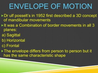


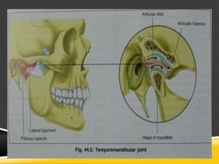


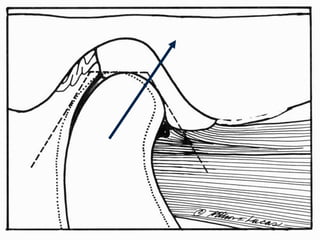
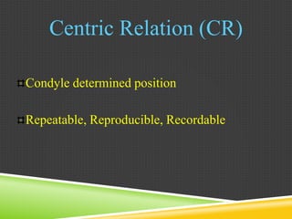
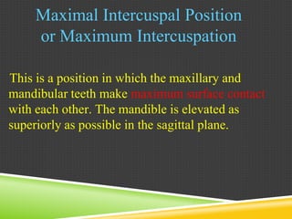


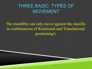

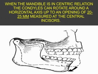

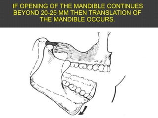
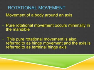

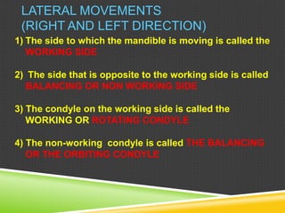


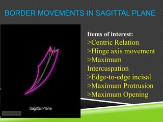

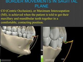
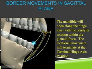


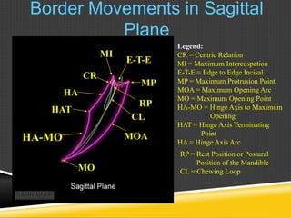


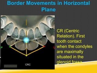
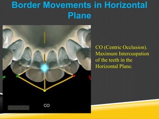
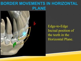


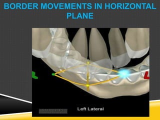


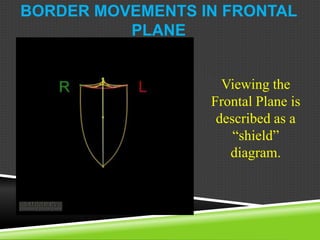
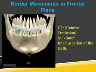


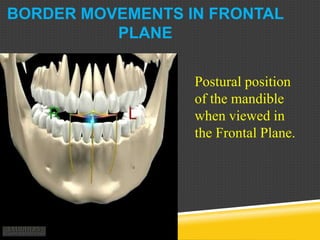


Dr. Ulf Posselt first described the envelope of motion in 1952 as a 3D concept to illustrate the possible movements of the mandible in all three planes of movement - sagittal, horizontal, and frontal. The envelope differs between individuals but maintains the same characteristic shape, with the superior surface determined by tooth contacts and the borders primarily determined by the TMJ anatomy and ligaments. The document then discusses reference positions like centric relation and maximum intercuspation, as well as types of mandibular movement including hinge, translational, and rotational. Border movements in the sagittal and horizontal planes are mapped out, illustrating positions like centric relation, maximum opening, and lateral excursions.


![occlusion mmmmmmmm- Copy [Autosaved].pptx](https://cdn.slidesharecdn.com/ss_thumbnails/occlusion-copyautosaved-240715160312-7fd34768-thumbnail.jpg?width=640&height=640&fit=bounds)


