Downloaded 284 times

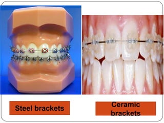
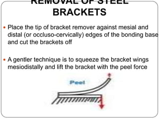
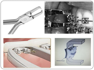
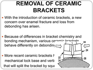
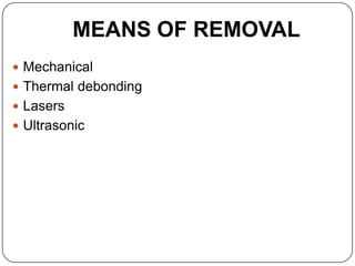
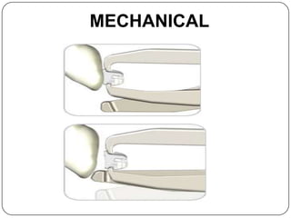
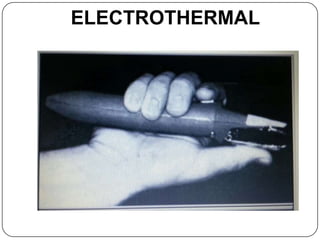
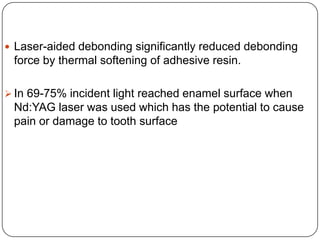
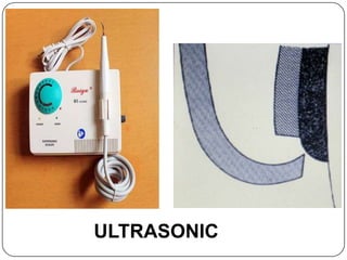
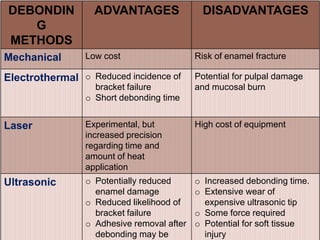
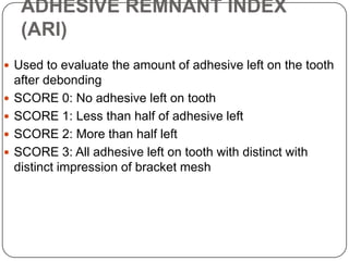
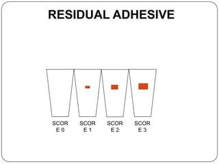
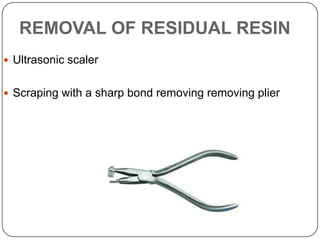
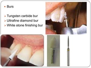
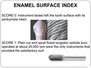

This document provides an overview of debonding procedures in orthodontics. It discusses the objective of restoring the tooth surface after removing attachments and adhesive resin. It describes different procedures for removing steel and ceramic brackets, as well as residual adhesive, using mechanical methods, electrothermal debonding, lasers, or ultrasonic tools. It compares the advantages and disadvantages of these debonding methods and discusses indices for evaluating the amount of remaining adhesive and surface changes to the enamel.