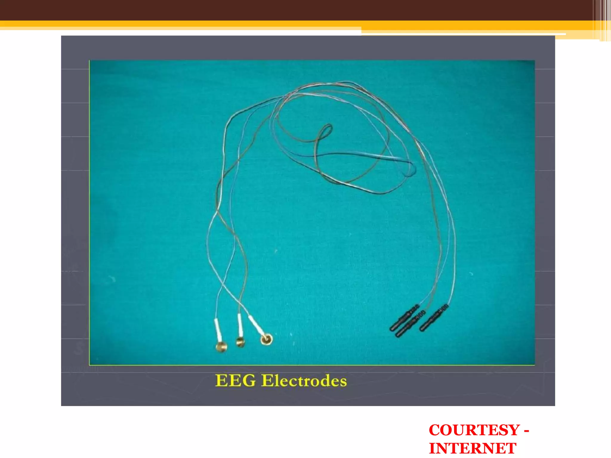
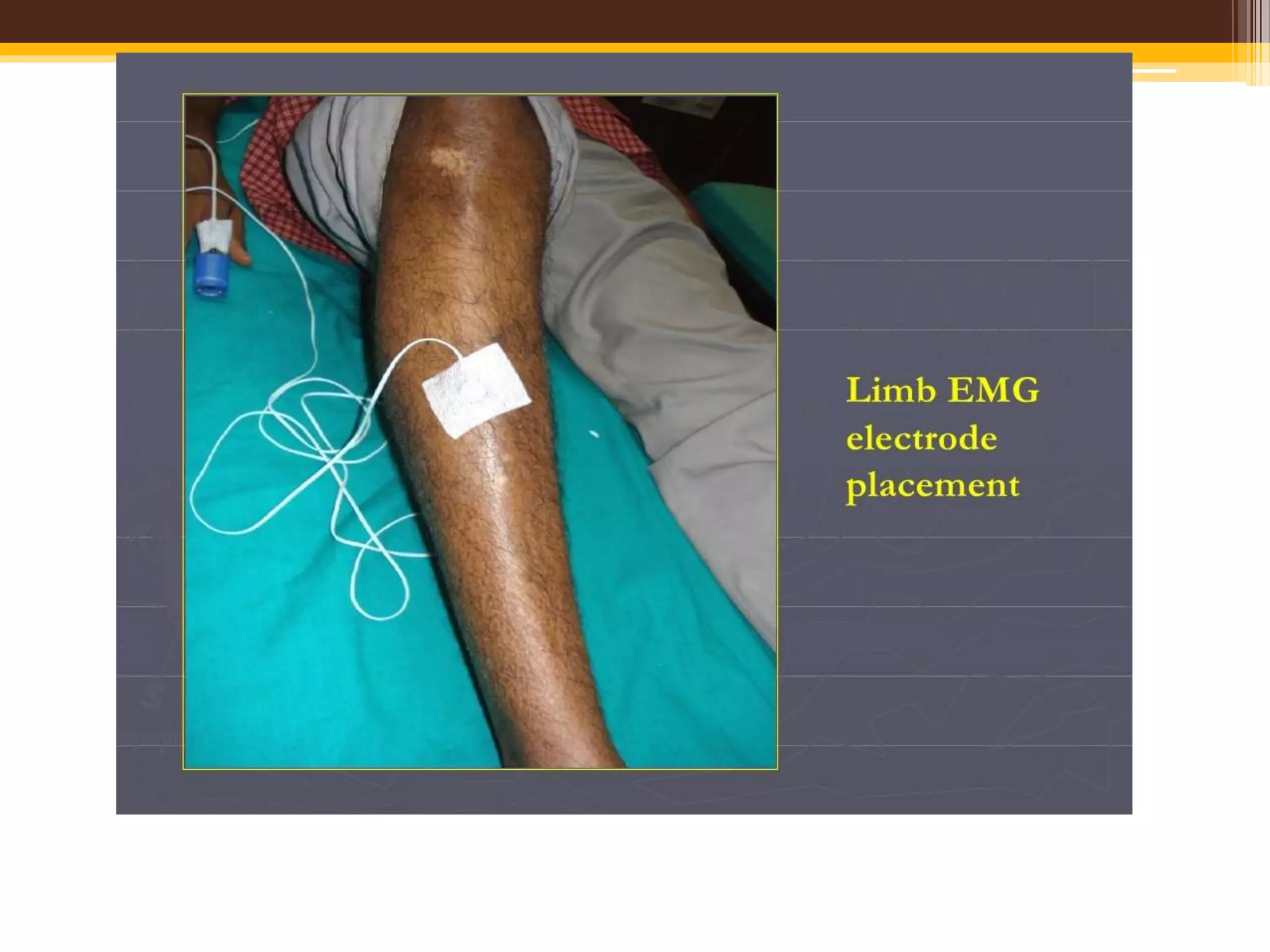
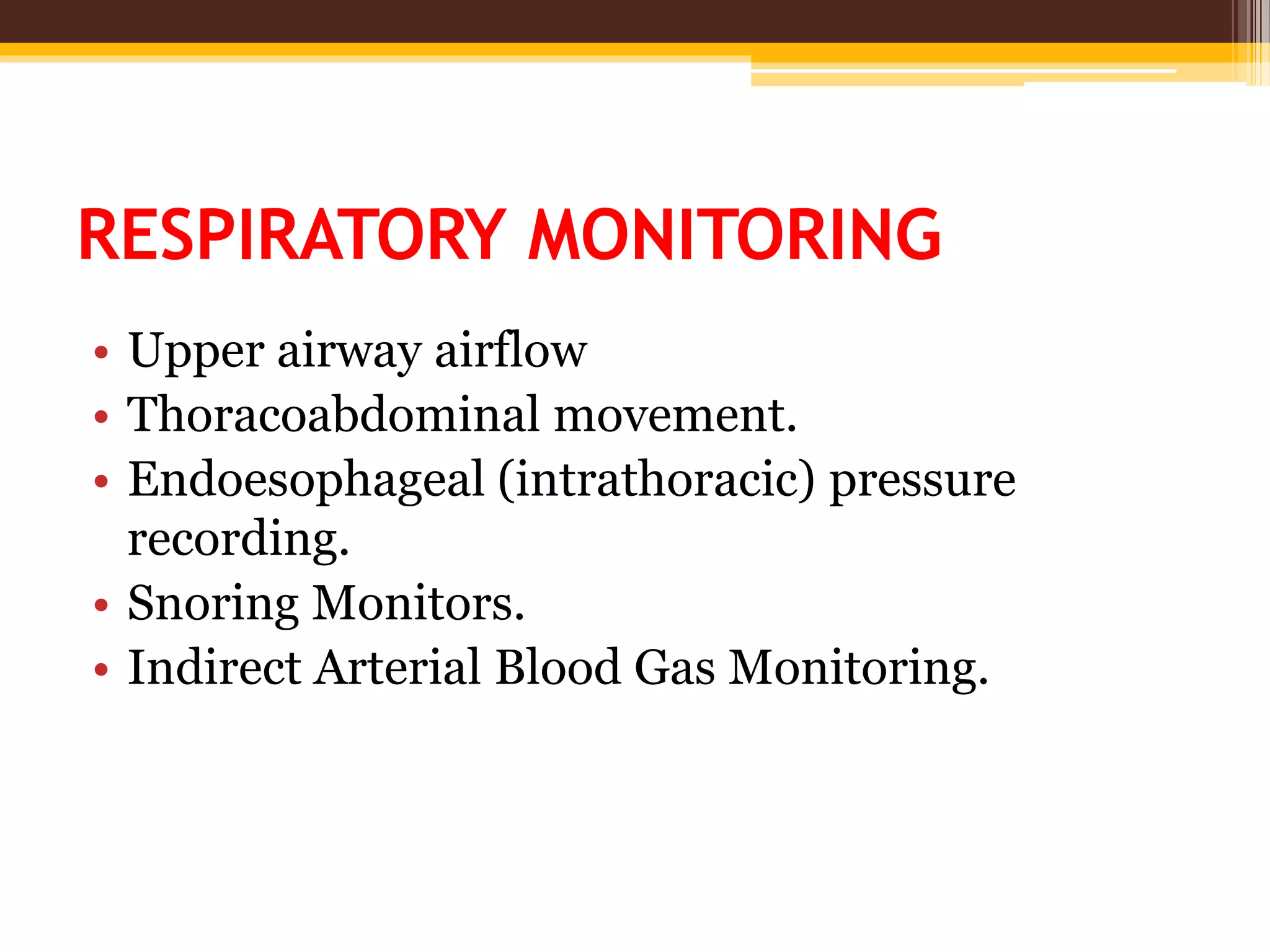
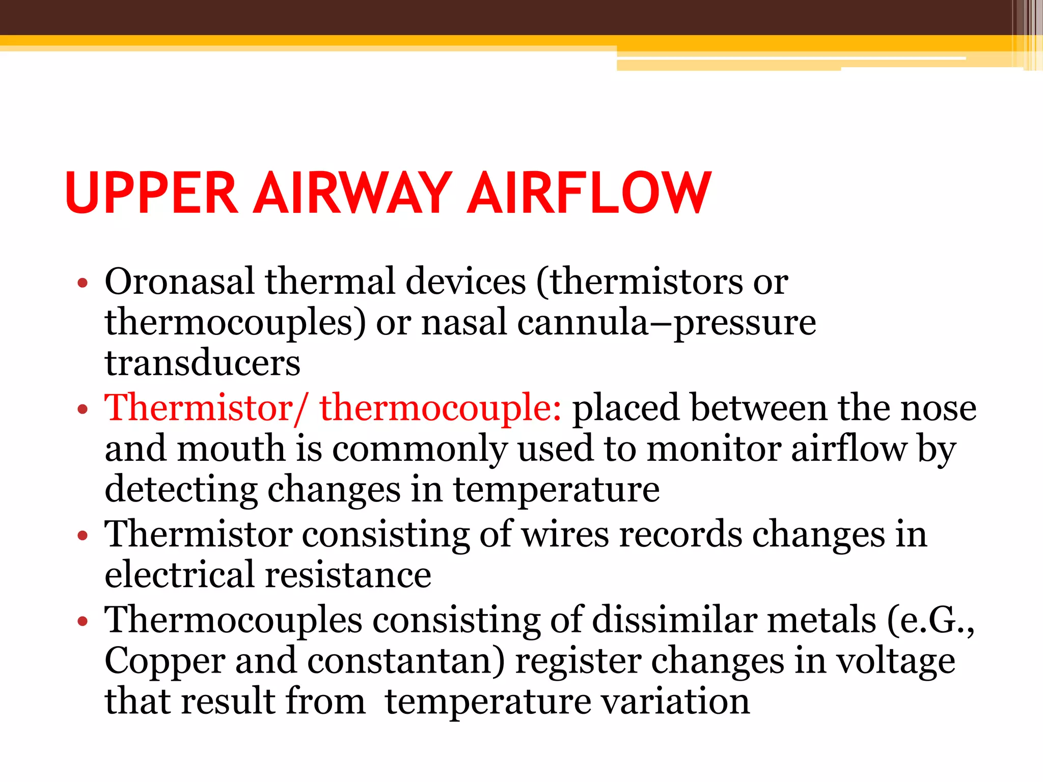
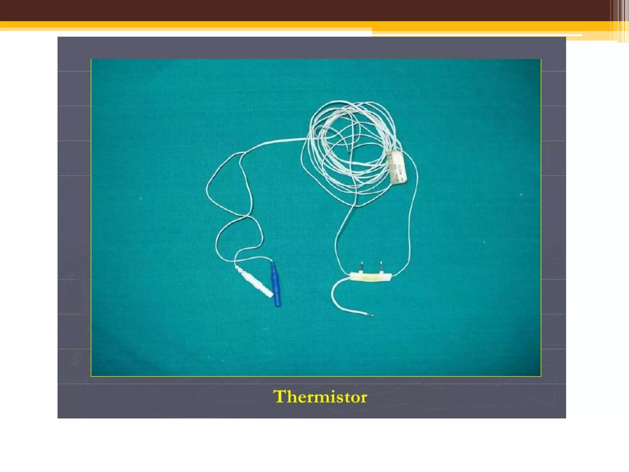
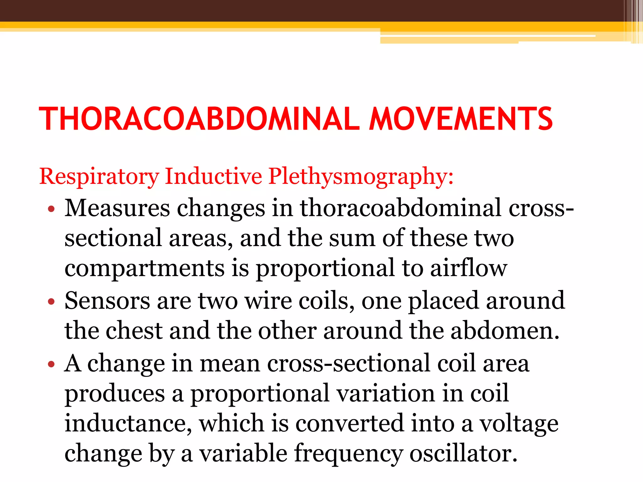
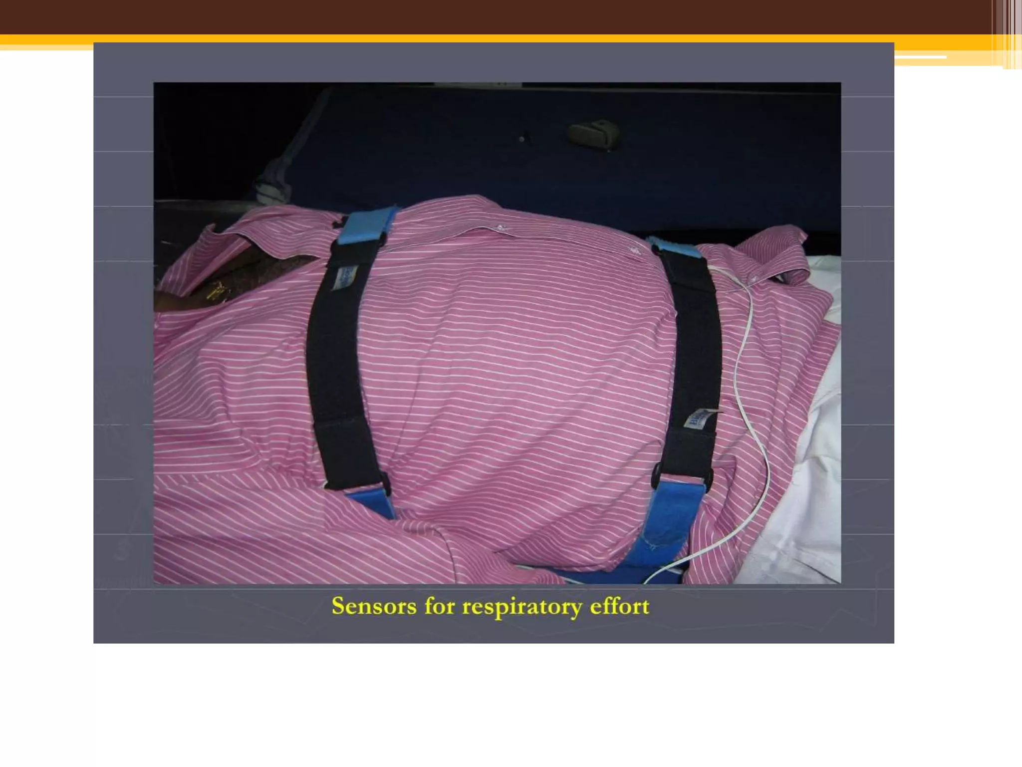
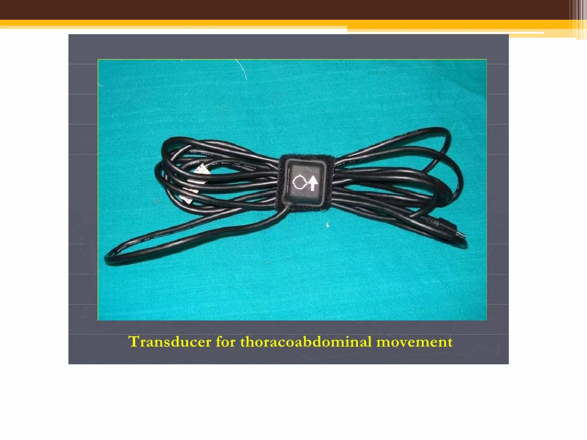
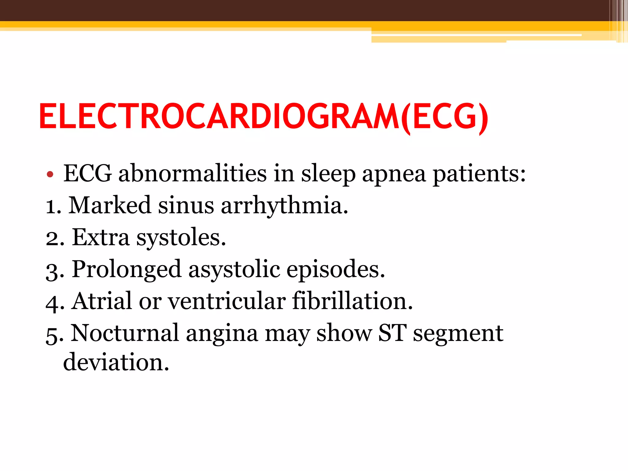
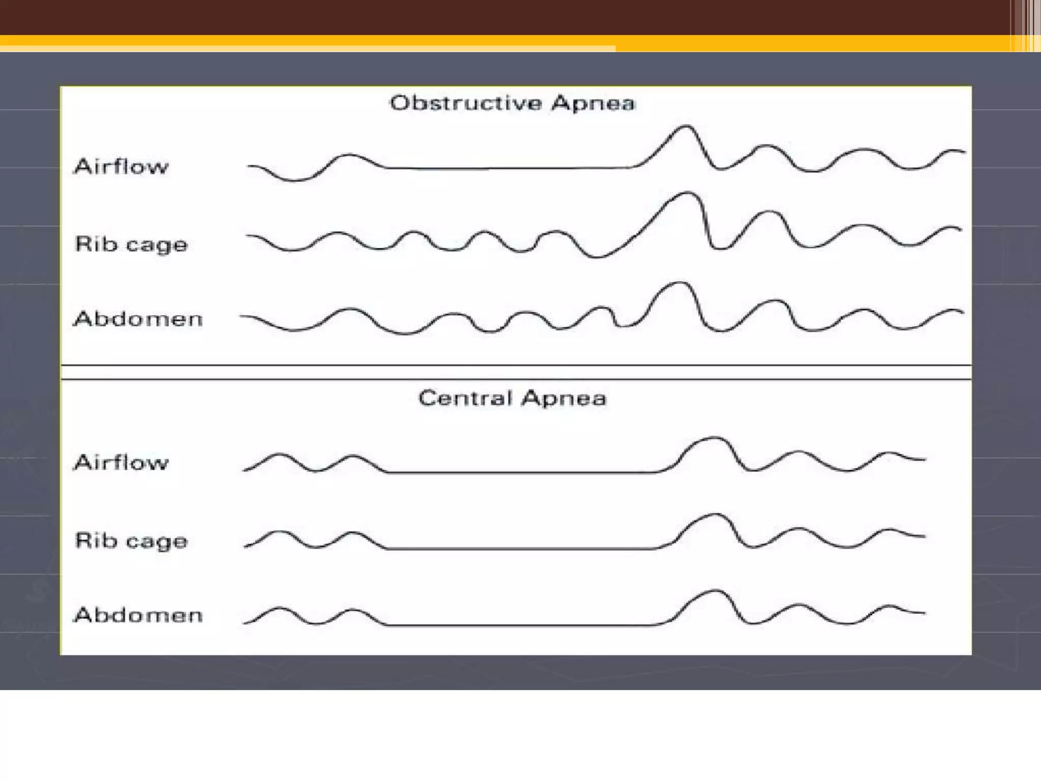
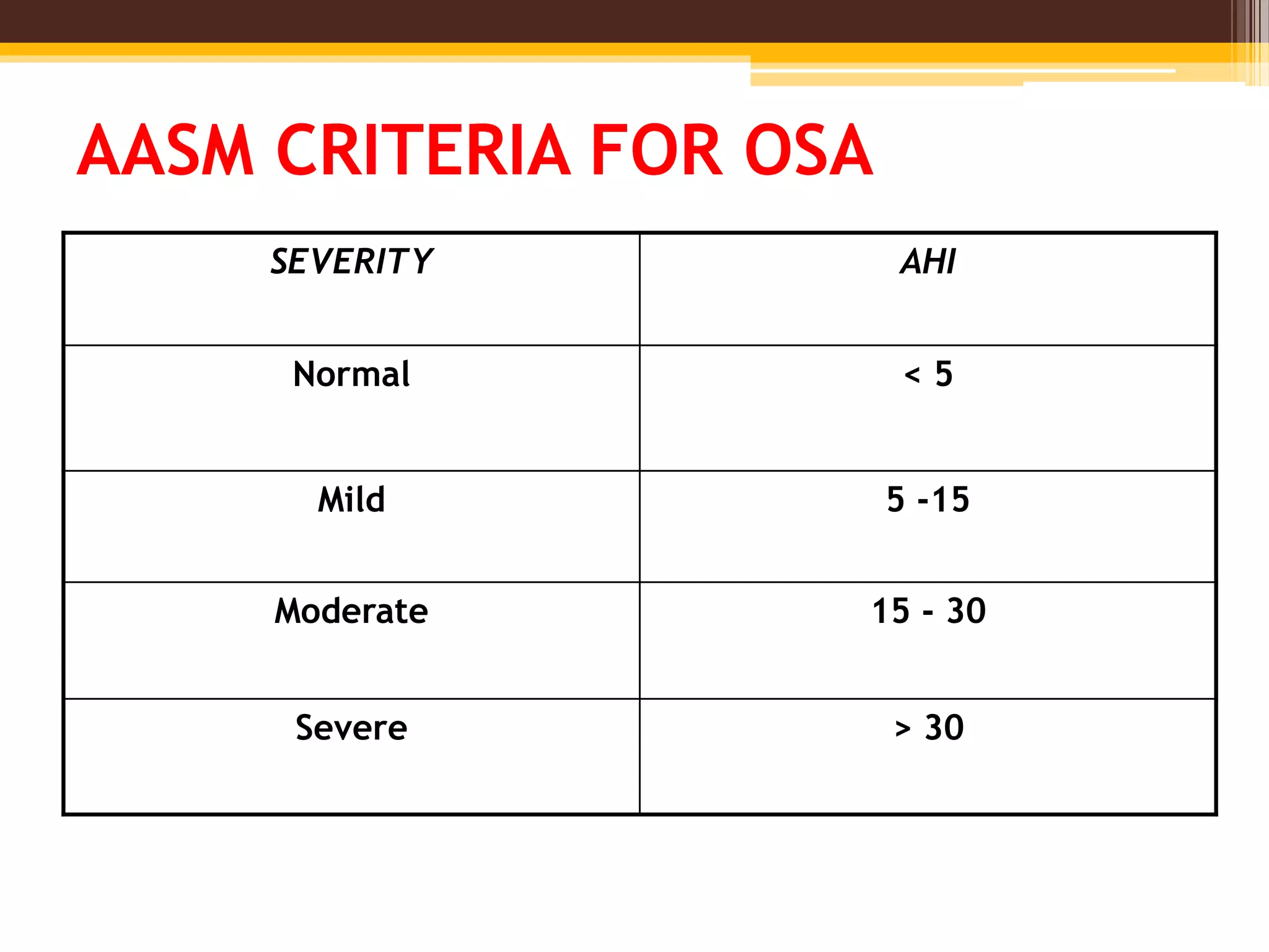
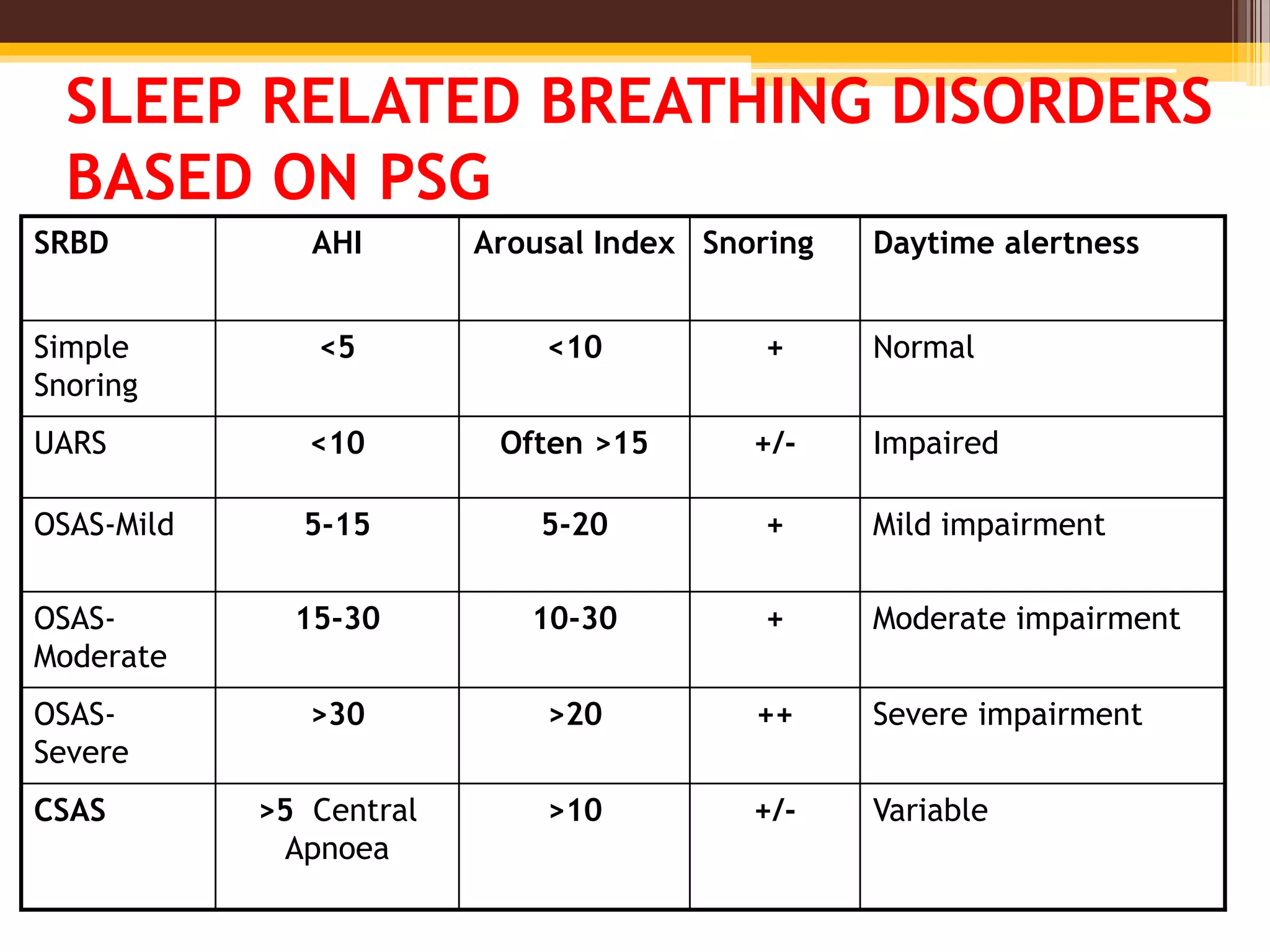
BASICS OF POLYSOMNOGRAPHY
Dr. Sunil Kumar Sharma
Senior Resident
Moderator
Dr. Dilip Maheshwari (DM)
Asso. Professor
Dept. of Neurology
GMC Kota
POLYSOMNOGRAPHY
• Introduction
• Indications
• Patient evaluation and instructions
• Components
• Derived Information
• Types
• Limitation
• Summmary
INTRODUCTION
• Term “Polysomnography” was proposed by
Holland ,Dement and Raynal (1974).
• The most commonly used test in the diagnosis
of obstructive sleep apnea syndrome (OSAS).
• Consists of a simultaneous recording of multiple
physiologic parameters related to sleep and
wakefulness.
INDICATIONS FOR POLYSOMNOGRAPHY
• Sleep related breathing disorders
• CPAP titration in OSA
• Assessment of treatment results- OSA
• Sleep related behavioral disorders
• Atypical or unusual parasomnias
• Narcolepsy
• Neuromuscular disorder & sleep related symptoms
• Paroxysmal arousal or seizure phenomenon
• Periodic Leg Movements of Sleep
• Parasomnias not responding to conventional
therapy
Initial Assessment
• Detailed medical & psychiatric history
• Medication, smoking, alcohol, activities on that day
Assessment of sleep:
• Sleep history for the last 24 hrs.
Sleep scoring systems
1. Stanford Sleepiness scale
2. Epworth Sleepiness scale
3. Pittsburgh Sleep Quality Index (PSQI)
RECORDING ROOM
• The study patient and the monitoring apparatus
and technologist are housed in adjacent rooms.
• Recording room should be as homely and
comfortable as possible.
• It should be sound-proofed and air-conditioned.
• Intercom system to interact with the patient.
• Rheostatically controlled lighting
• Toilet and restroom
COURTESY -
INTERNET
COURTESY -
INTERNET
INSTRUCTION TO THE PATIENT
• Reporting time: 1 hour prior to the usual time of
sleep.
• A prior visit to familiarize with the lab is useful.
• Patient should have had a relaxed day, without
daytime sleep.
• Headwash, adequate food is recommended.
• Abstain from caffeine in the afternoon and
evening of the day on which PSG is planned
• Avoid alcohol on the day of PSG
• Avoid strenuous exercise on the day of the PSG.
INSTRUCTION TO THE PATIENT
• Continue their usual medications on the night of the
PSG, including sleep aids.
• The medications should be recorded by the
technician so that the results can be optimally
interpreted.
• For patients who have a history of insomnia,
especially when sleeping in a new
environment, Zolpidem may be prescribed.
• Avoid stimulants, including medications for
narcolepsy.
• Avoid naps on the day of the sleep study.
COMPONENTS OF
POLYSOMNOGRAPHY
ELECTROENCEPHALOGRAPHY (EEG)
• To distinguish between wakefulness and the
various stages of sleep
• A minimum of three channels representing the
right frontal, central, and occipital electrodes
referenced to the contralateral mastoid electrode
is recommended (2007 AASM Manual for the Scoring of
Sleep and Associated Events)
COURTESY -
INTERNET
ELECTRO-OCULOGRAPHY (EOG)
• Two recommended electrodes are labeled E1 (1 cm
below the left outer canthus) and E2 (placed 1 cm
above the right outer canthus), both referenced to
the right mastoid.
• This allows simultaneous recording of both vertical
eye movements (such as blinking) and horizontal
eye movements (both slow and rapid).
• Documents the onset of rapid eye movement (REM)
sleep, and notes the presence of slow-rolling eye
movements that usually accompany the onset of
sleep.
ELECTROMYOGRAPHY (EMG)
• Help to determine sleep stage
• Help to diagnose and classify a variety of parasomnias
• Minimum components are chin EMG channels recording
activity from the mentalis and submental muscles (the
mylohyoid and anterior belly of the digastric) and
bilateral leg EMG channels recording activity from the
tibialis anterior muscles.
• Tonic EMG level in axial muscles usually decreases from
wakefulness through stages 1, 2, 3, and 4 of NREM sleep,
and is normally absent in REM sleep
EMG MONITORING
Utility of limb EMG:
• Periodic limb movements in sleep.
• Restless leg syndrome.
• Other movement disorders.
• To document the hand and arm gestures of REM
sleep behavior disorder.
• Record convulsive movements during nocturnal
epileptic seizures.
RESPIRATORY MONITORING
• Upper airway airflow
• Thoracoabdominal movement.
• Endoesophageal (intrathoracic) pressure
recording.
• Snoring Monitors.
• Indirect Arterial Blood Gas Monitoring.
UPPER AIRWAY AIRFLOW
• Oronasal thermal devices (thermistors or
thermocouples) or nasal cannula–pressure
transducers
• Thermistor/ thermocouple: placed between the nose
and mouth is commonly used to monitor airflow by
detecting changes in temperature
• Thermistor consisting of wires records changes in
electrical resistance
• Thermocouples consisting of dissimilar metals (e.G.,
Copper and constantan) register changes in voltage
that result from temperature variation
UPPER AIRWAY AIRFLOW
• Nasal prongs connected to a pressure transducer
detect inspiratory flow and may be the most
accurate method to identify subtle inspiratory flow
limitation – hypopneas
• An important limitation of nasal pressure
transducers is that they cannot detect mouth
breathing. To overcome this limitation, a thermistor
is usually added.
• Nasal pressure transducers are necessary for the
diagnosis of hypopneas and thermistors are
necessary for the diagnosis of apneas
THORACOABDOMINAL MOVEMENTS
Respiratory Inductive Plethysmography:
• Measures changes in thoracoabdominal cross-
sectional areas, and the sum of these two
compartments is proportional to airflow
• Sensors are two wire coils, one placed around
the chest and the other around the abdomen.
• A change in mean cross-sectional coil area
produces a proportional variation in coil
inductance, which is converted into a voltage
change by a variable frequency oscillator.
SNORING MONITORS
• Although snoring can be monitored by placing a
miniature microphone on the patient’s neck,
there is no accepted grading system to quantify
the intensity of this parameter.
• In practice the technologist’s notations as the
study is being recorded, as well as the
polysomnographer’s review of the audio as the
study is being read, provide a better estimation
of the degree of snoring
INTRATHORACIC PRESSURE MONITORING
Endoesophageal pressure probe:
▫ Most sensitive detection of heightened respiratory effort.
▫ An endoesophageal tube is passed nasally till the probe is
about 5 cm above the esophageal-gastric junction.
▫ Measures increases in intrathoracic negative pressure to
overcome increased upper airway resistance.
• Gold standard for measuring respiratory effort
• Not a routine practice , because of patient
discomfort and the technical skill required
CAPNOGRAPHY
• Capnography, or end-tidal CO2 (ETCO2),
monitoring detects the expired carbon dioxide
(CO2) level, which closely approximates intra-
alveolar CO2.
• An infrared analyzer over the nose and mouth
detects CO2 in the expired air, which
qualitatively measures the airflow
• Costly and therefore not used in most
laboratories
ABG
• An alternative to capnography is measurement
of the (Paco2) in the morning after their sleep
study to be compared to their waking Paco2
• Adults who have an increase in their paco2 in
sleep by 10 mm hg or more compared to an
awake supine paco2 have sleeprelated
hypoventilation
OXYGEN SATURATION
• Continuous oxygen saturation monitoring by
finger pulse oximetry is routine .
• PSG reports mention the time the patient spent
with an SpO2 below 90%.
ELECTROCARDIOGRAM(ECG)
• ECG abnormalities in sleep apnea patients:
1. Marked sinus arrhythmia.
2. Extra systoles.
3. Prolonged asystolic episodes.
4. Atrial or ventricular fibrillation.
5. Nocturnal angina may show ST segment
deviation.
BODY POSITION
• Some patients only have abnormalities when
sleeping in certain positions. Therefore, body
position (eg, supine, left lateral, right lateral,
prone) is monitored throughout the test using a
position sensor and/or video monitor.
OPTIONAL PARAMETERS
1)Esophageal pH
• Gastro-esophageal reflux of acidic stomach contents
into the lower esophagus may cause insomnia.
• pH probe is introduced nasally and swallowed to
about 5 cm above the esophageal sphincter.
2)Penile Tumescence
• Psychogenic Vs organic causes of impotence.
• In normal adult men, penile tumescence occurs
during REM sleep
• Psychogenic cases: Normal REM sleep-related
erections.
3)Core Body Temperature
INTERPRETATION OF PSG
• Total sleep time – The total sleep time (TST) is the total
duration of light sleep (stages N1 and N2), deep sleep
(stage N3), and rapid eye movement (REM) sleep
• Sleep efficiency – Sleep efficiency (SE) is the TST divided
by the total recording time (ie, the time in bed).
• Sleep stage percentage – The sleep stage percentage
(SSP) for a particular sleep stage is the duration of that
sleep stage divided by the TST.
• Sleep stage latency – The latency to any sleep stage is the
duration from sleep onset to the initiation of that sleep
stage.
• Arousals – Arousals range from full awakenings
to three-second transient
electroencephalography (EEG) shifts to a lighter
stage of sleep (alpha, theta, and/or frequencies
greater than 16 Hz, but not sleep spindles, with
at least 10 seconds of stable sleep preceding the
change).
• Arousals are generally counted and then divided
by the TST to give the number of arousals per
hour of sleep (ie, arousal index).
• Apnea is defined by the American Academy of Sleep
Medicine (AASM) as the cessation of airflow for at
least 10 seconds.
• Hypopnea is defined as decrease in airflow of ≥30 %
(by a valid measure of airflow) lasting ≥10 s,
associated with either ≥3 % desaturation from the
pre-event baseline or an arousal
• Respiratory effort–related arousal (RERA)
is an event characterized by increasing respiratory
effort for 10 seconds or longer leading to an arousal
from sleep but one that does not fulfill the criteria
for a hypopnea or apnea.
INDICES FOR SLEEP APNEA SYNDROMES
• Apnea-hypopnea index (AHI)
The AHI is defined as the average number of
episodes of apnea and hypopnea per hour.
• Respiratory disturbance index (RDI)
Defined as the average number of respiratory
disturbances (obstructive apneas, hypopneas,
and respiratory event–related arousals
[RERAs]) per hour.
AASM CRITERIA FOR OSA
SEVERITY AHI
Normal < 5
Mild 5 -15
Moderate 15 - 30
Severe > 30
SLEEP RELATED BREATHING DISORDERS
BASED ON PSG
SRBD AHI Arousal Index Snoring Daytime alertness
Simple
Snoring
<5 <10 + Normal
UARS <10 Often >15 +/- Impaired
OSAS-Mild 5-15 5-20 + Mild impairment
OSAS-
Moderate
15-30 10-30 + Moderate impairment
OSAS-
Severe
>30 >20 ++ Severe impairment
CSAS >5 Central
Apnoea
>10 +/- Variable
TYPES OF PSG
• Four types based on the number of parameters they
measure and the degree of attendance required.
• Level 1 devices - Traditional attended in-laboratory
PSGs
• Level 2 through 4 refer to home studies recorded by
portable devices with progressively fewer channels
measuring progressively fewer parameters
• Level II devices require a minimum of seven
channels, including EEG,EOG, chin EMG, ECG,
oximetry, airflow, and respiratory effort channels.
Thus they permit sleep scoring.
• Level III device: This device has a minimum of 4
channels, including ventilation or airflow (at least 2
channels of respiratory movement or airflow), heart
rate or ECG, and oxygen saturation.
• Level IV device: This type of device does not meet
requirements for other types, and many measure
only 1-2 parameters (eg, oxygen saturation or
airflow).
SPLIT NIGHT STUDIES
• Diagnosis of OSA is established during the first
portion of the study and the amount of positive
airway pressure that is necessary to prevent
upper airway collapse during sleep is
determined during the remaining portion.
LIMITATIONS OF PSG STUDY
▫ First Night Effect:
 Reduced sleep efficiency.
 Increased awakenings and arousals.
 Prolonged sleep and REM latency.
 Decreased percentage of REM and slow-wave sleep.
 Increased percentage of light sleep.
▫ Technology, technician, technique dependant.
▫ Episodic disorders may be missed: eg seizures,
parasomnias.
▫ Night- to- night variability: eg in case of apnea.
• VARIABILITY :
▫ Night to night variability makes it possible for a
single study to underestimate the severity of OSA
▫ Nasal patency, body position, or disruptive
environmental factors may all be important
factors in producing such variability.
▫ Therefore, it is reasonable to repeat the baseline
PSG if there is a strong clinical suspicion for OSA
CONCLUSION
• Attended, in-laboratory polysomnography (PSG)
is considered the gold standard diagnostic test
for obstructive sleep apnea (OSA)
• Diagnostic evaluation of suspected OSA,
titration of positive airway pressure therapy, and
assessment of the effectiveness of therapy are
the most common indications for PSG.

Polysomnography

  • 1.
    BASICS OF POLYSOMNOGRAPHY Dr.Sunil Kumar Sharma Senior Resident Moderator Dr. Dilip Maheshwari (DM) Asso. Professor Dept. of Neurology GMC Kota
  • 2.
    POLYSOMNOGRAPHY • Introduction • Indications •Patient evaluation and instructions • Components • Derived Information • Types • Limitation • Summmary
  • 3.
    INTRODUCTION • Term “Polysomnography”was proposed by Holland ,Dement and Raynal (1974). • The most commonly used test in the diagnosis of obstructive sleep apnea syndrome (OSAS). • Consists of a simultaneous recording of multiple physiologic parameters related to sleep and wakefulness.
  • 4.
    INDICATIONS FOR POLYSOMNOGRAPHY •Sleep related breathing disorders • CPAP titration in OSA • Assessment of treatment results- OSA • Sleep related behavioral disorders • Atypical or unusual parasomnias • Narcolepsy • Neuromuscular disorder & sleep related symptoms • Paroxysmal arousal or seizure phenomenon • Periodic Leg Movements of Sleep • Parasomnias not responding to conventional therapy
  • 5.
    Initial Assessment • Detailedmedical & psychiatric history • Medication, smoking, alcohol, activities on that day Assessment of sleep: • Sleep history for the last 24 hrs. Sleep scoring systems 1. Stanford Sleepiness scale 2. Epworth Sleepiness scale 3. Pittsburgh Sleep Quality Index (PSQI)
  • 6.
    RECORDING ROOM • Thestudy patient and the monitoring apparatus and technologist are housed in adjacent rooms. • Recording room should be as homely and comfortable as possible. • It should be sound-proofed and air-conditioned. • Intercom system to interact with the patient. • Rheostatically controlled lighting • Toilet and restroom
  • 7.
  • 8.
  • 9.
    INSTRUCTION TO THEPATIENT • Reporting time: 1 hour prior to the usual time of sleep. • A prior visit to familiarize with the lab is useful. • Patient should have had a relaxed day, without daytime sleep. • Headwash, adequate food is recommended. • Abstain from caffeine in the afternoon and evening of the day on which PSG is planned • Avoid alcohol on the day of PSG • Avoid strenuous exercise on the day of the PSG.
  • 10.
    INSTRUCTION TO THEPATIENT • Continue their usual medications on the night of the PSG, including sleep aids. • The medications should be recorded by the technician so that the results can be optimally interpreted. • For patients who have a history of insomnia, especially when sleeping in a new environment, Zolpidem may be prescribed. • Avoid stimulants, including medications for narcolepsy. • Avoid naps on the day of the sleep study.
  • 11.
  • 12.
    ELECTROENCEPHALOGRAPHY (EEG) • Todistinguish between wakefulness and the various stages of sleep • A minimum of three channels representing the right frontal, central, and occipital electrodes referenced to the contralateral mastoid electrode is recommended (2007 AASM Manual for the Scoring of Sleep and Associated Events)
  • 13.
  • 14.
    ELECTRO-OCULOGRAPHY (EOG) • Tworecommended electrodes are labeled E1 (1 cm below the left outer canthus) and E2 (placed 1 cm above the right outer canthus), both referenced to the right mastoid. • This allows simultaneous recording of both vertical eye movements (such as blinking) and horizontal eye movements (both slow and rapid). • Documents the onset of rapid eye movement (REM) sleep, and notes the presence of slow-rolling eye movements that usually accompany the onset of sleep.
  • 15.
    ELECTROMYOGRAPHY (EMG) • Helpto determine sleep stage • Help to diagnose and classify a variety of parasomnias • Minimum components are chin EMG channels recording activity from the mentalis and submental muscles (the mylohyoid and anterior belly of the digastric) and bilateral leg EMG channels recording activity from the tibialis anterior muscles. • Tonic EMG level in axial muscles usually decreases from wakefulness through stages 1, 2, 3, and 4 of NREM sleep, and is normally absent in REM sleep
  • 16.
    EMG MONITORING Utility oflimb EMG: • Periodic limb movements in sleep. • Restless leg syndrome. • Other movement disorders. • To document the hand and arm gestures of REM sleep behavior disorder. • Record convulsive movements during nocturnal epileptic seizures.
  • 18.
    RESPIRATORY MONITORING • Upperairway airflow • Thoracoabdominal movement. • Endoesophageal (intrathoracic) pressure recording. • Snoring Monitors. • Indirect Arterial Blood Gas Monitoring.
  • 19.
    UPPER AIRWAY AIRFLOW •Oronasal thermal devices (thermistors or thermocouples) or nasal cannula–pressure transducers • Thermistor/ thermocouple: placed between the nose and mouth is commonly used to monitor airflow by detecting changes in temperature • Thermistor consisting of wires records changes in electrical resistance • Thermocouples consisting of dissimilar metals (e.G., Copper and constantan) register changes in voltage that result from temperature variation
  • 20.
    UPPER AIRWAY AIRFLOW •Nasal prongs connected to a pressure transducer detect inspiratory flow and may be the most accurate method to identify subtle inspiratory flow limitation – hypopneas • An important limitation of nasal pressure transducers is that they cannot detect mouth breathing. To overcome this limitation, a thermistor is usually added. • Nasal pressure transducers are necessary for the diagnosis of hypopneas and thermistors are necessary for the diagnosis of apneas
  • 22.
    THORACOABDOMINAL MOVEMENTS Respiratory InductivePlethysmography: • Measures changes in thoracoabdominal cross- sectional areas, and the sum of these two compartments is proportional to airflow • Sensors are two wire coils, one placed around the chest and the other around the abdomen. • A change in mean cross-sectional coil area produces a proportional variation in coil inductance, which is converted into a voltage change by a variable frequency oscillator.
  • 26.
    SNORING MONITORS • Althoughsnoring can be monitored by placing a miniature microphone on the patient’s neck, there is no accepted grading system to quantify the intensity of this parameter. • In practice the technologist’s notations as the study is being recorded, as well as the polysomnographer’s review of the audio as the study is being read, provide a better estimation of the degree of snoring
  • 28.
    INTRATHORACIC PRESSURE MONITORING Endoesophagealpressure probe: ▫ Most sensitive detection of heightened respiratory effort. ▫ An endoesophageal tube is passed nasally till the probe is about 5 cm above the esophageal-gastric junction. ▫ Measures increases in intrathoracic negative pressure to overcome increased upper airway resistance. • Gold standard for measuring respiratory effort • Not a routine practice , because of patient discomfort and the technical skill required
  • 29.
    CAPNOGRAPHY • Capnography, orend-tidal CO2 (ETCO2), monitoring detects the expired carbon dioxide (CO2) level, which closely approximates intra- alveolar CO2. • An infrared analyzer over the nose and mouth detects CO2 in the expired air, which qualitatively measures the airflow • Costly and therefore not used in most laboratories
  • 30.
    ABG • An alternativeto capnography is measurement of the (Paco2) in the morning after their sleep study to be compared to their waking Paco2 • Adults who have an increase in their paco2 in sleep by 10 mm hg or more compared to an awake supine paco2 have sleeprelated hypoventilation
  • 31.
    OXYGEN SATURATION • Continuousoxygen saturation monitoring by finger pulse oximetry is routine . • PSG reports mention the time the patient spent with an SpO2 below 90%.
  • 32.
    ELECTROCARDIOGRAM(ECG) • ECG abnormalitiesin sleep apnea patients: 1. Marked sinus arrhythmia. 2. Extra systoles. 3. Prolonged asystolic episodes. 4. Atrial or ventricular fibrillation. 5. Nocturnal angina may show ST segment deviation.
  • 33.
    BODY POSITION • Somepatients only have abnormalities when sleeping in certain positions. Therefore, body position (eg, supine, left lateral, right lateral, prone) is monitored throughout the test using a position sensor and/or video monitor.
  • 34.
    OPTIONAL PARAMETERS 1)Esophageal pH •Gastro-esophageal reflux of acidic stomach contents into the lower esophagus may cause insomnia. • pH probe is introduced nasally and swallowed to about 5 cm above the esophageal sphincter. 2)Penile Tumescence • Psychogenic Vs organic causes of impotence. • In normal adult men, penile tumescence occurs during REM sleep • Psychogenic cases: Normal REM sleep-related erections. 3)Core Body Temperature
  • 35.
    INTERPRETATION OF PSG •Total sleep time – The total sleep time (TST) is the total duration of light sleep (stages N1 and N2), deep sleep (stage N3), and rapid eye movement (REM) sleep • Sleep efficiency – Sleep efficiency (SE) is the TST divided by the total recording time (ie, the time in bed). • Sleep stage percentage – The sleep stage percentage (SSP) for a particular sleep stage is the duration of that sleep stage divided by the TST. • Sleep stage latency – The latency to any sleep stage is the duration from sleep onset to the initiation of that sleep stage.
  • 36.
    • Arousals –Arousals range from full awakenings to three-second transient electroencephalography (EEG) shifts to a lighter stage of sleep (alpha, theta, and/or frequencies greater than 16 Hz, but not sleep spindles, with at least 10 seconds of stable sleep preceding the change). • Arousals are generally counted and then divided by the TST to give the number of arousals per hour of sleep (ie, arousal index).
  • 37.
    • Apnea isdefined by the American Academy of Sleep Medicine (AASM) as the cessation of airflow for at least 10 seconds. • Hypopnea is defined as decrease in airflow of ≥30 % (by a valid measure of airflow) lasting ≥10 s, associated with either ≥3 % desaturation from the pre-event baseline or an arousal • Respiratory effort–related arousal (RERA) is an event characterized by increasing respiratory effort for 10 seconds or longer leading to an arousal from sleep but one that does not fulfill the criteria for a hypopnea or apnea.
  • 38.
    INDICES FOR SLEEPAPNEA SYNDROMES • Apnea-hypopnea index (AHI) The AHI is defined as the average number of episodes of apnea and hypopnea per hour. • Respiratory disturbance index (RDI) Defined as the average number of respiratory disturbances (obstructive apneas, hypopneas, and respiratory event–related arousals [RERAs]) per hour.
  • 40.
    AASM CRITERIA FOROSA SEVERITY AHI Normal < 5 Mild 5 -15 Moderate 15 - 30 Severe > 30
  • 41.
    SLEEP RELATED BREATHINGDISORDERS BASED ON PSG SRBD AHI Arousal Index Snoring Daytime alertness Simple Snoring <5 <10 + Normal UARS <10 Often >15 +/- Impaired OSAS-Mild 5-15 5-20 + Mild impairment OSAS- Moderate 15-30 10-30 + Moderate impairment OSAS- Severe >30 >20 ++ Severe impairment CSAS >5 Central Apnoea >10 +/- Variable
  • 42.
    TYPES OF PSG •Four types based on the number of parameters they measure and the degree of attendance required. • Level 1 devices - Traditional attended in-laboratory PSGs • Level 2 through 4 refer to home studies recorded by portable devices with progressively fewer channels measuring progressively fewer parameters
  • 43.
    • Level IIdevices require a minimum of seven channels, including EEG,EOG, chin EMG, ECG, oximetry, airflow, and respiratory effort channels. Thus they permit sleep scoring. • Level III device: This device has a minimum of 4 channels, including ventilation or airflow (at least 2 channels of respiratory movement or airflow), heart rate or ECG, and oxygen saturation. • Level IV device: This type of device does not meet requirements for other types, and many measure only 1-2 parameters (eg, oxygen saturation or airflow).
  • 44.
    SPLIT NIGHT STUDIES •Diagnosis of OSA is established during the first portion of the study and the amount of positive airway pressure that is necessary to prevent upper airway collapse during sleep is determined during the remaining portion.
  • 45.
    LIMITATIONS OF PSGSTUDY ▫ First Night Effect:  Reduced sleep efficiency.  Increased awakenings and arousals.  Prolonged sleep and REM latency.  Decreased percentage of REM and slow-wave sleep.  Increased percentage of light sleep. ▫ Technology, technician, technique dependant. ▫ Episodic disorders may be missed: eg seizures, parasomnias. ▫ Night- to- night variability: eg in case of apnea.
  • 46.
    • VARIABILITY : ▫Night to night variability makes it possible for a single study to underestimate the severity of OSA ▫ Nasal patency, body position, or disruptive environmental factors may all be important factors in producing such variability. ▫ Therefore, it is reasonable to repeat the baseline PSG if there is a strong clinical suspicion for OSA
  • 47.
    CONCLUSION • Attended, in-laboratorypolysomnography (PSG) is considered the gold standard diagnostic test for obstructive sleep apnea (OSA) • Diagnostic evaluation of suspected OSA, titration of positive airway pressure therapy, and assessment of the effectiveness of therapy are the most common indications for PSG.