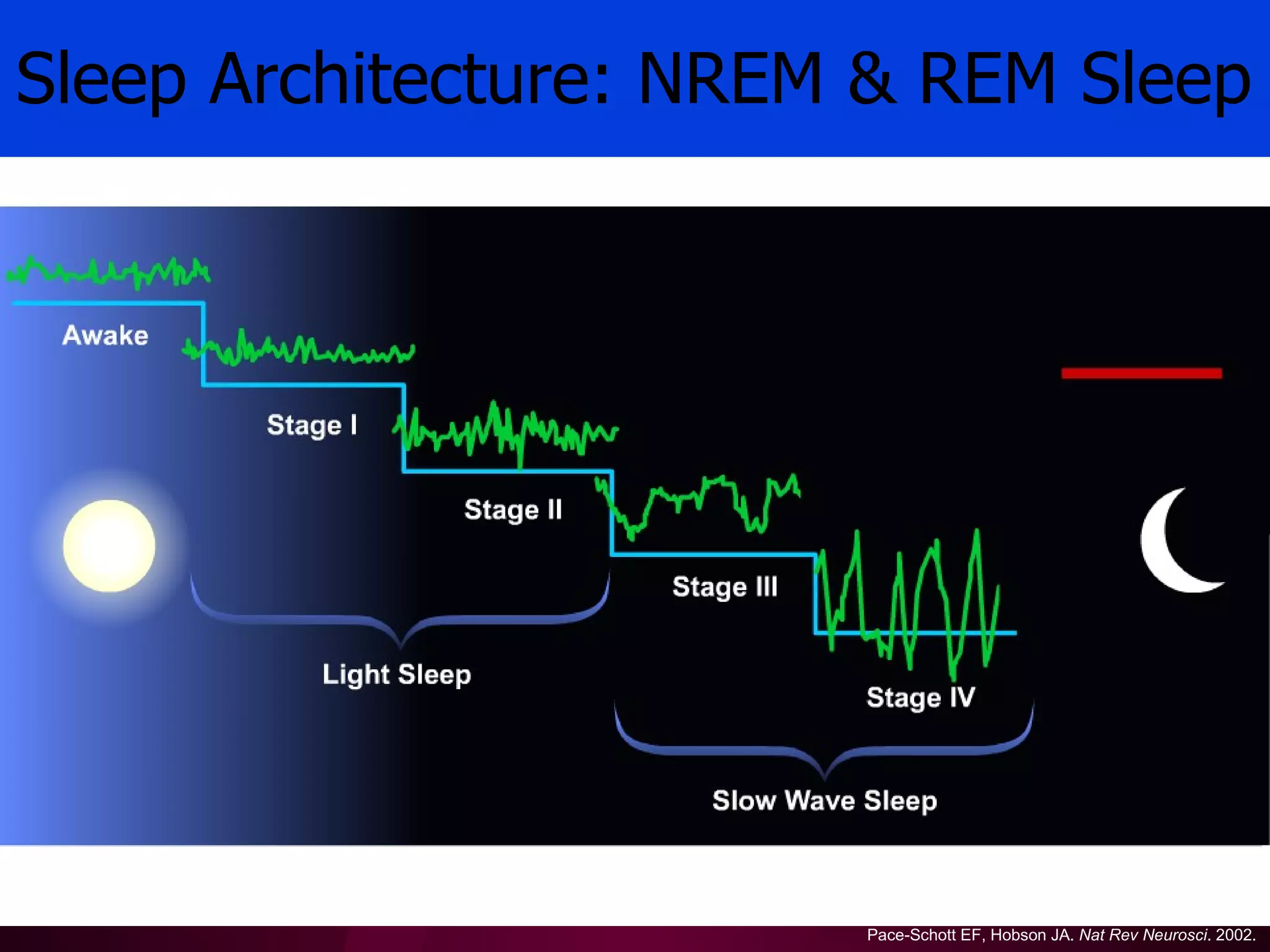
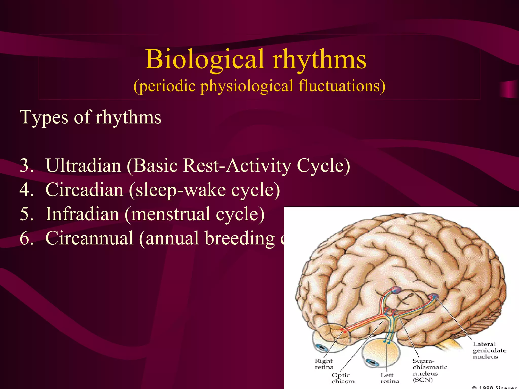
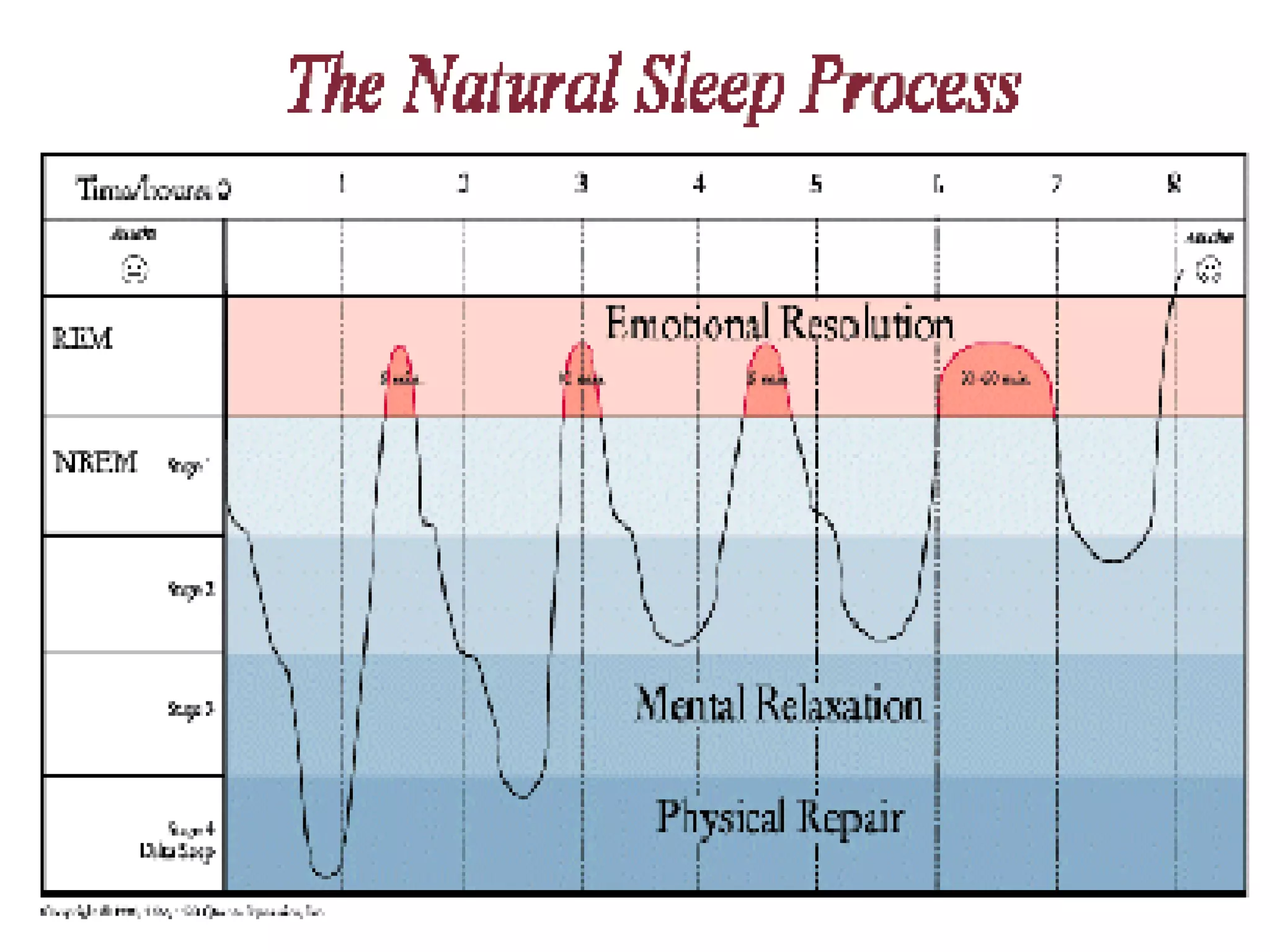
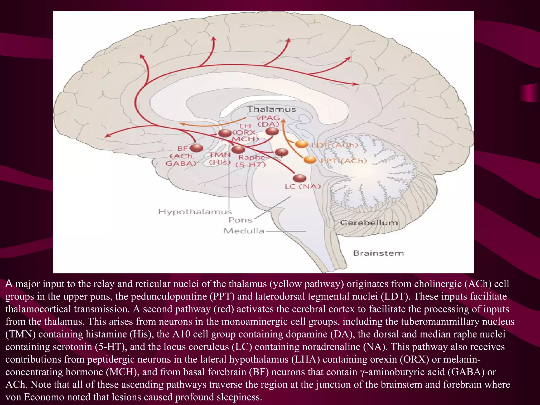
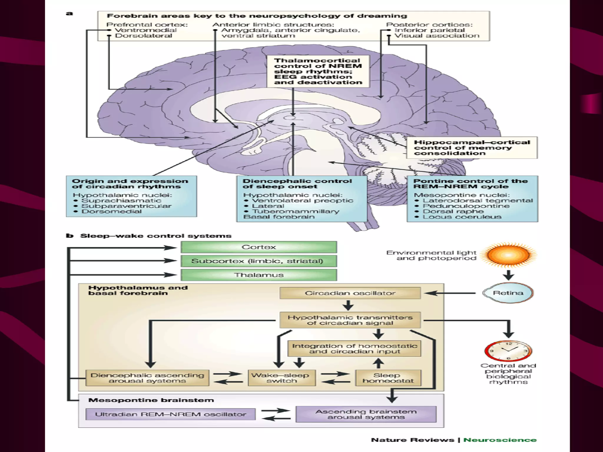
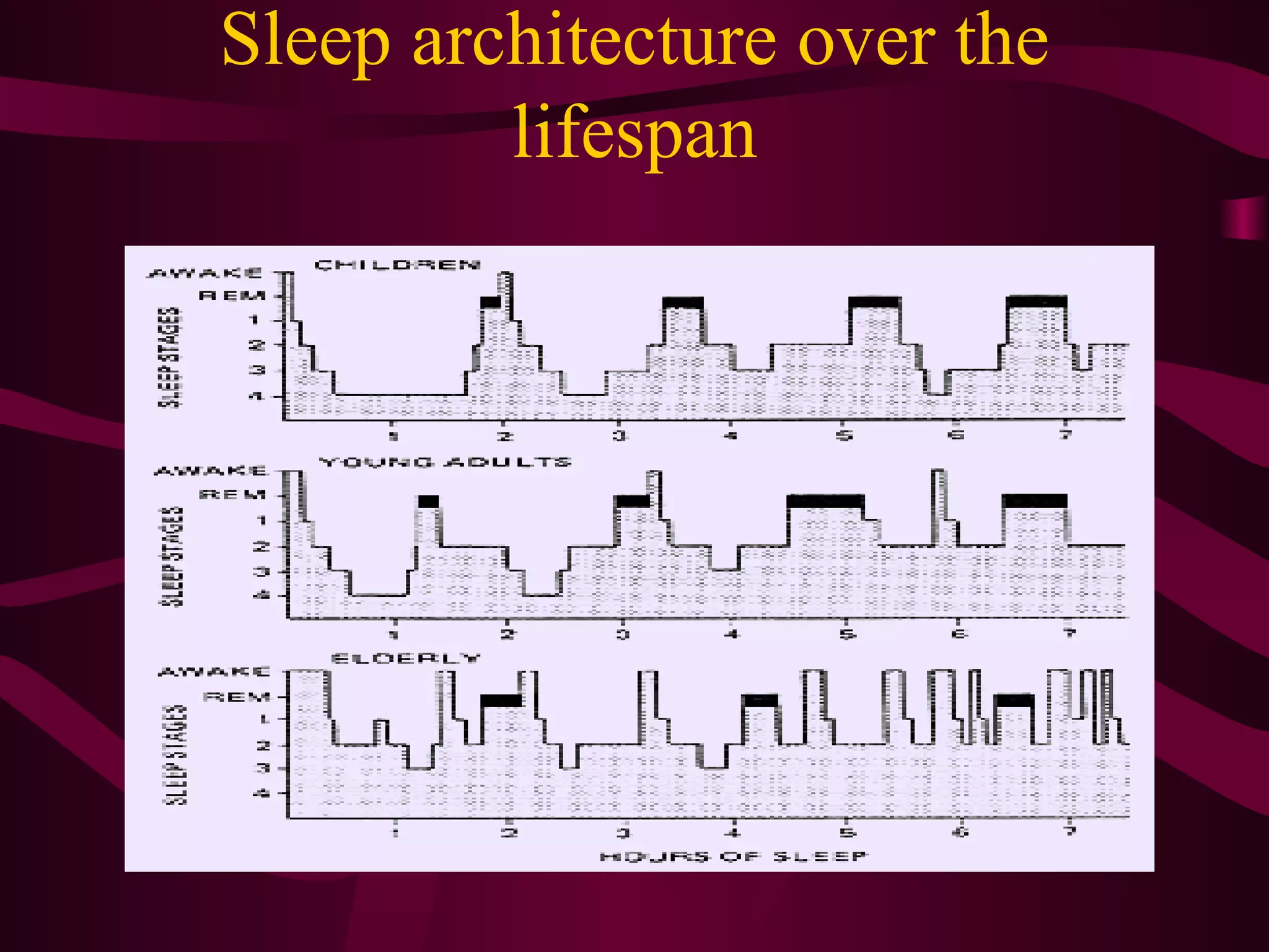
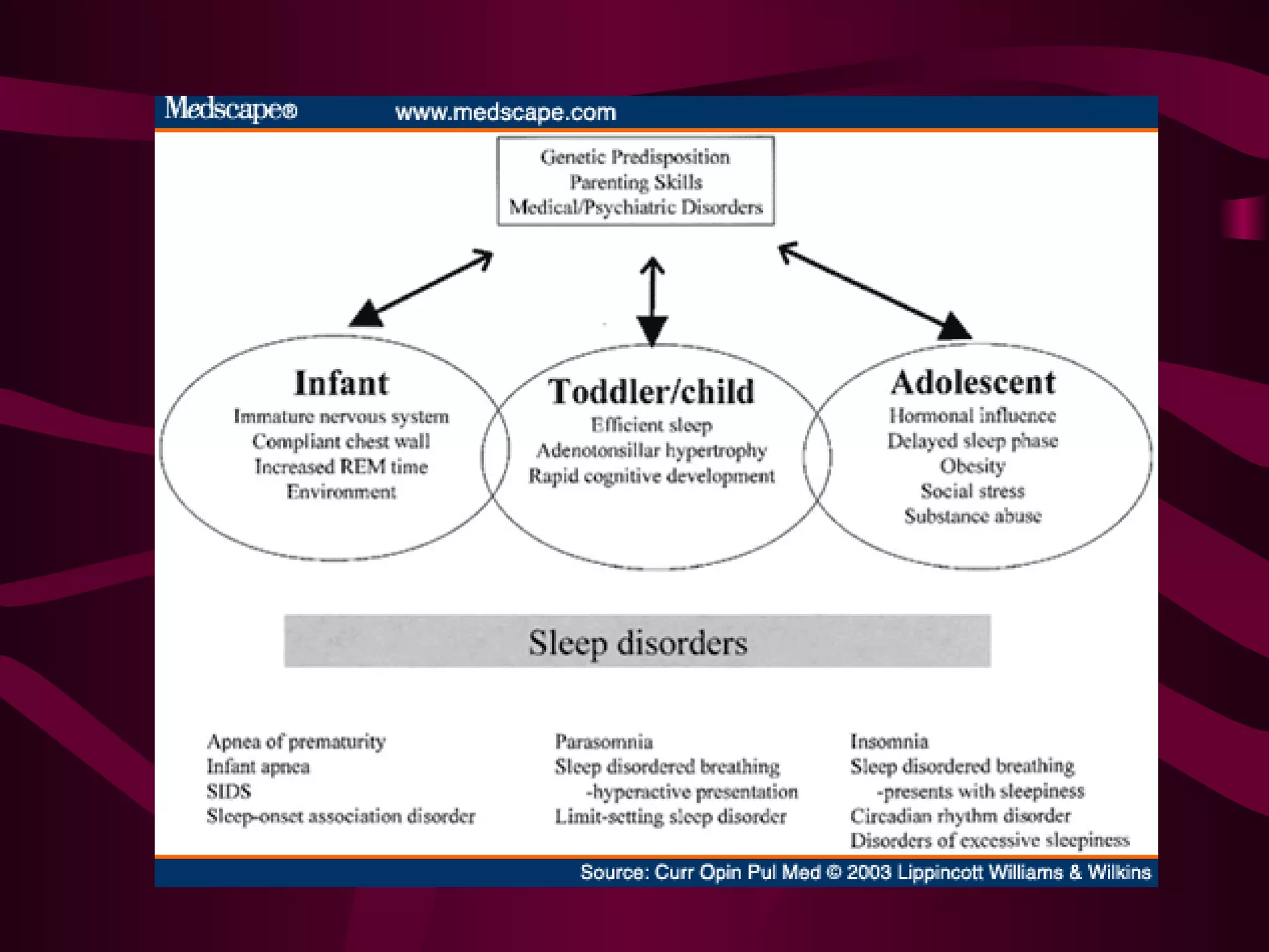
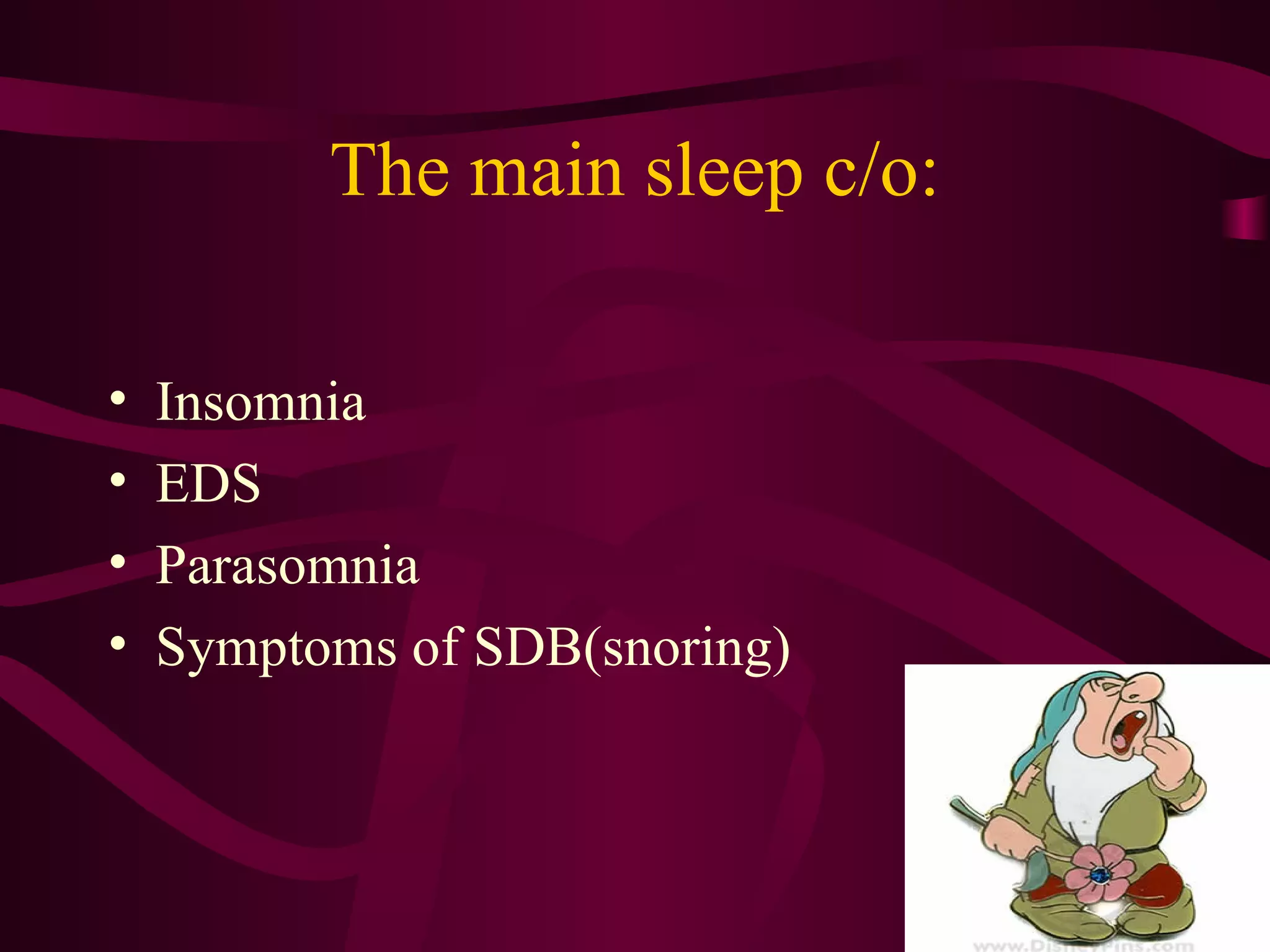
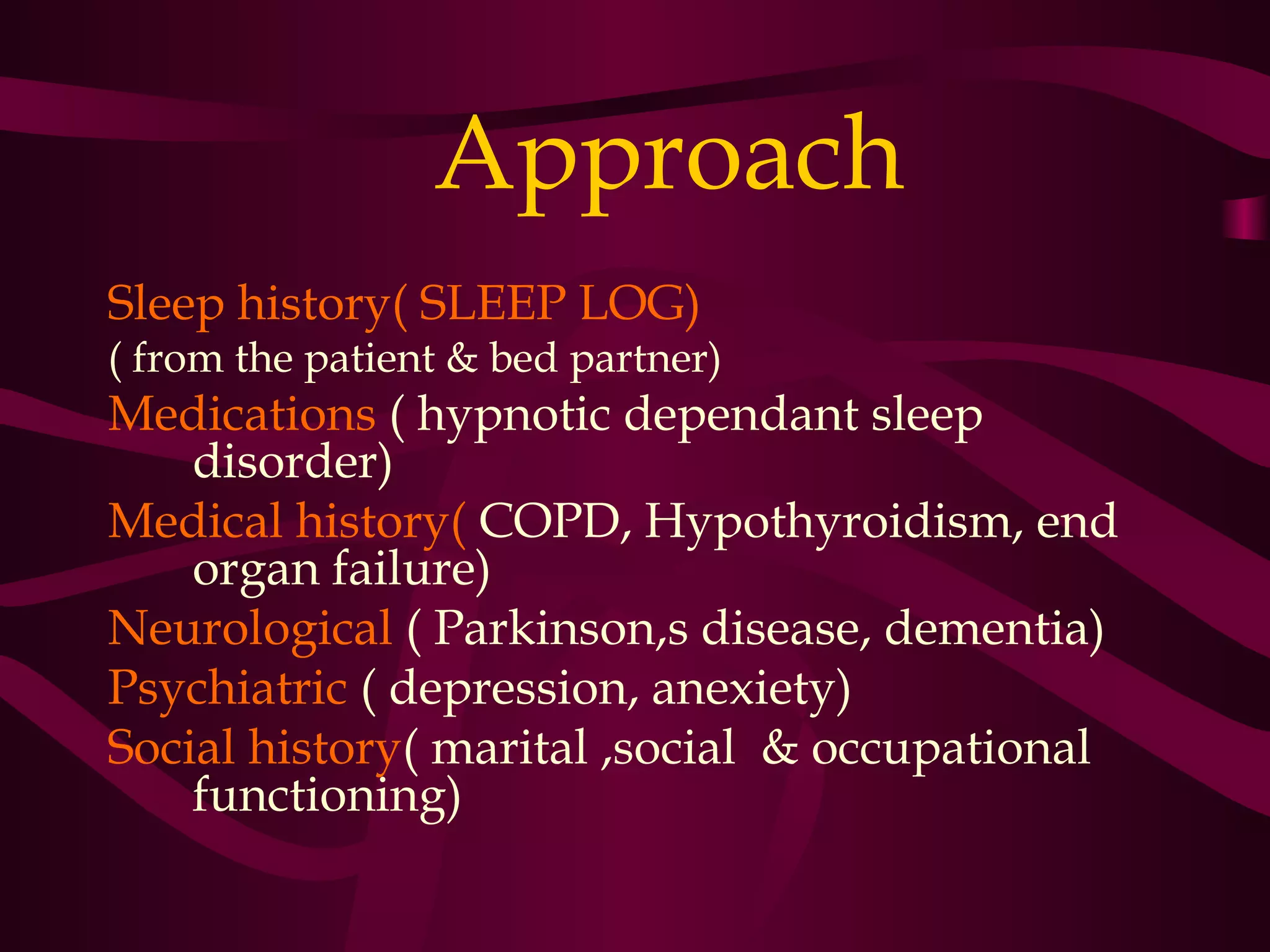
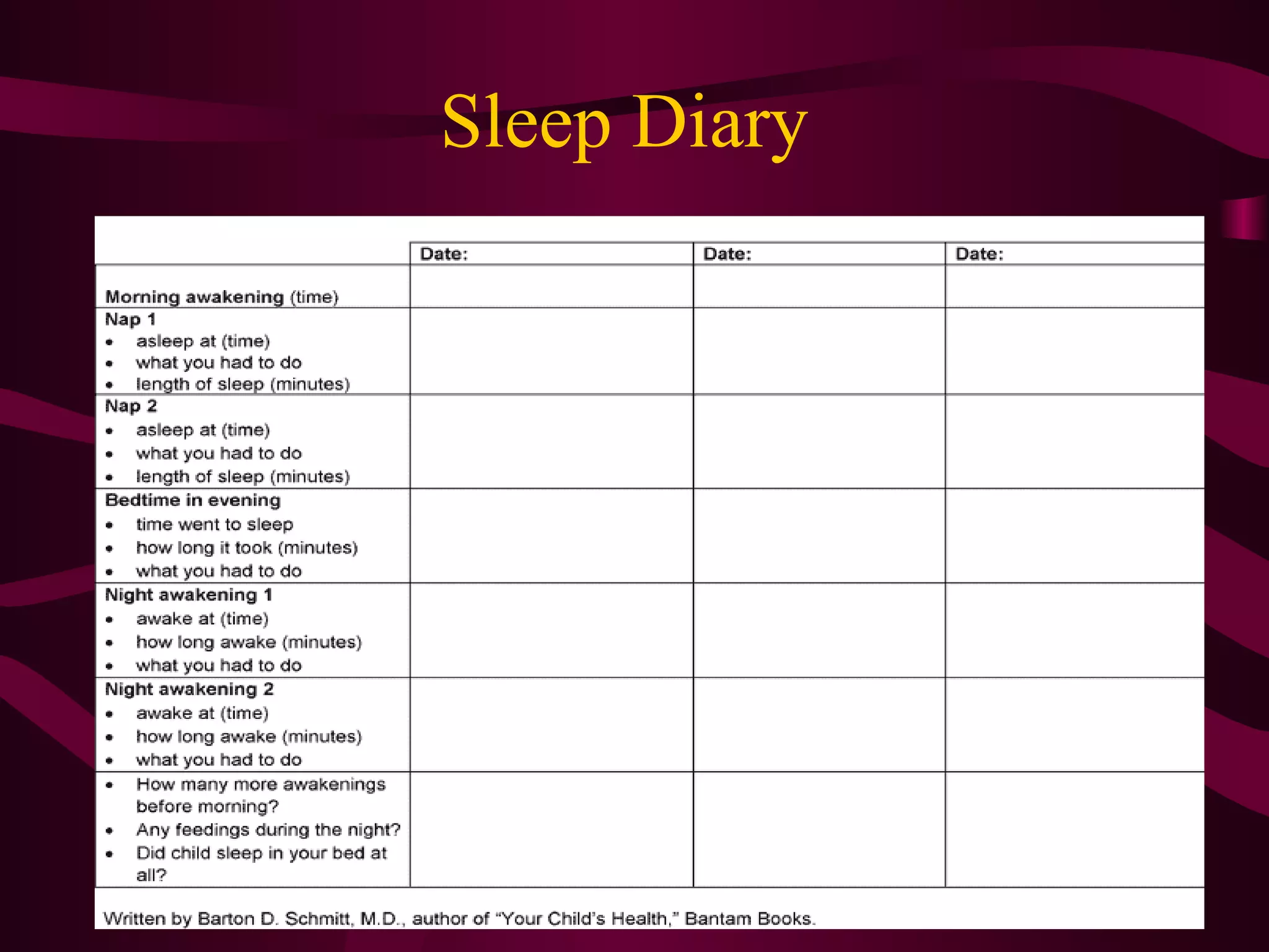
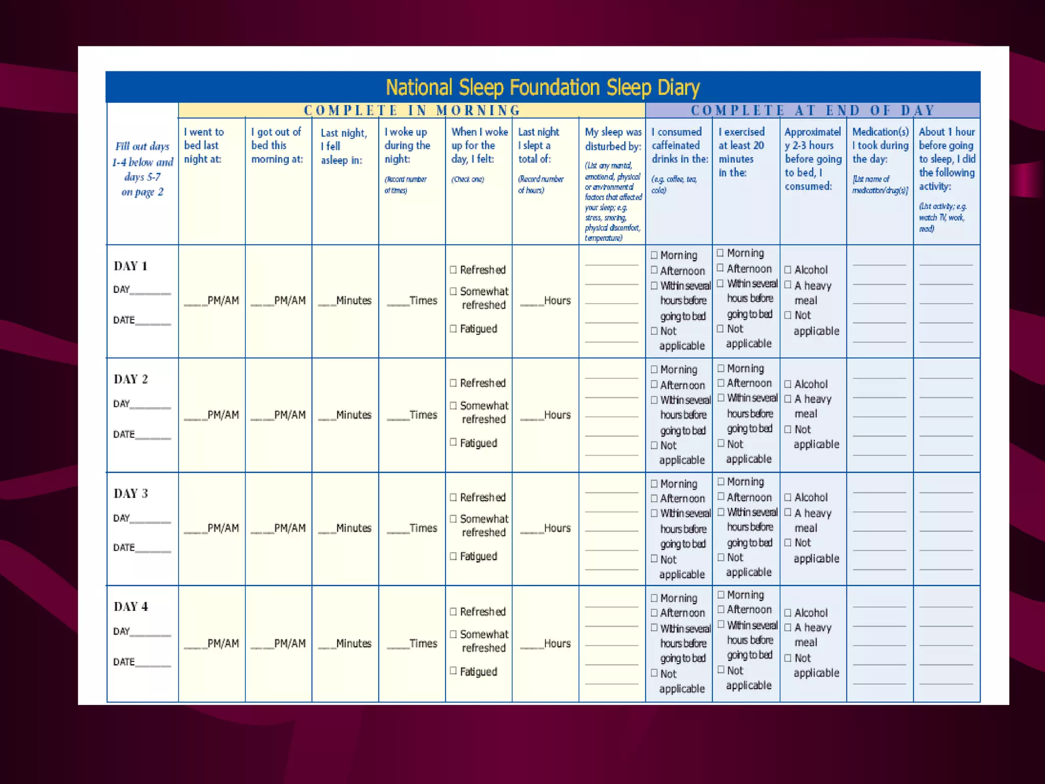
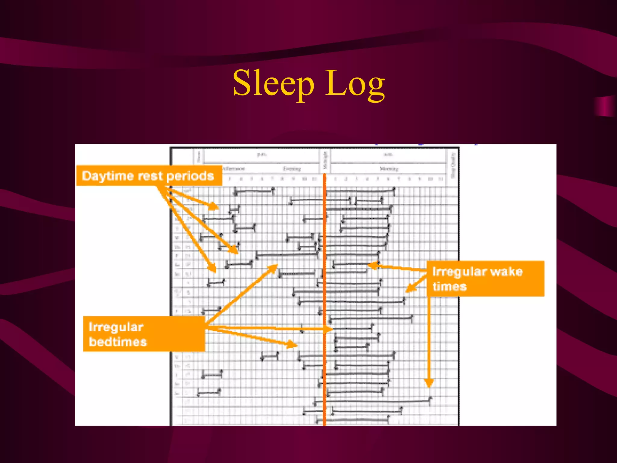
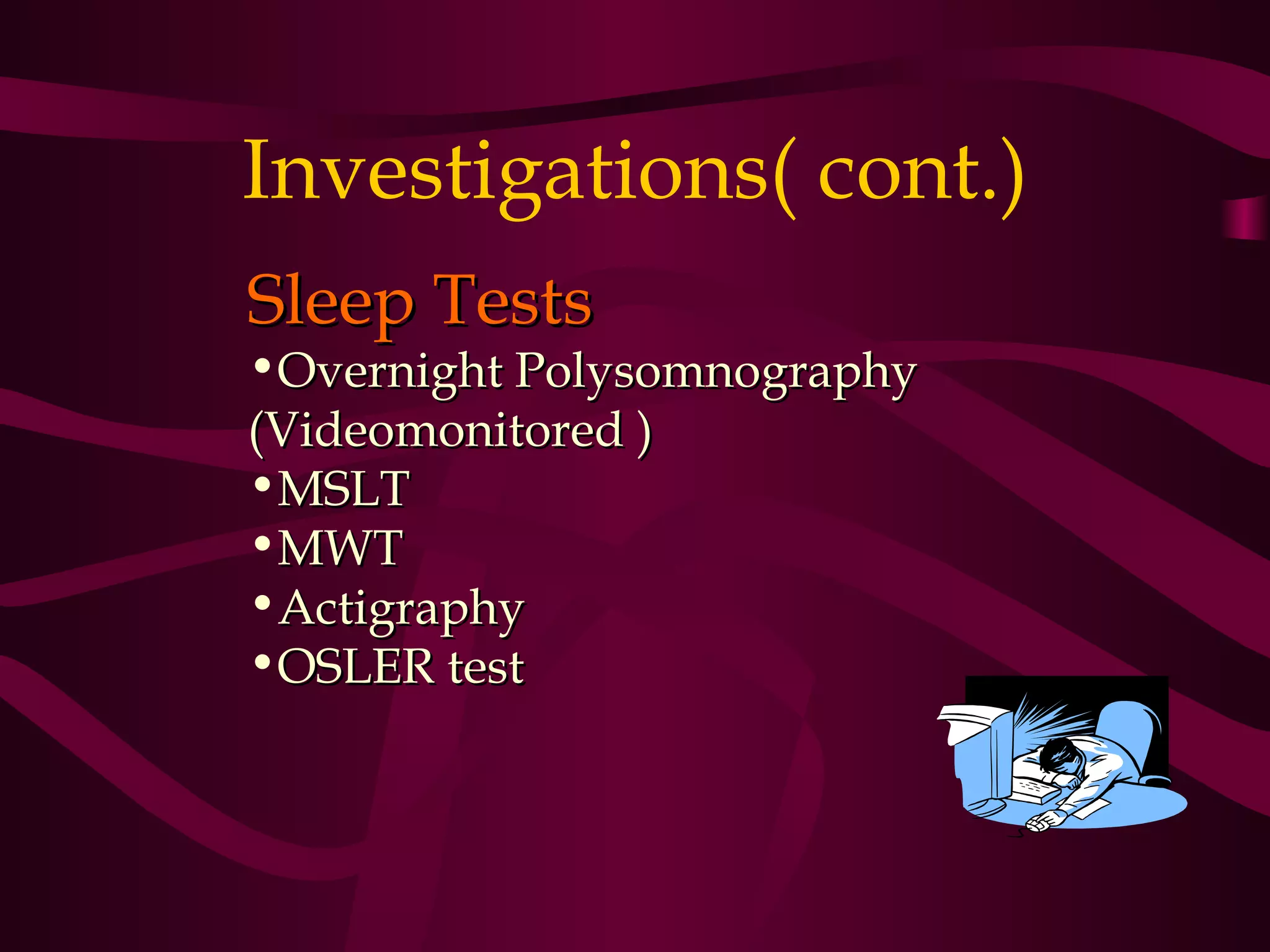
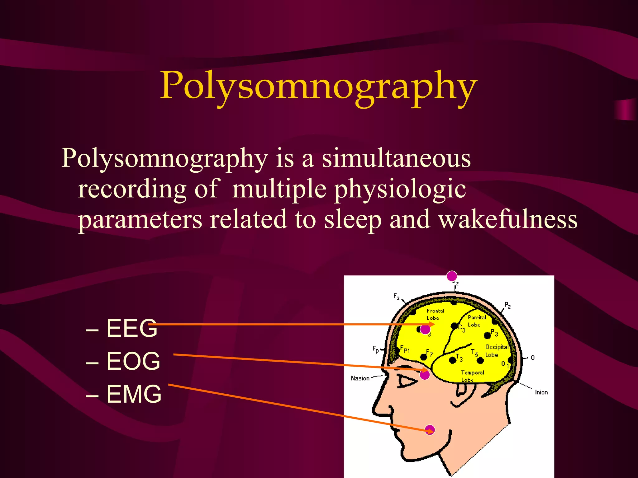
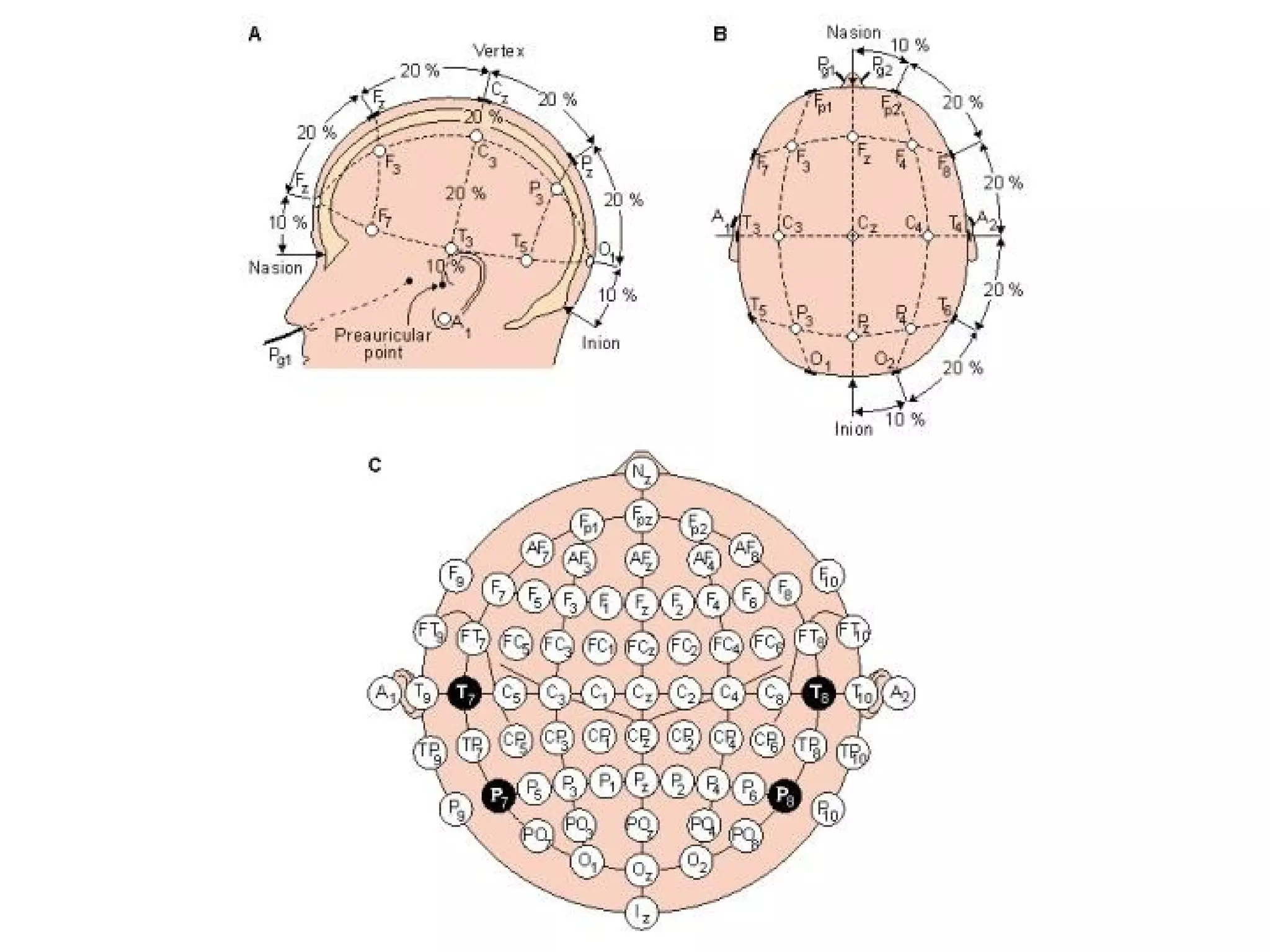
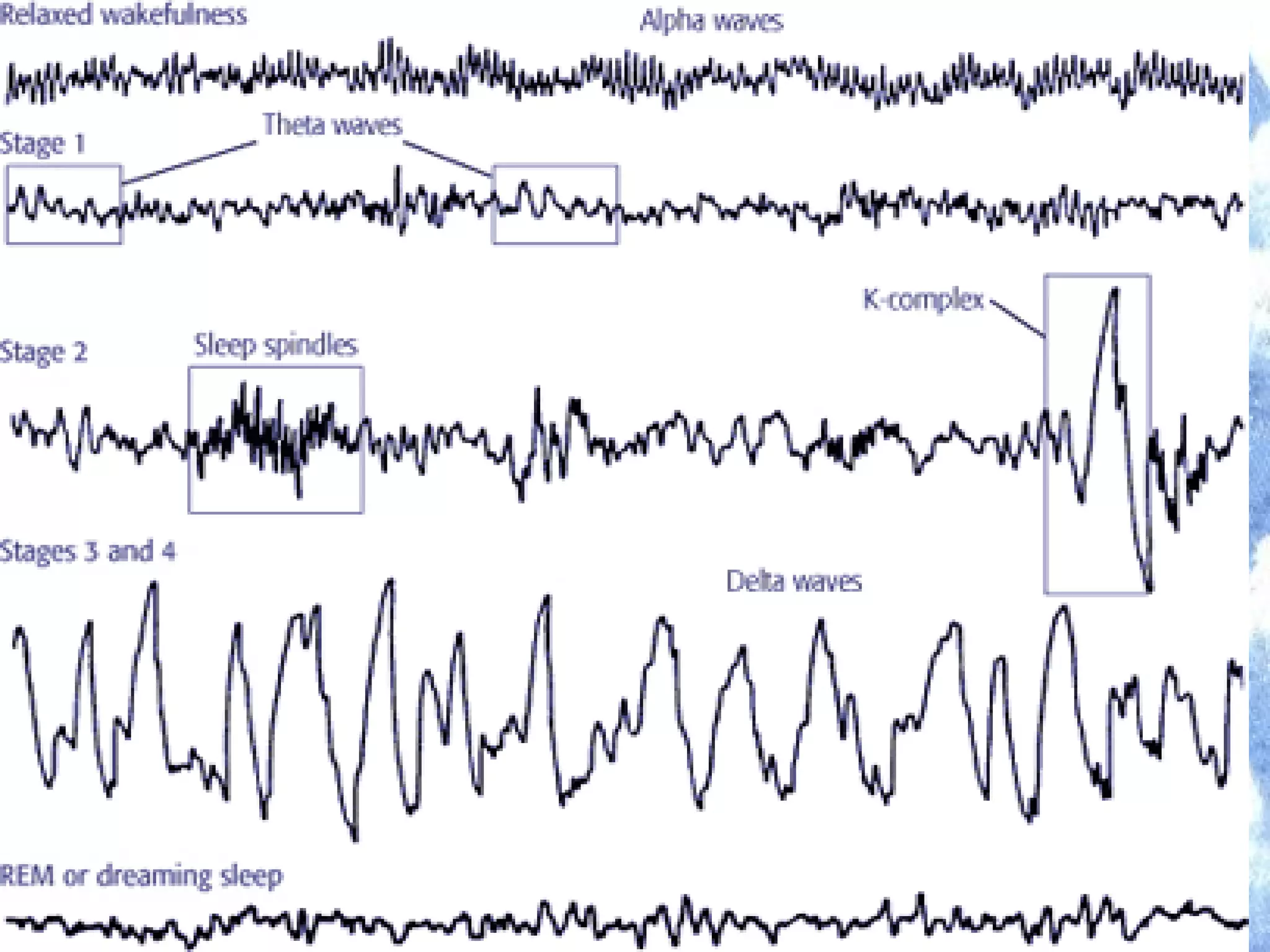
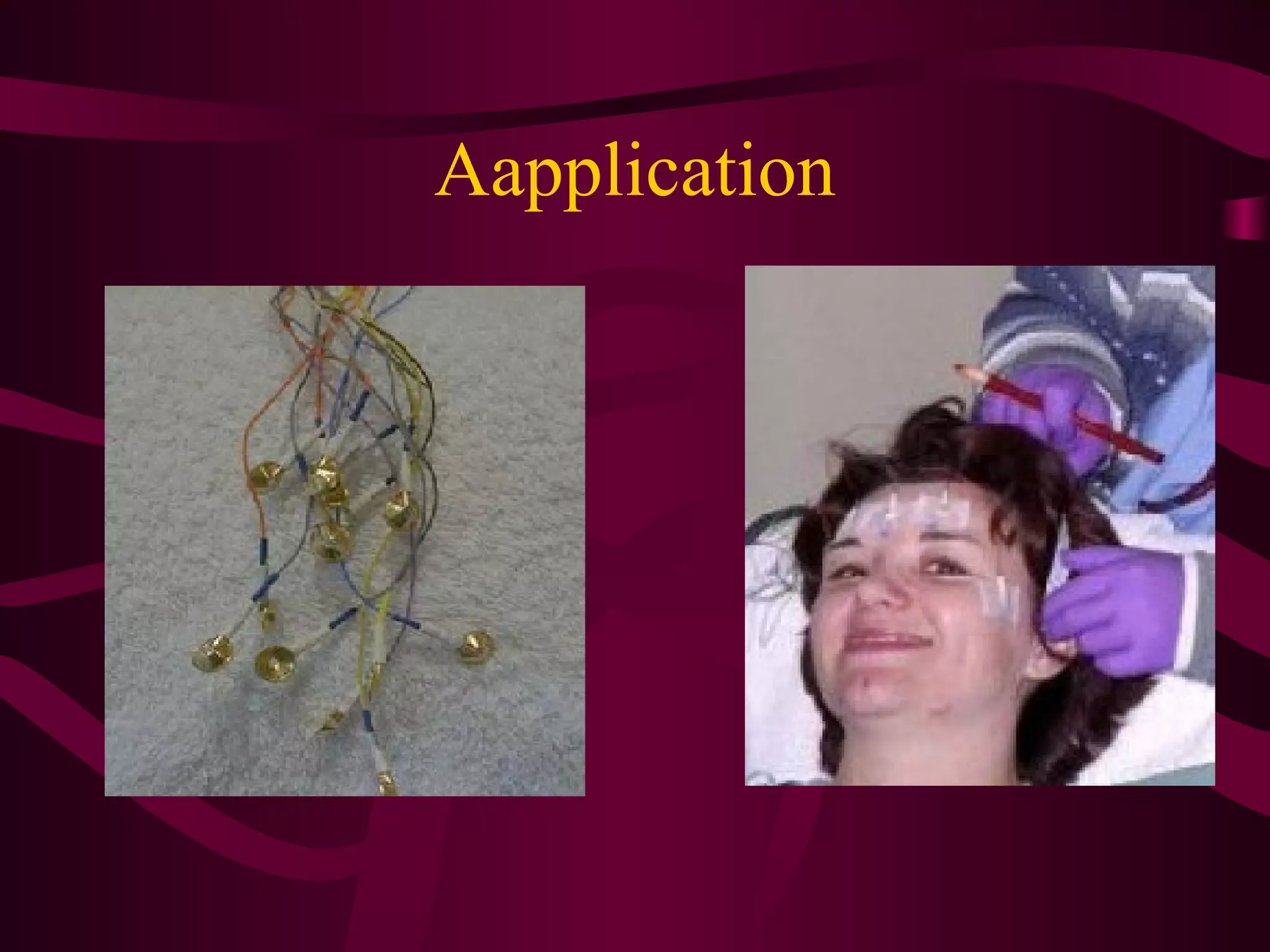
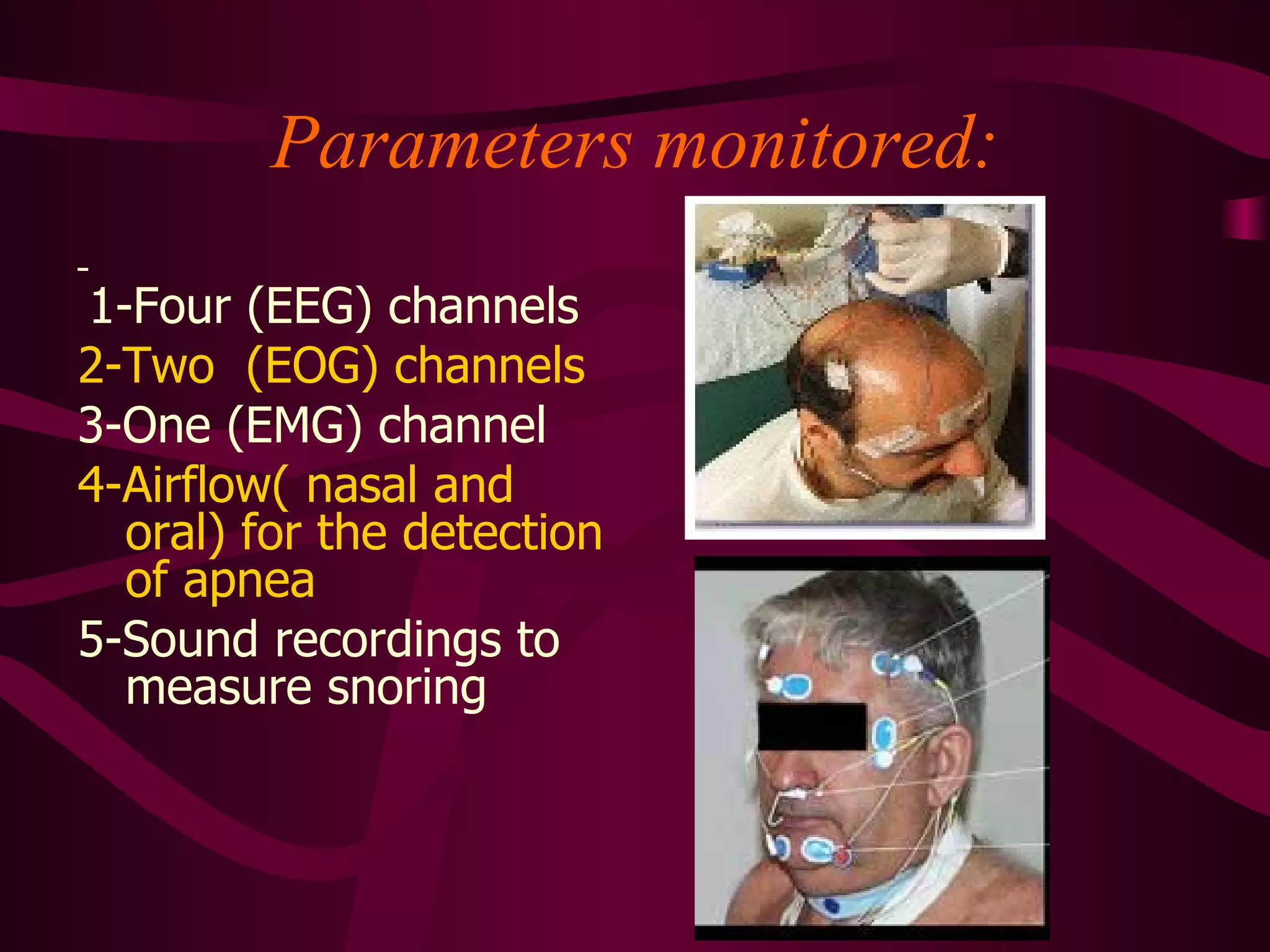
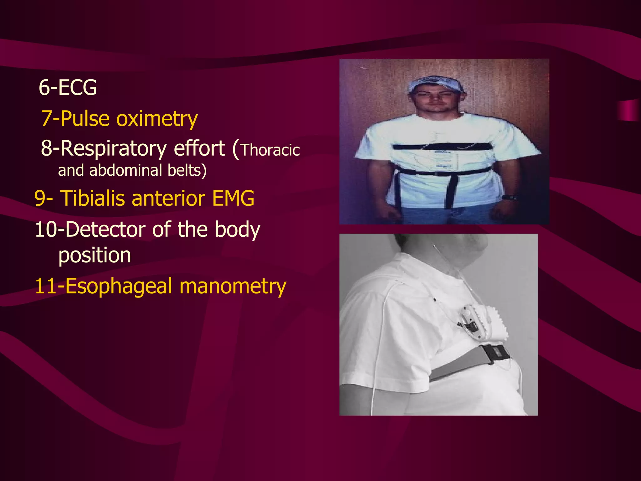
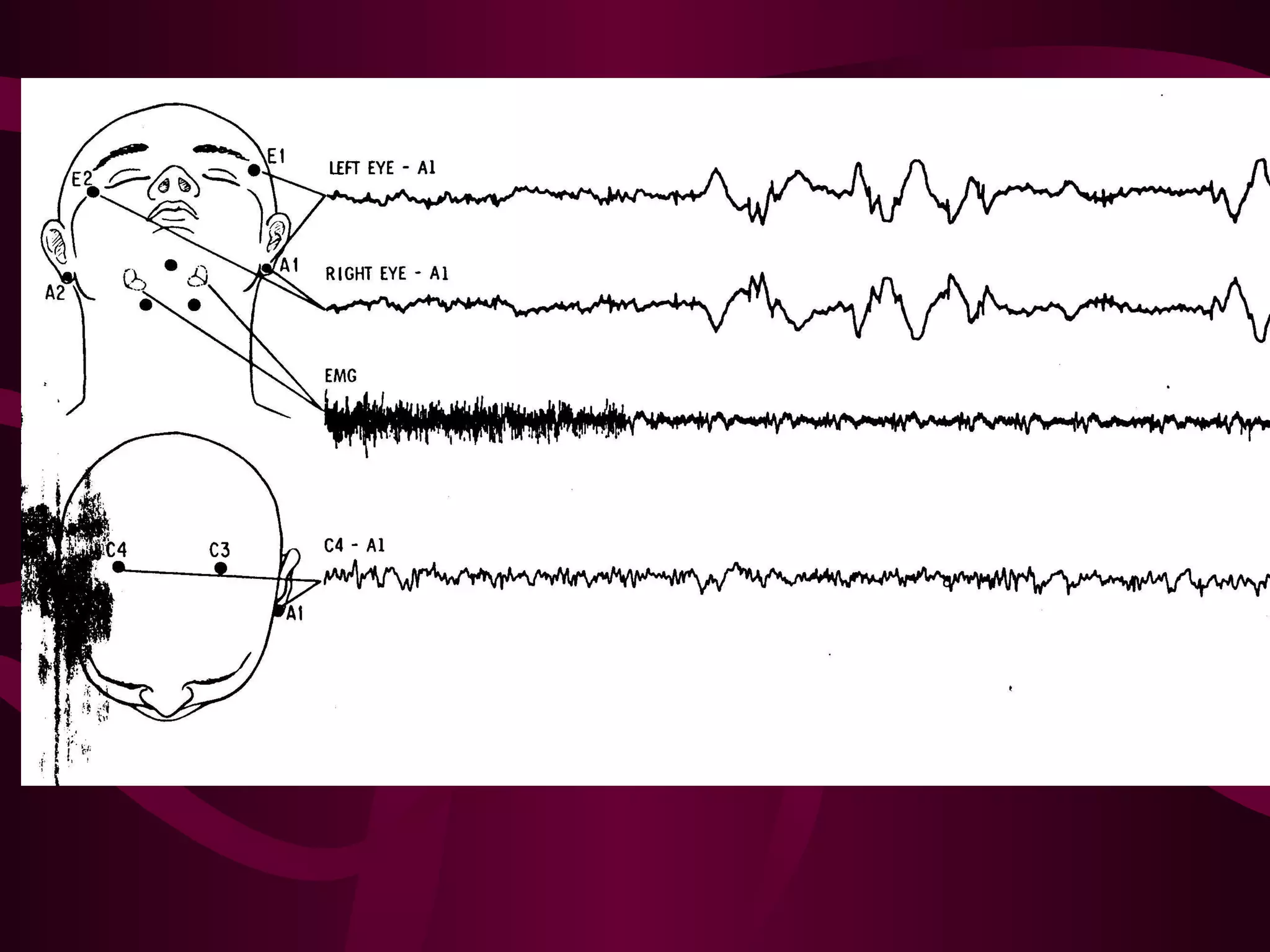
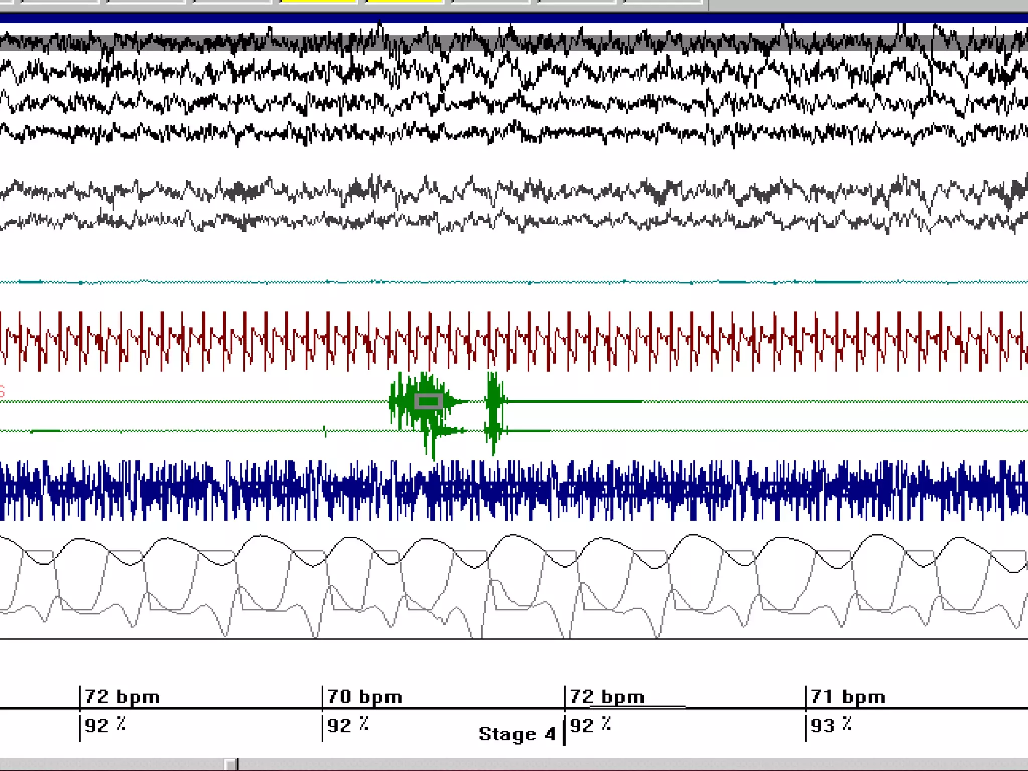
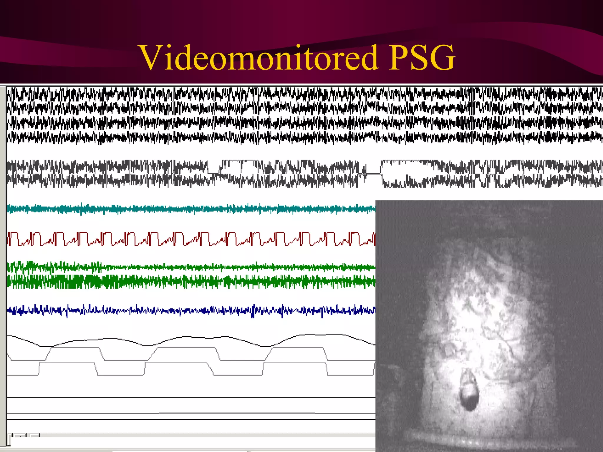
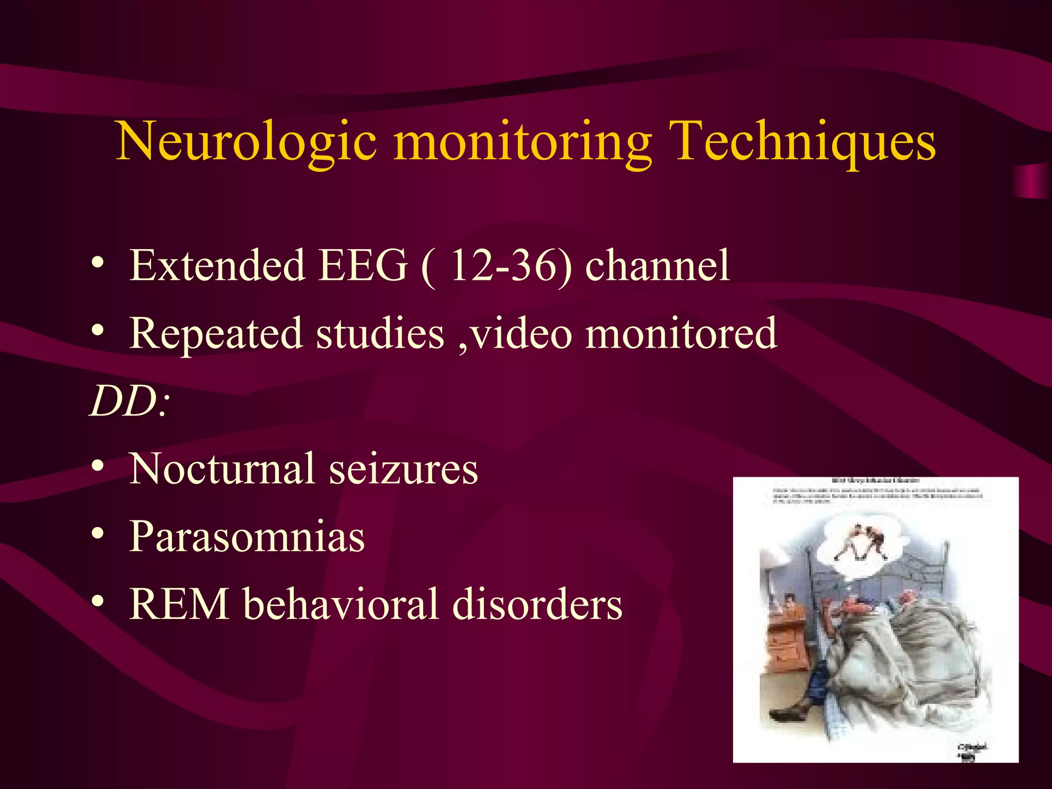
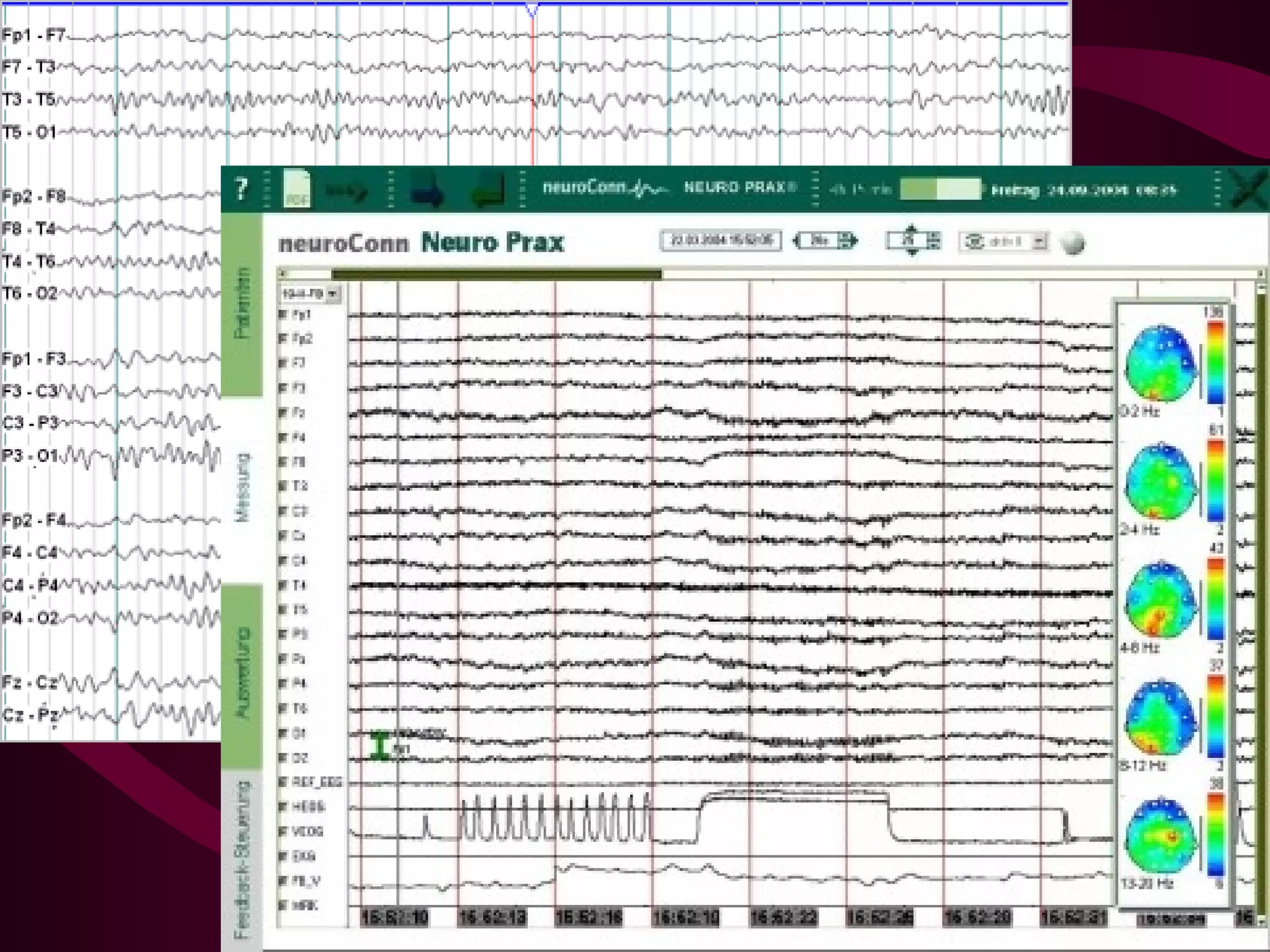
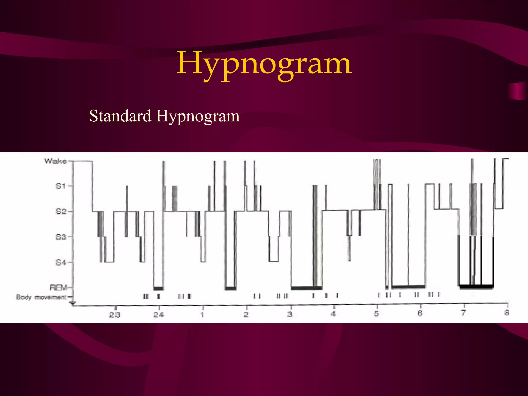
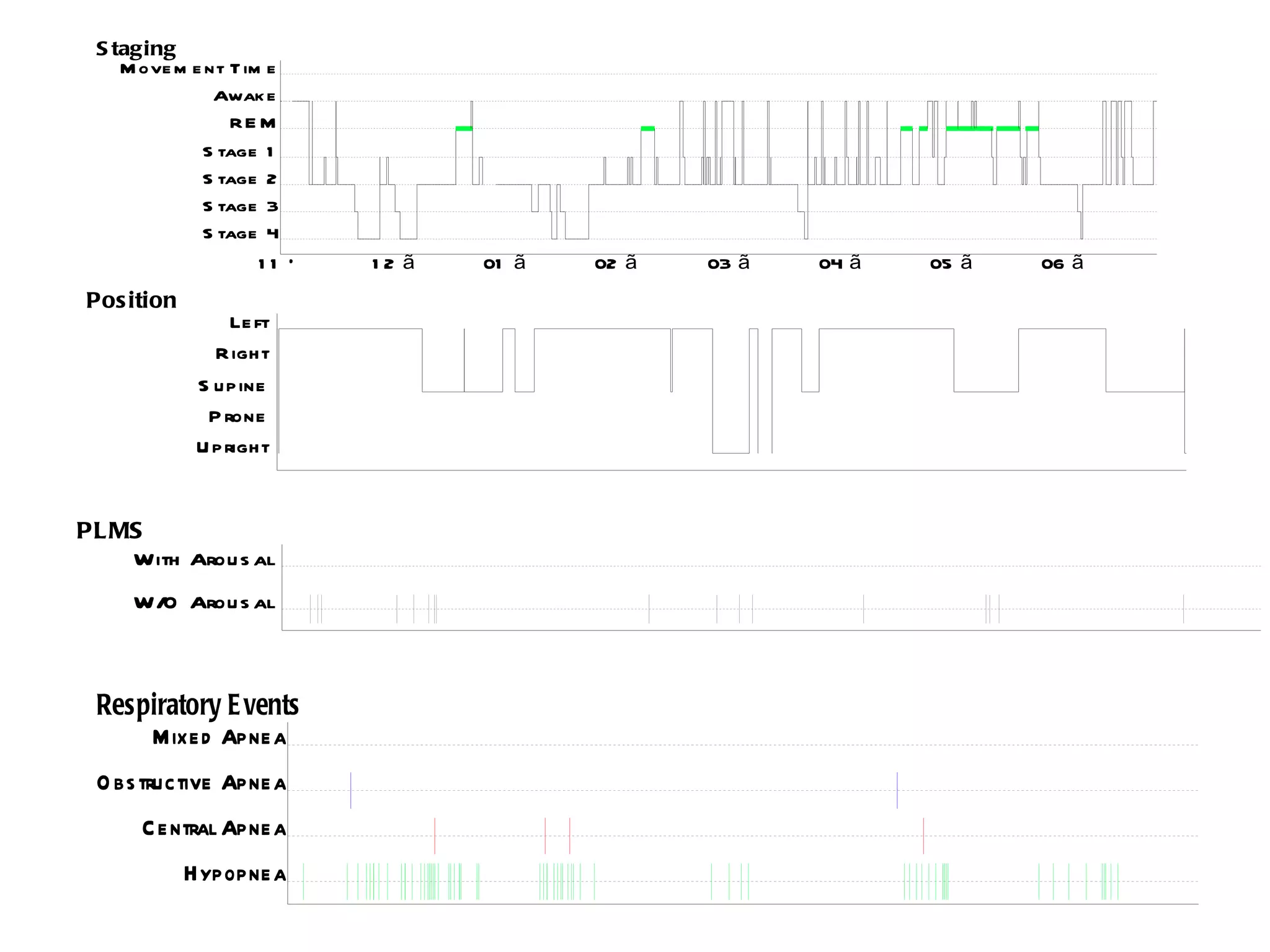
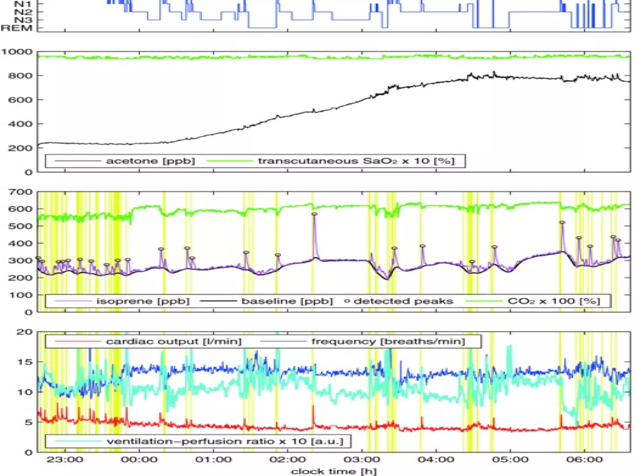
Investigations
  of sleep disorders
                    BY

           Dr.Jaidaa Mekky
    Lecturer of Neuropsychiatry
     Sleep Medicine Consultant
Member of the American Academy of Neurology
  Member of the American Academy of Sleep
                 Medicine
         Faculty of Medicine
        Alexandria University
Background
• One-half to one-third of life asleep
• Sleep medicine relatively new field
• Sleep is a co-morbidity in a long list of
  diseases
• It was mentioned in the holy Quran 9
  times, describing the sleep fnctions and
  stages
    ‫وقال إن أبقراط قال: إذا كان النوم فى المراض المزمنة يس ّب وجعً، فذلك‬
          ‫ا‬     ‫ب‬
   ‫. من علمات الموت‬
Milestones
• 1837 – Dickens – describes
  overweight/hypersomnolent boy in the
  Posthumous Papers of the Pickwick Club (term
  “pickwickian” used by Osler)
• 1875 – Caton – EEG in dogs
• 1928 – Berger – Human EEG alpha waves
• 1937 – Loomis – EEG Sleep stages
      described
Milestones
•   1953 – Aserinsky & Kleitman – REM sleep
•   1970s – Polysomnography
•   1972 – Guilleminault – coins term OSA
•   1990 – International Classification of Sleep
             Disorders
Sleep Physiology
• What is Sleep?
  – “a reversible behavioral state of perceptual
    disengagement from and unresponsiveness to
    the environment”
• 75% in Non-REM sleep
• 25% REM sleep – muscle atonia,
  autonomic   activation
Sleep Architecture: NREM & REM Sleep


                        Pace-Schott EF, Hobson JA. Nat Rev Neurosci. 2002.
Biological rhythms
               (periodic physiological fluctuations)
Types of rhythms

3.   Ultradian (Basic Rest-Activity Cycle)
4.   Circadian (sleep-wake cycle)
5.   Infradian (menstrual cycle)
6.   Circannual (annual breeding cycles)
A major input to the relay and reticular nuclei of the thalamus (yellow pathway) originates from cholinergic (ACh) cell
groups in the upper pons, the pedunculopontine (PPT) and laterodorsal tegmental nuclei (LDT). These inputs facilitate
thalamocortical transmission. A second pathway (red) activates the cerebral cortex to facilitate the processing of inputs
from the thalamus. This arises from neurons in the monoaminergic cell groups, including the tuberomammillary nucleus
(TMN) containing histamine (His), the A10 cell group containing dopamine (DA), the dorsal and median raphe nuclei
containing serotonin (5-HT), and the locus coeruleus (LC) containing noradrenaline (NA). This pathway also receives
contributions from peptidergic neurons in the lateral hypothalamus (LHA) containing orexin (ORX) or melanin-
concentrating hormone (MCH), and from basal forebrain (BF) neurons that contain γ-aminobutyric acid (GABA) or
ACh. Note that all of these ascending pathways traverse the region at the junction of the brainstem and forebrain where
von Economo noted that lesions caused profound sleepiness.
Sleep architecture over the
         lifespan
The main sleep c/o:

•   Insomnia
•   EDS
•   Parasomnia
•   Symptoms of SDB(snoring)
Approach
Sleep history( SLEEP LOG)
( from the patient & bed partner)
Medications ( hypnotic dependant sleep
    disorder)
Medical history( COPD, Hypothyroidism, end
    organ failure)
Neurological ( Parkinson,s disease, dementia)
Psychiatric ( depression, anexiety)
Social history( marital ,social & occupational
    functioning)
Sleep Diary
Sleep Log
Epworth sleepiness scale
Situation                                                         Chance of dozing

Sitting and reading . . . . . . . . . . . . . . . . . . . . . . . . . . . . . . . . . . . . . .
Watching TV . . . . . . . . . . . . . . . . . . . . . . . . . . . . . . . . . . . . . . . . . . .
Sitting, inactive in a public place (e.g. a theatre or a meeting) . . . . . . .
As a passenger in a car for an hour without a break . . . . . . . . . . . . . . .
Lying down to rest in the afternoon when circumstances permit . . . . .
Sitting and talking to someone . . . . . . . . . . . . . . . . . . . . . . . . . . . . . .
Sitting quietly after a lunch without alcohol . . . . . . . . . . . . . . . . . . . . .
In a car, while stopped for a few minutes in the traffic

Total . . . . . . . . . . . . . . . . . . . . . . . .


                  :Score

                  Normal range 0-10 
                  Borderline 10-12
                  pathological 12-24
Examination
• Body habitus( obesity, poor hygiene)
• Neck( circumference ,thyroid etc.)
• Craniofascial abnormalities (retrognathia,
  craniosynsitosis)
• Otolaryngeal examination( nasal mucosa,
  tongue ,uvula)
• Pulmonary & cardiac examination
• Neurological & Psychiatric assessment
Mallampati classification
Investigations
Laboratory Tests:
Thyroid function
Serum ferritin, hemoglobin
HLA typing(HLA DQB1*0602 )
Toxocological screening
Liver & kidney function
Investigations( cont.)
Neurologic Assessment:
Videomonitored EEG
Imaging Studies:
 CT& / MRI
Assessment of the upper airway

•   Fluoroscopy
•   Nasopharyngoscopy
•   Cephalometry
•   CT, Volumetric reconstruction
•   MRI
Investigations( cont.)
Sleep Tests
•Overnight Polysomnography
(Videomonitored )
•MSLT
•MWT
•Actigraphy
•OSLER test
Polysomnography
Polysomnography is a simultaneous
 recording of multiple physiologic
 parameters related to sleep and wakefulness


  – EEG
  – EOG
  – EMG
Aapplication
Parameters monitored:

1-Four (EEG) channels
2-Two  (EOG) channels 
3-One (EMG) channel
4-Airflow( nasal and 
  oral) for the detection 
  of apnea 
5-Sound recordings to 
  measure snoring

      
6-ECG
 7-Pulse oximetry 
 8-Respiratory effort (Thoracic 
  and abdominal belts)
9- Tibialis anterior EMG
10-Detector of the body 
   position 
11-Esophageal manometry
Videomonitored PSG
Neurologic monitoring Techniques

• Extended EEG ( 12-36) channel
• Repeated studies ,video monitored
DD:
• Nocturnal seizures
• Parasomnias
• REM behavioral disorders
Hypnogram
Standard Hypnogram
S taging
    M ove m e nt T im e
               Awak e
                 RE M
             S tage 1
             S tage 2
             S tage 3
             S tage 4
                    11 Õ   12 ã   01 ã   02 ã   03 ã   04 ã   05 ã   06 ã
Pos ition
                 Le ft
               R ight
             S upine
              P rone
             U pright


PL MS
    With Arou s al
     W/O Arou s al


 Respiratory E vents
     Mixe d Apne a
 O bs tructive Apne a
      C e ntral Apne a
            Hypopne a
The main data presented in PSG are:

• 1) Total sleep time, wake time, total recording time;
• 2) Sleep efficiency (total sleep time/total recording time);
• 3) Latency for sleep onset, latency for REM sleep and other sleep
  stages.
• 4) Duration (in minutes) and proportion of total-sleep-time sleep
  stages (5) Frequency of apneas and hypopneas per hour of sleep
• 6) Saturation values and events of oxyhemoglobin desaturation
• 7) Total number and index of periodic lower limb movements per hour
  of sleep.
• 8) Total number and index of micro-arousals per hour of sleep and
  their relationship with breathing events or lower limb movements;
• 9)Esophageal ph anormalities
• 10)Penile tumecence
Portable PSG
MSLT -MWT
• 4-6 naps with 2h interval
• Parameters monitored:
•   EEG(4channel)
•   EOG
•   Chin EMG
•   ECG
•   Respiratory flow (if needed)
•   Microphone (if needed)
GERD
Actigraphy
Actigraphy
• Cost efficient
• Records motor movements
• Aallows estimates for several days,
  avoiding the sampling error of NPSG
• It gives an idea about TST,SL, Nocturnal
  arousals
• It is superior to sleep log
Uses:
• Used in assessment of Insomnia
• Useful in children and old age
• Circadian rhythm disorders
• Epidemiologic sleep studies
Limitations:
• It is not standardized yet for diagnosing
  PLMS,SDB or RBD.
Osler test
The Oxford Sleep Resistance Test
Sleep tight

Inv sleep 2012

  • 2.
    Investigations ofsleep disorders BY Dr.Jaidaa Mekky Lecturer of Neuropsychiatry Sleep Medicine Consultant Member of the American Academy of Neurology Member of the American Academy of Sleep Medicine Faculty of Medicine Alexandria University
  • 3.
    Background • One-half toone-third of life asleep • Sleep medicine relatively new field • Sleep is a co-morbidity in a long list of diseases • It was mentioned in the holy Quran 9 times, describing the sleep fnctions and stages  ‫وقال إن أبقراط قال: إذا كان النوم فى المراض المزمنة يس ّب وجعً، فذلك‬ ‫ا‬ ‫ب‬ ‫. من علمات الموت‬
  • 4.
    Milestones • 1837 –Dickens – describes overweight/hypersomnolent boy in the Posthumous Papers of the Pickwick Club (term “pickwickian” used by Osler) • 1875 – Caton – EEG in dogs • 1928 – Berger – Human EEG alpha waves • 1937 – Loomis – EEG Sleep stages described
  • 5.
    Milestones • 1953 – Aserinsky & Kleitman – REM sleep • 1970s – Polysomnography • 1972 – Guilleminault – coins term OSA • 1990 – International Classification of Sleep Disorders
  • 6.
    Sleep Physiology • Whatis Sleep? – “a reversible behavioral state of perceptual disengagement from and unresponsiveness to the environment” • 75% in Non-REM sleep • 25% REM sleep – muscle atonia, autonomic activation
  • 7.
    Sleep Architecture: NREM & REM Sleep Pace-Schott EF, Hobson JA. Nat Rev Neurosci. 2002.
  • 8.
    Biological rhythms (periodic physiological fluctuations) Types of rhythms 3. Ultradian (Basic Rest-Activity Cycle) 4. Circadian (sleep-wake cycle) 5. Infradian (menstrual cycle) 6. Circannual (annual breeding cycles)
  • 10.
    A major inputto the relay and reticular nuclei of the thalamus (yellow pathway) originates from cholinergic (ACh) cell groups in the upper pons, the pedunculopontine (PPT) and laterodorsal tegmental nuclei (LDT). These inputs facilitate thalamocortical transmission. A second pathway (red) activates the cerebral cortex to facilitate the processing of inputs from the thalamus. This arises from neurons in the monoaminergic cell groups, including the tuberomammillary nucleus (TMN) containing histamine (His), the A10 cell group containing dopamine (DA), the dorsal and median raphe nuclei containing serotonin (5-HT), and the locus coeruleus (LC) containing noradrenaline (NA). This pathway also receives contributions from peptidergic neurons in the lateral hypothalamus (LHA) containing orexin (ORX) or melanin- concentrating hormone (MCH), and from basal forebrain (BF) neurons that contain γ-aminobutyric acid (GABA) or ACh. Note that all of these ascending pathways traverse the region at the junction of the brainstem and forebrain where von Economo noted that lesions caused profound sleepiness.
  • 12.
  • 14.
    The main sleepc/o: • Insomnia • EDS • Parasomnia • Symptoms of SDB(snoring)
  • 19.
    Approach Sleep history( SLEEPLOG) ( from the patient & bed partner) Medications ( hypnotic dependant sleep disorder) Medical history( COPD, Hypothyroidism, end organ failure) Neurological ( Parkinson,s disease, dementia) Psychiatric ( depression, anexiety) Social history( marital ,social & occupational functioning)
  • 20.
  • 22.
  • 23.
    Epworth sleepiness scale Situation Chance of dozing Sitting and reading . . . . . . . . . . . . . . . . . . . . . . . . . . . . . . . . . . . . . . Watching TV . . . . . . . . . . . . . . . . . . . . . . . . . . . . . . . . . . . . . . . . . . . Sitting, inactive in a public place (e.g. a theatre or a meeting) . . . . . . . As a passenger in a car for an hour without a break . . . . . . . . . . . . . . . Lying down to rest in the afternoon when circumstances permit . . . . . Sitting and talking to someone . . . . . . . . . . . . . . . . . . . . . . . . . . . . . . Sitting quietly after a lunch without alcohol . . . . . . . . . . . . . . . . . . . . . In a car, while stopped for a few minutes in the traffic Total . . . . . . . . . . . . . . . . . . . . . . . . :Score Normal range 0-10  Borderline 10-12 pathological 12-24
  • 24.
    Examination • Body habitus(obesity, poor hygiene) • Neck( circumference ,thyroid etc.) • Craniofascial abnormalities (retrognathia, craniosynsitosis) • Otolaryngeal examination( nasal mucosa, tongue ,uvula) • Pulmonary & cardiac examination • Neurological & Psychiatric assessment
  • 26.
  • 27.
    Investigations Laboratory Tests: Thyroid function Serumferritin, hemoglobin HLA typing(HLA DQB1*0602 ) Toxocological screening Liver & kidney function
  • 28.
  • 29.
    Assessment of theupper airway • Fluoroscopy • Nasopharyngoscopy • Cephalometry • CT, Volumetric reconstruction • MRI
  • 33.
    Investigations( cont.) Sleep Tests •OvernightPolysomnography (Videomonitored ) •MSLT •MWT •Actigraphy •OSLER test
  • 34.
    Polysomnography Polysomnography is asimultaneous recording of multiple physiologic parameters related to sleep and wakefulness – EEG – EOG – EMG
  • 41.
  • 42.
    Parameters monitored: 1-Four (EEG) channels 2-Two  (EOG) channels  3-One (EMG) channel 4-Airflow( nasal and  oral) for the detection  of apnea  5-Sound recordings to  measure snoring       
  • 43.
  • 46.
  • 47.
    Neurologic monitoring Techniques •Extended EEG ( 12-36) channel • Repeated studies ,video monitored DD: • Nocturnal seizures • Parasomnias • REM behavioral disorders
  • 49.
  • 50.
    S taging M ove m e nt T im e Awak e RE M S tage 1 S tage 2 S tage 3 S tage 4 11 Õ 12 ã 01 ã 02 ã 03 ã 04 ã 05 ã 06 ã Pos ition Le ft R ight S upine P rone U pright PL MS With Arou s al W/O Arou s al Respiratory E vents Mixe d Apne a O bs tructive Apne a C e ntral Apne a Hypopne a
  • 52.
    The main datapresented in PSG are: • 1) Total sleep time, wake time, total recording time; • 2) Sleep efficiency (total sleep time/total recording time); • 3) Latency for sleep onset, latency for REM sleep and other sleep stages. • 4) Duration (in minutes) and proportion of total-sleep-time sleep stages (5) Frequency of apneas and hypopneas per hour of sleep • 6) Saturation values and events of oxyhemoglobin desaturation • 7) Total number and index of periodic lower limb movements per hour of sleep. • 8) Total number and index of micro-arousals per hour of sleep and their relationship with breathing events or lower limb movements; • 9)Esophageal ph anormalities • 10)Penile tumecence
  • 53.
  • 54.
    MSLT -MWT • 4-6naps with 2h interval • Parameters monitored: • EEG(4channel) • EOG • Chin EMG • ECG • Respiratory flow (if needed) • Microphone (if needed)
  • 55.
  • 56.
  • 58.
    Actigraphy • Cost efficient •Records motor movements • Aallows estimates for several days, avoiding the sampling error of NPSG • It gives an idea about TST,SL, Nocturnal arousals • It is superior to sleep log
  • 59.
    Uses: • Used inassessment of Insomnia • Useful in children and old age • Circadian rhythm disorders • Epidemiologic sleep studies Limitations: • It is not standardized yet for diagnosing PLMS,SDB or RBD.
  • 60.
    Osler test The OxfordSleep Resistance Test
  • 61.

Editor's Notes

  • #8 NREM sleep is divided into 4 stages. Stages 1 and 2 are characterized by low arousal thresholds and are considered light sleep. Stages 3 and 4 are characterized by high arousal thresholds and are considered deep or “slow wave” sleep. All 4 of these stages can be differentiated by electroencephalography (EEG). Stage 1 is marked by low voltage, mixed frequency EEG. Stage 2 is marked by the presence of K complexes and sleep spindles on EEG recordings. Both stages 3 and 4 are marked by delta waves, which have a voltage of 75 microvolts or more and a frequency range of 0.5 to 4 hertz, but differ in composition. Stage 3 is defined as sleep consisting of 20-50% delta waves and stage 4 as sleep consisting of more than 50% delta waves. REM sleep is characterized by minimal movement, low muscle tone, activation of cortical activity, and rapid eye movements accompanied by vivid dreams. [Text: Comella, p. 18-B; Pace-Schott, p. 600. Figure: Pace-Schott, p. 600] Comella CL, Walters AS, Hening WA. Sleep and wakefulness. In: Goetz CG, Pappert EJ, eds. Textbook of Clinical Neurology . Philadelphia: WB Saunders Company; 1999:18-27. Pace-Schott EF, Hobson JA. The neurobiology of sleep: genetics, cellular physiology and subcortical networks. Nat Rev Neurosci . 2002;3:591-605. Scammell TE. The regulation of sleep and circadian rhythms. Sleep Med Alert. 2004;8:1-6.