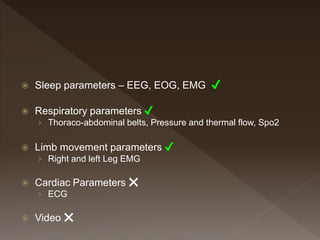
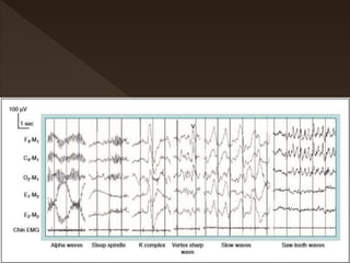
 Sleep parameters – EEG, EOG, EMG
 Respiratory parameters
› Thoraco-abdominal belts, Pressure and thermal flow, Spo2
 Limb movement parameters
› Leg EMG, Arm EMG
 Cardiac Parameters
› ECG
 Video
 Sleep parameters – EEG, EOG, EMG ✔
 Respiratory parameters ✔
› Thoraco-abdominal belts, Pressure and thermal flow, Spo2
 Limb movement parameters ✔
› Right and left Leg EMG
 Cardiac Parameters ❌
› ECG
 Video ❌
 R&K – 1968
 AASM – 2007
↺
 Sleep parameters – EEG, EOG, EMG ✔
 Respiratory parameters
› Thoraco-abdominal belts, Pressure and thermal
flow, Spo2
 Limb movement parameters
› Right and left Leg EMG
N1
2-5%
N2
45-55%
N3
20-25%
REM
20-25%
Duration of sleep Satages
N1
N2
N3
REM
First Half Second Half
 Scoring is according to AASM standards
 Sleep is evaluated in 30 second block called
Epoch
 Respiratory events are scored in 2 minutes or
in 5 Minutes
 Each 30 Sec Epoch is assigned the sleep
stage that occupies the greatest portion of the
epoch
 Measured in the number of “waves” or cycles
per second (cps) or hertz (Hz)
 Categories include
› Alpha : 8 – 13
› Theta : 4 – 7.9
› Delta :0.5 – 3.9
 Slow wave 0.5 – 2 amplitude of >75 µV,
› Beta : > 13
 Microsleep
 K complex
› K complex activity is greatest in frontal derivations (also central >
occipital). A K complex is said to be associated with an arousal if the
arousal commences no more than 1 second after the K complex.
 Sleep spindles
› are bursts of activity with a frequency range of 11 to 16 Hz
(usually 12–14) with a duration of 0.5 sec or greater
(usually 0.5–1.5 sec). The term spindle is used because
the shape of sleep spindle burst is often like that of a yarn
spindle
› Sleep spindles arise from thalamocortical oscillations. The
reticular nucleus of the thalamus is responsible for
generating sleep spindles
If there is uncertainty about whether activity is a burst of alpha
activity or a sleep spindle, one can display a 10-second window
(see Fig. 1–3) and actually count the deflections (waves) per
second.
 Score arousal during any stage of sleep if
there is an abrupt shift of EEG frequency
including alpha, theta, and/or frequencies >
16 Hz (but not spindles) that lasts at least 3
seconds, with at least 10 seconds of stable
sleep preceding the change.
 Subjects must be asleep, defined as > 10
continuous seconds of the indication of any
stage of sleep, before an EEG arousal can be
scored
 The EEG frequency shift must be 3 seconds
or greater in duration to be scored as an
arousal.
 A minimum of 10 continuous seconds of
intervening sleep is necessary to score a
second arousal
 Arousals are scored in REM sleep only when
accompanied by at least 1 second concurrent
increases in submental EMG amplitude.
 Arousals cannot be scored based on
changes in submental EMG amplitude alone.
 Respiratory scoring
 Apnea ✔
 Hypopnea ✔
 RERA / UARS ✔
 Hypoventilation
 Cheyne-Stokes breathing
Score a respiratory event as an apnea when BOTH of the
following criteria are met:
a. There is a drop in the peak signal excursion by ≥90% of pre-
event baseline using an oronasal thermal sensor (diagnostic
study), PAP device flow (titration study) or an alternative
apnea sensor (diagnostic study).
b. The duration of the ≥90% drop in sensor signal is ≥10
seconds.
 180 events – TST 6 Hrs
 180 events !!
 2 events at a time
1. Insomnia
2. Sleep-related breathing disorders
3. Central Disorders of hypersomnolence
4. Circadian rhythm sleep –wake disorders
5. Parasomnias
6. Sleep related movements disorders
7. Other sleep disorders
The International Classification of Sleep Disorders: Diagnostic and Coding Manual American Academy of Sleep Medicine. 2014;3rd ed Rev. ed. Darien IL
1. Insomnia
2. Sleep-related breathing disorders
3. Central Disorders of hypersomnolence
4. Circadian rhythm sleep –wake disorders
5. Parasomnias
6. Sleep related movements disorders
7. Other sleep disorders
 RLS
 PLMD
 Bruxism
The International Classification of Sleep Disorders: Diagnostic and Coding Manual American Academy of Sleep Medicine. 2014;3rd ed Rev. ed. Darien IL
 Leg EMG -AC amplifiers with surface electrodes
 Impedance less than 10 kilo-ohms (less than 5 kohms is preferred).
 The recommended low and high filter display settings are 10 hertz
(Hz) and 100 Hz,
 Use of a 60-Hz notch filter is not recommended.
 Move the left and right legs (wiggle toes) is part of the biocalibration
series.
 Separate EMG electrodes - long axis of the belly of the anterior
tibialis muscle
 2 to 3 cmH2O apart or one third the length of the anterior tibialis
muscle, whichever is shorter.
Yes It is a LM
Sleep Scoring New.pptx

Sleep Scoring New.pptx

  • 3.
     Sleep parameters– EEG, EOG, EMG  Respiratory parameters › Thoraco-abdominal belts, Pressure and thermal flow, Spo2  Limb movement parameters › Leg EMG, Arm EMG  Cardiac Parameters › ECG  Video
  • 4.
     Sleep parameters– EEG, EOG, EMG ✔  Respiratory parameters ✔ › Thoraco-abdominal belts, Pressure and thermal flow, Spo2  Limb movement parameters ✔ › Right and left Leg EMG  Cardiac Parameters ❌ › ECG  Video ❌
  • 6.
     R&K –1968  AASM – 2007
  • 7.
  • 8.
     Sleep parameters– EEG, EOG, EMG ✔  Respiratory parameters › Thoraco-abdominal belts, Pressure and thermal flow, Spo2  Limb movement parameters › Right and left Leg EMG
  • 9.
  • 12.
  • 15.
     Scoring isaccording to AASM standards  Sleep is evaluated in 30 second block called Epoch  Respiratory events are scored in 2 minutes or in 5 Minutes  Each 30 Sec Epoch is assigned the sleep stage that occupies the greatest portion of the epoch
  • 17.
     Measured inthe number of “waves” or cycles per second (cps) or hertz (Hz)  Categories include › Alpha : 8 – 13 › Theta : 4 – 7.9 › Delta :0.5 – 3.9  Slow wave 0.5 – 2 amplitude of >75 µV, › Beta : > 13
  • 24.
  • 34.
     K complex ›K complex activity is greatest in frontal derivations (also central > occipital). A K complex is said to be associated with an arousal if the arousal commences no more than 1 second after the K complex.
  • 36.
     Sleep spindles ›are bursts of activity with a frequency range of 11 to 16 Hz (usually 12–14) with a duration of 0.5 sec or greater (usually 0.5–1.5 sec). The term spindle is used because the shape of sleep spindle burst is often like that of a yarn spindle › Sleep spindles arise from thalamocortical oscillations. The reticular nucleus of the thalamus is responsible for generating sleep spindles
  • 37.
    If there isuncertainty about whether activity is a burst of alpha activity or a sleep spindle, one can display a 10-second window (see Fig. 1–3) and actually count the deflections (waves) per second.
  • 61.
     Score arousalduring any stage of sleep if there is an abrupt shift of EEG frequency including alpha, theta, and/or frequencies > 16 Hz (but not spindles) that lasts at least 3 seconds, with at least 10 seconds of stable sleep preceding the change.
  • 62.
     Subjects mustbe asleep, defined as > 10 continuous seconds of the indication of any stage of sleep, before an EEG arousal can be scored
  • 64.
     The EEGfrequency shift must be 3 seconds or greater in duration to be scored as an arousal.
  • 66.
     A minimumof 10 continuous seconds of intervening sleep is necessary to score a second arousal
  • 68.
     Arousals arescored in REM sleep only when accompanied by at least 1 second concurrent increases in submental EMG amplitude.
  • 70.
     Arousals cannotbe scored based on changes in submental EMG amplitude alone.
  • 73.
  • 79.
     Apnea ✔ Hypopnea ✔  RERA / UARS ✔  Hypoventilation  Cheyne-Stokes breathing
  • 81.
    Score a respiratoryevent as an apnea when BOTH of the following criteria are met: a. There is a drop in the peak signal excursion by ≥90% of pre- event baseline using an oronasal thermal sensor (diagnostic study), PAP device flow (titration study) or an alternative apnea sensor (diagnostic study). b. The duration of the ≥90% drop in sensor signal is ≥10 seconds.
  • 97.
     180 events– TST 6 Hrs
  • 98.
  • 99.
     2 eventsat a time
  • 100.
    1. Insomnia 2. Sleep-relatedbreathing disorders 3. Central Disorders of hypersomnolence 4. Circadian rhythm sleep –wake disorders 5. Parasomnias 6. Sleep related movements disorders 7. Other sleep disorders The International Classification of Sleep Disorders: Diagnostic and Coding Manual American Academy of Sleep Medicine. 2014;3rd ed Rev. ed. Darien IL
  • 101.
    1. Insomnia 2. Sleep-relatedbreathing disorders 3. Central Disorders of hypersomnolence 4. Circadian rhythm sleep –wake disorders 5. Parasomnias 6. Sleep related movements disorders 7. Other sleep disorders  RLS  PLMD  Bruxism The International Classification of Sleep Disorders: Diagnostic and Coding Manual American Academy of Sleep Medicine. 2014;3rd ed Rev. ed. Darien IL
  • 102.
     Leg EMG-AC amplifiers with surface electrodes  Impedance less than 10 kilo-ohms (less than 5 kohms is preferred).  The recommended low and high filter display settings are 10 hertz (Hz) and 100 Hz,  Use of a 60-Hz notch filter is not recommended.  Move the left and right legs (wiggle toes) is part of the biocalibration series.  Separate EMG electrodes - long axis of the belly of the anterior tibialis muscle  2 to 3 cmH2O apart or one third the length of the anterior tibialis muscle, whichever is shorter.
  • 107.

Editor's Notes

  • #78 Normal Respiration during sleep: See that flow in the thermistor and cannula is regular, rhythmic, and has equal amplitude across the epoch. Chest and abdomen RIP belts show in phase movement and effort sum (RIP-Sum) does not show any change in amplitude. Oxygen saturation is maintained throughout the epoch and microphone does not show any signal.