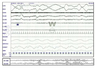
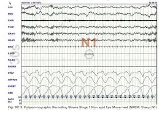
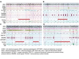
BASIC OF POLYSOMNOGRAPHY
Dr. Shaitan Singh
Senior Resident Neurology
POLYSOMNOGRAPHY
• Introduction
• Indications
• Patient evaluation and instructions
• Components
• Derived Information
• Types
• Limitation
• Summary
INTRODUCTION
• Term “Polysomnography” was proposed by Holland ,Dement and Raynal
(1974).
• The most commonly used test in the diagnosis of obstructive sleep apnea
syndrome (OSAS).
INDICATIONS FOR POLYSOMNOGRAPHY
• Sleep related breathing disorders
• CPAP titration in OSA
• Assessment of treatment results- OSA
• Sleep related behavioural disorders
• Narcolepsy
• Paroxysmal arousal or seizure phenomenon
• Periodic Leg Movements of Sleep
Initial Assessment
• Detailed medical & psychiatric history
• Medication, smoking, alcohol, activities on that day
RECORDING ROOM
• The study patient and the monitoring apparatus and technologist are
housed in adjacent rooms.
• Recording room should be as homely and comfortable as possible.
• It should be sound-proofed and air-conditioned.
• Intercom system to interact with the patient.
INSTRUCTION TO THE PATIENT
• Reporting time: 1 hour prior to the usual time of sleep.
• A prior visit to familiarize with the lab is useful.
• Patient should have had a relaxed day, without daytime sleep.
• Head wash, adequate food is recommended.
• Abstain from caffeine in the afternoon and evening of the day on
which PSG is planned
• Avoid alcohol on the day of PSG
• Avoid strenuous exercise on the day of the PSG.
INSTRUCTION TO THE PATIENT
Contiue..
• Continue their usual medications on the night of the PSG, including
sleep aids.
• The medications should be recorded by the technician so that the
results can be optimally interpreted.
• For patients who have a history of insomnia, especially when sleeping
in a new environment, Zolpidem may be prescribed.
• Avoid stimulants, including medications for narcolepsy.
• Avoid naps on the day of the sleep study.
Parameters to be reported in Polysomnography
• Berry RB, Brooks R, Gamaldo CE, et al.; for the American Academy of Sleep Medicine. The AASM Manual for the Scoring of Sleep and Associated Events: Rules, Terminology and
Technical Specifications. Version 2.6. Darien, IL: American Academy of Sleep Medicine; 2020.
ELECTROENCEPHALOGRAPHY (EEG)
• To distinguish between wakefulness and the various stages of sleep
• A minimum of three channels representing the right frontal, central,
and occipital electrodes referenced to the contra-lateral mastoid
electrode is recommended (2007 AASM Manual for the Scoring of
Sleep and Associated Events)
EEG Electrode Placement – International 10-20
system
Recommended:
a. F4-M1
b. C4-M1
c. O2-M1
Backup:
a. F3-M2
b. C3-M2
c. O1-M2
Berry RB, Brooks R, Gamaldo CE, et al.; for the American Academy of Sleep Medicine. The AASM Manual for the Scoring of Sleep and
Associated Events: Rules, Terminology and Technical Specifications. Version 2.6. Darien, IL: American Academy of Sleep Medicine; 2020.
Electroencephalographic Activity during
Wakefulness and Sleep
EEG wave patterns are used to differentiate wake and sleep states and
classify sleep stages:
(1) Beta activity
(2) Alpha activity
(3) Theta activity
(4) Delta activity
(5) Vertex sharp waves
(6) Sleep spindles
(7) K complex
ELECTRO-OCULOGRAPHY (EOG)
• Two recommended electrodes are labelled E1 (1 cm below the
left outer canthus) and E2 (placed 1 cm above the right outer
canthus), both referenced to the right mastoid.
• This allows simultaneous recording of both vertical eye
movements (such as blinking) and horizontal eye movements
(both slow and rapid).
• Documents the onset of rapid eye movement (REM) sleep, and
notes the presence of slow-rolling eye movements that usually
accompany the onset of sleep.
EOG Electrode Placement
Recommended: E1-M2 and E2-M2
Acceptable: E1-Fpz and E2-Fpz
Berry RB, Brooks R, Gamaldo CE, et al.; for the American Academy of Sleep Medicine. The AASM Manual for the Scoring of Sleep and
Associated Events: Rules, Terminology and Technical Specifications. Version 2.6. Darien, IL: American Academy of Sleep Medicine; 2020.
Eye Movements Pattern Definitions
• Eye blinks: Conjugate vertical eye movements at a frequency of 0.5–2 Hz present in
wakefulness with the eyes open or closed.
• Reading eye movements: Trains of conjugate eye movements consisting of a slow
phase followed by a rapid phase in the opposite direction as the subject reads.
• Slow eye movements: Conjugate, fairly regular, sinusoidal eye movements with an
initial deflection lasting > 500 msec.
• Rapid eye movements (REMs): Conjugate, irregular, sharply peaked eye movements
with an initial deflection usually lasting < 500 msec. Whereas rapid eye movements are
characteristic of stage R sleep, they may also be seen in wakefulness with eyes open (as
patients look around the room)
ELECTROMYOGRAPHY (EMG)
• Help to determine sleep stage
• Help to diagnose and classify a variety of parasomnia
• Minimum components are chin EMG channels recording activity from the
mentalis and submental muscles (the mylohyoid and anterior belly of the
digastric).
• bilateral leg EMG channels recording activity from the tibialis anterior
muscles.
EMG MONITORING continue..
Utility of limb EMG:
• Periodic limb movements in sleep.
• Restless leg syndrome.
• Other movement disorders.
• To document the hand and arm gestures of REM sleep behaviour
disorder.
• Record convulsive movements during nocturnal epileptic seizures.
RESPIRATORY MONITORING
• Upper airway airflow
• Thoraco-abdominal movement.
• Snoring Monitors.
• Indirect Arterial Blood Gas Monitoring.
• Endoesophageal (intrathoracic) pressure recording.
UPPER AIRWAY AIRFLOW
• Oronasal thermal devices (thermistors or thermocouples) or nasal
cannula–pressure transducers.
• Thermistor/ thermocouple: placed between the nose and mouth is
commonly used to monitor airflow by detecting changes in temperature.
UPPER AIRWAY AIRFLOW
• Nasal prongs connected to a pressure transducer detect inspiratory flow and
may be the most accurate method to identify subtle inspiratory flow limitation –
hypopneas
• An important limitation of nasal pressure transducers is that they cannot detect
mouth breathing. To overcome this limitation, a thermistor is usually added.
• Nasal pressure transducers are necessary for the diagnosis of hypopneas and
thermistors are necessary for the diagnosis of apnea.
THORACOABDOMINAL MOVEMENTS
Respiratory Inductive Plethysmography:
•Measures changes in thoracoabdominal cross-sectional
areas, and the sum of these two compartments is
proportional to airflow.
•Sensors are two wire coils, one placed around the chest
and the other around the abdomen.
•A change in mean cross-sectional coil area produces a
proportional variation in coil inductance, which is
converted into a voltage change by a variable frequency
oscillator.
SNORING MONITORS
• Although snoring can be monitored by placing a miniature microphone
(acoustic sensor, piezoelectric sensor ) on the patient’s neck, there is no
accepted grading system to quantify the intensity of this parameter.
INTRATHORACIC PRESSURE MONITORING
Endoesophageal pressure probe:
• Most sensitive detection of heightened respiratory effort.
• An endoesophageal tube is passed nasally till the probe is
about 5 cm above the esophageal-gastric junction.
• Measures increases in intrathoracic negative pressure to
overcome increased upper airway resistance.
• Gold standard for measuring respiratory effort
• Not a routine practice , because of patient discomfort and the
technical skill required
CAPNOGRAPHY
• Capnography, or end-tidal CO2 (ETCO2), monitoring detects the
expired carbon dioxide (CO2) level, which closely approximates intra-
alveolar CO2.
• An infrared analyzer over the nose and mouth detects CO2 in the
expired air, which qualitatively measures the airflow.
• Costly and therefore not used in most laboratories
ABG
• An alternative to capnography is measurement of the (Paco2) in the
morning after their sleep study to be compared to their waking
Paco2.
• Adults who have an increase in their paco2 in sleep by 10 mm hg or
more compared to an awake supine paco2 have sleep related
hypoventilation
OXYGEN SATURATION
• Continuous oxygen saturation monitoring by finger pulse oximetry is
routine .
• PSG reports mention the time the patient spent with an SpO2 below
90%.
Respiratory Parameters to be Reported
Occurrence of snoring - OPTIONAL
Berry RB, Brooks R, Gamaldo CE, et al.; for the American Academy of Sleep Medicine. The AASM Manual for the Scoring of Sleep and
Associated Events: Rules, Terminology and Technical Specifications. Version 2.6. Darien, IL: American Academy of Sleep Medicine; 2020.
Respiratory Rules – Respiratory Effort
For monitoring respiratory effort, use one of the following :
• Esophageal manometry (GOLD STANDARD) (RECOMMENDED)
• Dual thoracoabdominal RIP belts (COMMON) (RECOMMENDED)
A standard polysomnographic montage
CHIN = chin electromyogram; EEG = electroencephalogram; EFFORT = chest and abdomen movement;
EKG = electrocardiogram; EOG = electrooculogram; HR: heart rate; LEG = right and left anterior tibialis
electromyogram; O2 SAT = oxygen saturation; PAP PT = pressure transducer measured by positive airway
pressure device; SNORE: snore signal; TCCO2 = transcutaneous carbon dioxide.
Periodic Limb Movement Disorder
1.Repetitive, often stereotyped movements.
2.Usually noted in legs and consisting of extension of great toe, dorsiflexion
of ankle, and flexion of knee and hip; sometimes seen in arms.
3.Periodic or quasi-periodic at an average interval of 20–40sec(range,5–90
sec) with a duration of 0.5–10 sec and as part of at least 4 consecutive
movements.
4. Prevalence increases with age.
5.Seen in at least 80% of patients with restless legs syndrome.
6.Occurs predominantly during NREM Sleep.
The duration of the leg movement must be 0.5 to 10 seconds. The period length for two consecutive
movements to be considered a periodic limb movement must be at least 10 seconds and no more than 90
seconds
ELECTROCARDIOGRAM(ECG)
• ECG abnormalities in sleep apnea patients:
1. Marked sinus arrhythmia.
3. Prolonged asystolic episodes.
4. Atrial or ventricular fibrillation.
5. Nocturnal angina may show ST segment deviation.
Cardiac Events
BODY POSITION
• Some patients only have abnormalities when sleeping in certain positions.
Therefore, body position (eg, supine, left lateral, right lateral, prone) is
monitored throughout the test using a position sensor and/or video
monitor.
TYPES OF PSG
INTERPRETATION OF PSG
• Total sleep time – The total sleep time (TST) is the total duration of light
sleep (stages N1 and N2), deep sleep (stage N3), and rapid eye
movement (REM) sleep
• Sleep efficiency – Sleep efficiency (SE) is the TST divided by the total
recording time (ie, the time in bed).
• Sleep stage percentage – The sleep stage percentage (SSP) for a
particular sleep stage is the duration of that sleep stage divided by the
TST.
• Sleep stage latency – The latency to any sleep stage is the duration from
sleep onset to the initiation of that sleep stage.
• Arousals – Arousals range from full awakenings to three-
second transient electroencephalography (EEG) shifts to a
lighter stage of sleep (alpha and/or frequencies greater than
16 Hz, but not sleep spindles, with at least 10 seconds of
stable sleep preceding the change).
• Arousals are generally counted and then divided by the TST
to give the number of arousals per hour of sleep (ie, arousal
index).
• Apnea is defined by the American Academy of Sleep
Medicine (AASM) as the cessation of airflow for at least 10
seconds.
• Hypopnea is defined as decrease in airflow of ≥30 % (by a
valid measure of airflow) lasting ≥10 s, associated with ≥3 %
desaturation from the pre-event baseline or an arousal
• Respiratory effort–related arousal (RERA)
is an event characterized by increasing respiratory effort for
10 seconds or longer leading to an arousal from sleep but
one that does not fulfill the criteria for a hypopnea or
apnea.
INDICES FOR SLEEP APNEA SYNDROMES
• Apnea-hypopnea index (AHI)
The AHI is defined as the average number of episodes of apnea
and hypopnea per hour.
• Respiratory disturbance index (RDI)
Defined as the average number of respiratory disturbances
(obstructive apneas, hypopneas, and respiratory event–related
arousals [RERAs]) per hour.
AASM CRITERIA FOR OSA
SEVERITY AHI
Normal < 5
Mild 5 -14.99
Moderate 15 – 29.99
Severe > 30
SLEEP RELATED BREATHING DISORDERS BASED ON PSG
SRBD AHI Arousal Index Snoring Daytime alertness
Simple
Snoring
<5 <10 + Normal
UARS <10 Often >15 +/- Impaired
OSAS-Mild 5-15 5-20 + Mild impairment
OSAS-
Moderate
15-30 10-30 + Moderate impairment
OSAS-
Severe
>30 >20 ++ Severe impairment
CSAS >5 Central
Apnoea
>10 +/- Variable
SPLIT NIGHT STUDIES
• Diagnosis of OSA is established during the first portion of the study
and the amount of positive airway pressure that is necessary to
prevent upper airway collapse during sleep is determined during the
remaining portion.
LIMITATIONS OF PSG STUDY
• First Night Effect:
• Reduced sleep efficiency.
• Increased awakenings and arousals.
• Prolonged sleep and REM latency.
• Decreased percentage of REM and slow-wave sleep.
• Increased percentage of light sleep.
• Technology, technician, technique dependant.
• Episodic disorders may be missed: eg seizures, parasomnias.
• Night- to- night variability: eg in case of apnea.
•VARIABILITY:
• Night to night variability makes it possible for a single study to
underestimate the severity of OSA
• Nasal patency, body position, or disruptive environmental
factors may all be important factors in producing such
variability.
• Therefore, it is reasonable to repeat the baseline PSG if there
is a strong clinical suspicion for OSA
CONCLUSION
• Attended, in-laboratory polysomnography (PSG) is considered the
gold standard diagnostic test for obstructive sleep apnea (OSA)
• Diagnostic evaluation of suspected OSA, titration of positive
airway pressure therapy, and assessment of the effectiveness of
therapy are the most common indications for PSG.
Thank You.

basic of polysomnography presentation.pptx

  • 1.
    BASIC OF POLYSOMNOGRAPHY Dr.Shaitan Singh Senior Resident Neurology
  • 2.
    POLYSOMNOGRAPHY • Introduction • Indications •Patient evaluation and instructions • Components • Derived Information • Types • Limitation • Summary
  • 3.
    INTRODUCTION • Term “Polysomnography”was proposed by Holland ,Dement and Raynal (1974). • The most commonly used test in the diagnosis of obstructive sleep apnea syndrome (OSAS).
  • 4.
    INDICATIONS FOR POLYSOMNOGRAPHY •Sleep related breathing disorders • CPAP titration in OSA • Assessment of treatment results- OSA • Sleep related behavioural disorders • Narcolepsy • Paroxysmal arousal or seizure phenomenon • Periodic Leg Movements of Sleep
  • 5.
    Initial Assessment • Detailedmedical & psychiatric history • Medication, smoking, alcohol, activities on that day
  • 6.
    RECORDING ROOM • Thestudy patient and the monitoring apparatus and technologist are housed in adjacent rooms. • Recording room should be as homely and comfortable as possible. • It should be sound-proofed and air-conditioned. • Intercom system to interact with the patient.
  • 7.
    INSTRUCTION TO THEPATIENT • Reporting time: 1 hour prior to the usual time of sleep. • A prior visit to familiarize with the lab is useful. • Patient should have had a relaxed day, without daytime sleep. • Head wash, adequate food is recommended. • Abstain from caffeine in the afternoon and evening of the day on which PSG is planned • Avoid alcohol on the day of PSG • Avoid strenuous exercise on the day of the PSG.
  • 8.
    INSTRUCTION TO THEPATIENT Contiue.. • Continue their usual medications on the night of the PSG, including sleep aids. • The medications should be recorded by the technician so that the results can be optimally interpreted. • For patients who have a history of insomnia, especially when sleeping in a new environment, Zolpidem may be prescribed. • Avoid stimulants, including medications for narcolepsy. • Avoid naps on the day of the sleep study.
  • 9.
    Parameters to bereported in Polysomnography • Berry RB, Brooks R, Gamaldo CE, et al.; for the American Academy of Sleep Medicine. The AASM Manual for the Scoring of Sleep and Associated Events: Rules, Terminology and Technical Specifications. Version 2.6. Darien, IL: American Academy of Sleep Medicine; 2020.
  • 10.
    ELECTROENCEPHALOGRAPHY (EEG) • Todistinguish between wakefulness and the various stages of sleep • A minimum of three channels representing the right frontal, central, and occipital electrodes referenced to the contra-lateral mastoid electrode is recommended (2007 AASM Manual for the Scoring of Sleep and Associated Events)
  • 11.
    EEG Electrode Placement– International 10-20 system Recommended: a. F4-M1 b. C4-M1 c. O2-M1 Backup: a. F3-M2 b. C3-M2 c. O1-M2 Berry RB, Brooks R, Gamaldo CE, et al.; for the American Academy of Sleep Medicine. The AASM Manual for the Scoring of Sleep and Associated Events: Rules, Terminology and Technical Specifications. Version 2.6. Darien, IL: American Academy of Sleep Medicine; 2020.
  • 12.
    Electroencephalographic Activity during Wakefulnessand Sleep EEG wave patterns are used to differentiate wake and sleep states and classify sleep stages: (1) Beta activity (2) Alpha activity (3) Theta activity (4) Delta activity (5) Vertex sharp waves (6) Sleep spindles (7) K complex
  • 13.
    ELECTRO-OCULOGRAPHY (EOG) • Tworecommended electrodes are labelled E1 (1 cm below the left outer canthus) and E2 (placed 1 cm above the right outer canthus), both referenced to the right mastoid. • This allows simultaneous recording of both vertical eye movements (such as blinking) and horizontal eye movements (both slow and rapid). • Documents the onset of rapid eye movement (REM) sleep, and notes the presence of slow-rolling eye movements that usually accompany the onset of sleep.
  • 14.
    EOG Electrode Placement Recommended:E1-M2 and E2-M2 Acceptable: E1-Fpz and E2-Fpz Berry RB, Brooks R, Gamaldo CE, et al.; for the American Academy of Sleep Medicine. The AASM Manual for the Scoring of Sleep and Associated Events: Rules, Terminology and Technical Specifications. Version 2.6. Darien, IL: American Academy of Sleep Medicine; 2020.
  • 15.
    Eye Movements PatternDefinitions • Eye blinks: Conjugate vertical eye movements at a frequency of 0.5–2 Hz present in wakefulness with the eyes open or closed. • Reading eye movements: Trains of conjugate eye movements consisting of a slow phase followed by a rapid phase in the opposite direction as the subject reads. • Slow eye movements: Conjugate, fairly regular, sinusoidal eye movements with an initial deflection lasting > 500 msec. • Rapid eye movements (REMs): Conjugate, irregular, sharply peaked eye movements with an initial deflection usually lasting < 500 msec. Whereas rapid eye movements are characteristic of stage R sleep, they may also be seen in wakefulness with eyes open (as patients look around the room)
  • 16.
    ELECTROMYOGRAPHY (EMG) • Helpto determine sleep stage • Help to diagnose and classify a variety of parasomnia • Minimum components are chin EMG channels recording activity from the mentalis and submental muscles (the mylohyoid and anterior belly of the digastric). • bilateral leg EMG channels recording activity from the tibialis anterior muscles.
  • 17.
    EMG MONITORING continue.. Utilityof limb EMG: • Periodic limb movements in sleep. • Restless leg syndrome. • Other movement disorders. • To document the hand and arm gestures of REM sleep behaviour disorder. • Record convulsive movements during nocturnal epileptic seizures.
  • 18.
    RESPIRATORY MONITORING • Upperairway airflow • Thoraco-abdominal movement. • Snoring Monitors. • Indirect Arterial Blood Gas Monitoring. • Endoesophageal (intrathoracic) pressure recording.
  • 19.
    UPPER AIRWAY AIRFLOW •Oronasal thermal devices (thermistors or thermocouples) or nasal cannula–pressure transducers. • Thermistor/ thermocouple: placed between the nose and mouth is commonly used to monitor airflow by detecting changes in temperature.
  • 20.
    UPPER AIRWAY AIRFLOW •Nasal prongs connected to a pressure transducer detect inspiratory flow and may be the most accurate method to identify subtle inspiratory flow limitation – hypopneas • An important limitation of nasal pressure transducers is that they cannot detect mouth breathing. To overcome this limitation, a thermistor is usually added. • Nasal pressure transducers are necessary for the diagnosis of hypopneas and thermistors are necessary for the diagnosis of apnea.
  • 21.
    THORACOABDOMINAL MOVEMENTS Respiratory InductivePlethysmography: •Measures changes in thoracoabdominal cross-sectional areas, and the sum of these two compartments is proportional to airflow. •Sensors are two wire coils, one placed around the chest and the other around the abdomen. •A change in mean cross-sectional coil area produces a proportional variation in coil inductance, which is converted into a voltage change by a variable frequency oscillator.
  • 23.
    SNORING MONITORS • Althoughsnoring can be monitored by placing a miniature microphone (acoustic sensor, piezoelectric sensor ) on the patient’s neck, there is no accepted grading system to quantify the intensity of this parameter.
  • 24.
    INTRATHORACIC PRESSURE MONITORING Endoesophagealpressure probe: • Most sensitive detection of heightened respiratory effort. • An endoesophageal tube is passed nasally till the probe is about 5 cm above the esophageal-gastric junction. • Measures increases in intrathoracic negative pressure to overcome increased upper airway resistance. • Gold standard for measuring respiratory effort • Not a routine practice , because of patient discomfort and the technical skill required
  • 25.
    CAPNOGRAPHY • Capnography, orend-tidal CO2 (ETCO2), monitoring detects the expired carbon dioxide (CO2) level, which closely approximates intra- alveolar CO2. • An infrared analyzer over the nose and mouth detects CO2 in the expired air, which qualitatively measures the airflow. • Costly and therefore not used in most laboratories
  • 26.
    ABG • An alternativeto capnography is measurement of the (Paco2) in the morning after their sleep study to be compared to their waking Paco2. • Adults who have an increase in their paco2 in sleep by 10 mm hg or more compared to an awake supine paco2 have sleep related hypoventilation
  • 27.
    OXYGEN SATURATION • Continuousoxygen saturation monitoring by finger pulse oximetry is routine . • PSG reports mention the time the patient spent with an SpO2 below 90%.
  • 28.
    Respiratory Parameters tobe Reported Occurrence of snoring - OPTIONAL Berry RB, Brooks R, Gamaldo CE, et al.; for the American Academy of Sleep Medicine. The AASM Manual for the Scoring of Sleep and Associated Events: Rules, Terminology and Technical Specifications. Version 2.6. Darien, IL: American Academy of Sleep Medicine; 2020.
  • 29.
    Respiratory Rules –Respiratory Effort For monitoring respiratory effort, use one of the following : • Esophageal manometry (GOLD STANDARD) (RECOMMENDED) • Dual thoracoabdominal RIP belts (COMMON) (RECOMMENDED)
  • 30.
  • 33.
    CHIN = chinelectromyogram; EEG = electroencephalogram; EFFORT = chest and abdomen movement; EKG = electrocardiogram; EOG = electrooculogram; HR: heart rate; LEG = right and left anterior tibialis electromyogram; O2 SAT = oxygen saturation; PAP PT = pressure transducer measured by positive airway pressure device; SNORE: snore signal; TCCO2 = transcutaneous carbon dioxide.
  • 35.
    Periodic Limb MovementDisorder 1.Repetitive, often stereotyped movements. 2.Usually noted in legs and consisting of extension of great toe, dorsiflexion of ankle, and flexion of knee and hip; sometimes seen in arms. 3.Periodic or quasi-periodic at an average interval of 20–40sec(range,5–90 sec) with a duration of 0.5–10 sec and as part of at least 4 consecutive movements. 4. Prevalence increases with age. 5.Seen in at least 80% of patients with restless legs syndrome. 6.Occurs predominantly during NREM Sleep.
  • 36.
    The duration ofthe leg movement must be 0.5 to 10 seconds. The period length for two consecutive movements to be considered a periodic limb movement must be at least 10 seconds and no more than 90 seconds
  • 37.
    ELECTROCARDIOGRAM(ECG) • ECG abnormalitiesin sleep apnea patients: 1. Marked sinus arrhythmia. 3. Prolonged asystolic episodes. 4. Atrial or ventricular fibrillation. 5. Nocturnal angina may show ST segment deviation.
  • 38.
  • 39.
    BODY POSITION • Somepatients only have abnormalities when sleeping in certain positions. Therefore, body position (eg, supine, left lateral, right lateral, prone) is monitored throughout the test using a position sensor and/or video monitor.
  • 40.
  • 41.
    INTERPRETATION OF PSG •Total sleep time – The total sleep time (TST) is the total duration of light sleep (stages N1 and N2), deep sleep (stage N3), and rapid eye movement (REM) sleep • Sleep efficiency – Sleep efficiency (SE) is the TST divided by the total recording time (ie, the time in bed). • Sleep stage percentage – The sleep stage percentage (SSP) for a particular sleep stage is the duration of that sleep stage divided by the TST. • Sleep stage latency – The latency to any sleep stage is the duration from sleep onset to the initiation of that sleep stage.
  • 42.
    • Arousals –Arousals range from full awakenings to three- second transient electroencephalography (EEG) shifts to a lighter stage of sleep (alpha and/or frequencies greater than 16 Hz, but not sleep spindles, with at least 10 seconds of stable sleep preceding the change). • Arousals are generally counted and then divided by the TST to give the number of arousals per hour of sleep (ie, arousal index).
  • 43.
    • Apnea isdefined by the American Academy of Sleep Medicine (AASM) as the cessation of airflow for at least 10 seconds. • Hypopnea is defined as decrease in airflow of ≥30 % (by a valid measure of airflow) lasting ≥10 s, associated with ≥3 % desaturation from the pre-event baseline or an arousal • Respiratory effort–related arousal (RERA) is an event characterized by increasing respiratory effort for 10 seconds or longer leading to an arousal from sleep but one that does not fulfill the criteria for a hypopnea or apnea.
  • 44.
    INDICES FOR SLEEPAPNEA SYNDROMES • Apnea-hypopnea index (AHI) The AHI is defined as the average number of episodes of apnea and hypopnea per hour. • Respiratory disturbance index (RDI) Defined as the average number of respiratory disturbances (obstructive apneas, hypopneas, and respiratory event–related arousals [RERAs]) per hour.
  • 45.
    AASM CRITERIA FOROSA SEVERITY AHI Normal < 5 Mild 5 -14.99 Moderate 15 – 29.99 Severe > 30
  • 46.
    SLEEP RELATED BREATHINGDISORDERS BASED ON PSG SRBD AHI Arousal Index Snoring Daytime alertness Simple Snoring <5 <10 + Normal UARS <10 Often >15 +/- Impaired OSAS-Mild 5-15 5-20 + Mild impairment OSAS- Moderate 15-30 10-30 + Moderate impairment OSAS- Severe >30 >20 ++ Severe impairment CSAS >5 Central Apnoea >10 +/- Variable
  • 47.
    SPLIT NIGHT STUDIES •Diagnosis of OSA is established during the first portion of the study and the amount of positive airway pressure that is necessary to prevent upper airway collapse during sleep is determined during the remaining portion.
  • 48.
    LIMITATIONS OF PSGSTUDY • First Night Effect: • Reduced sleep efficiency. • Increased awakenings and arousals. • Prolonged sleep and REM latency. • Decreased percentage of REM and slow-wave sleep. • Increased percentage of light sleep. • Technology, technician, technique dependant. • Episodic disorders may be missed: eg seizures, parasomnias. • Night- to- night variability: eg in case of apnea.
  • 49.
    •VARIABILITY: • Night tonight variability makes it possible for a single study to underestimate the severity of OSA • Nasal patency, body position, or disruptive environmental factors may all be important factors in producing such variability. • Therefore, it is reasonable to repeat the baseline PSG if there is a strong clinical suspicion for OSA
  • 50.
    CONCLUSION • Attended, in-laboratorypolysomnography (PSG) is considered the gold standard diagnostic test for obstructive sleep apnea (OSA) • Diagnostic evaluation of suspected OSA, titration of positive airway pressure therapy, and assessment of the effectiveness of therapy are the most common indications for PSG.
  • 51.

Editor's Notes

  • #3 Polysomnography (PSG) is a comprehensive test used in sleep to diagnose various sleep disorders by recording multiple physiological parameters during sleep.
  • #5 Over night polysomnography followed by MSLT for diagnosis of narcolepsy
  • #6 If any medication which altered sleep cycle. Stop before 2 weeks or 5 time of half life of drug Sleep scoring systems Stanford Sleepiness scale  Epworth Sleepiness scale Pittsburgh Sleep Quality Index (PSQI)
  • #18 Tonic EMG level in axial muscles usually decreases from wakefulness through stages 1, 2, 3, and 4 of NREM sleep, and is normally absent in REM sleep
  • #21 Thermistor consisting of wires records changes in electrical resistance. Thermocouples consisting of dissimilar metals (e.G., Copper and constantan) register changes in voltage that result from temperature variation
  • #25 In practice the technologist’s notations as the study is being recorded, as well as the polysomnographer’s review of the audio as the study is being read, provide a better estimation of the degree of snoring
  • #32 The typical montage includes: electrooculography (LOC and ROC), EEG of the bilateral frontal (F), central (C), and occipital (O) regions, ECG, bilateral surface EMG of the anterior tibialis, snore microphone signal (SNORE), nasal pressure (PTAF) and nasal-oral thermistor airflow (FLOW) signals, respiratory inductance plethysmography of the chest and abdomen (ie, respiratory effort signals, CHEST and ABD), and pulse oximetry (ie, oxygen saturation, SaO2
  • #34 The electroencephalograms (EEG) portion of the diagram demonstrates posterior dominant 10-Hz alpha rhythm intermixed with a small amount of low-amplitude beta rhythms. Waking eye movements are seen in the electrooculogram (EOG) in the left (LOC) and right (ROC) electrodes. Stage W is defined by the presence of alpha rhythm noted in the occipital leads (★) increased chin EMG tone (CHIN electrode) rapid eye movements of wakefulness (LOC, ROC). Stage W is scored when more than 50% of the epoch is composed on alpha EEG frequency over the occipital region. Submental EMG depicts relatively high tone and will reflect the high-amplitude muscle contractions and occasionally, movement artifacts. The EOG channels will show eye blinks
  • #35 The epoch demonstrates a decrease of alpha activity to less than 50% and low-amplitude beta and theta activities. Alpha rhythm is best visualized in the posterior regions of the head (O2-M1), appearing when closing the eyes and relaxing (at the beginning of the epoch), and disappearing when opening the eyes (at the end of the epoch). Stage N1 sleep is scored if the alpha rhythm is attenuated or replaced by low-amplitude, mixed-frequency activity (4–7 Hz) for more than half of the epoch. The EMG shows less activity than in wake, but the transition is gradual. During stage N1 sleep, breathing becomes shallow, heart rate becomes regular, blood pressure falls, and the patient exhibits little or no body movement. The monitored patient is still easily awakened and might even deny having slept
  • #36 Scoring of respiratory events on polysomnography. The red bars indicate event duration. A, Obstructive apnea with greater than or equal to 90% reduction in thermistor signal (THERM) with persistent effort. B, Obstructive hypopnea with greater than 30% reduction in pressure transducer (PT) with greater than or equal to 3% oxygen desaturation or arousal. C, Central apnea with greater than or equal to 90% reduction in PT signal with absent effort. D, Mixed apnea with greater than or equal to 90% reduction in PT signal with initially absent effort followed by resumption of effort.
  • #43 Four types based on the number of parameters they measure and the degree of attendance required.