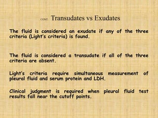
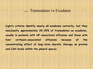
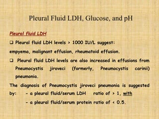
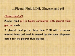
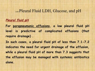
It is about interpretation of information delivered as results of pleural fluid exams, including physical characteristics, differentiation between transudates and exudates, cell counts, culture and cytology.
A simplified description of ascitic fluid analysis. Aim of the presentation is to give a very clear understanding about the analysis of ascities.
Presentation will help the medical residents diagnose the cause of fluid accumulation in abdomen and thus will guide to adopt the appropriate pathway to solve the issue.
It is fluid which is present in the pleural cavity of
lungs b/w parietal pleura n visceral pleura.
The pleural cavity is a potential space lined by
mesothelium of the visceral n parietal pleura.
D dimer test and sample collection procedure anjalatchi
Normally D-dimer levels are undetectable or detectable at very low levels, but they rise sharply when the body breaks down clots. D-dimer tests help in ruling out pulmonary embolisms in hospitalised Covid-19 patients
It is fluid which is present
in the pericardial cavity of
heart b/w parietal pericardium n visceral pericardium.
The pericardial cavity is a
potential space lined by
mesothelium of the visceral n parietal pericardium.
It is fluid which is present in
the abdominal cavity.
The peritoneal cavity is a potential
space lined by mesothelium of the
visceral n parietal peritoneum.
Apparently a lengthy presentation actually very good for junior physicians as it covers all aspects of assessment, diagnosis and treatment of pleural effusion
Pleural effusion is an accumulation of fluid in the pleural cavity
between the lining of the lungs and the thoracic cavity (i.e., the visceral
and parietal pleurae
).
ARTIFICIAL INTELLIGENCE IN HEALTHCARE.pdfAnujkumaranit
Artificial intelligence (AI) refers to the simulation of human intelligence processes by machines, especially computer systems. It encompasses tasks such as learning, reasoning, problem-solving, perception, and language understanding. AI technologies are revolutionizing various fields, from healthcare to finance, by enabling machines to perform tasks that typically require human intelligence.
HOT NEW PRODUCT! BIG SALES FAST SHIPPING NOW FROM CHINA!! EU KU DB BK substit...GL Anaacs
Contact us if you are interested:
Email / Skype : kefaya1771@gmail.com
Threema: PXHY5PDH
New BATCH Ku !!! MUCH IN DEMAND FAST SALE EVERY BATCH HAPPY GOOD EFFECT BIG BATCH !
Contact me on Threema or skype to start big business!!
Hot-sale products:
NEW HOT EUTYLONE WHITE CRYSTAL!!
5cl-adba precursor (semi finished )
5cl-adba raw materials
ADBB precursor (semi finished )
ADBB raw materials
APVP powder
5fadb/4f-adb
Jwh018 / Jwh210
Eutylone crystal
Protonitazene (hydrochloride) CAS: 119276-01-6
Flubrotizolam CAS: 57801-95-3
Metonitazene CAS: 14680-51-4
Payment terms: Western Union,MoneyGram,Bitcoin or USDT.
Deliver Time: Usually 7-15days
Shipping method: FedEx, TNT, DHL,UPS etc.Our deliveries are 100% safe, fast, reliable and discreet.
Samples will be sent for your evaluation!If you are interested in, please contact me, let's talk details.
We specializes in exporting high quality Research chemical, medical intermediate, Pharmaceutical chemicals and so on. Products are exported to USA, Canada, France, Korea, Japan,Russia, Southeast Asia and other countries.
The prostate is an exocrine gland of the male mammalian reproductive system
It is a walnut-sized gland that forms part of the male reproductive system and is located in front of the rectum and just below the urinary bladder
Function is to store and secrete a clear, slightly alkaline fluid that constitutes 10-30% of the volume of the seminal fluid that along with the spermatozoa, constitutes semen
A healthy human prostate measures (4cm-vertical, by 3cm-horizontal, 2cm ant-post ).
It surrounds the urethra just below the urinary bladder. It has anterior, median, posterior and two lateral lobes
It’s work is regulated by androgens which are responsible for male sex characteristics
Generalised disease of the prostate due to hormonal derangement which leads to non malignant enlargement of the gland (increase in the number of epithelial cells and stromal tissue)to cause compression of the urethra leading to symptoms (LUTS
Tom Selleck Health: A Comprehensive Look at the Iconic Actor’s Wellness Journeygreendigital
Tom Selleck, an enduring figure in Hollywood. has captivated audiences for decades with his rugged charm, iconic moustache. and memorable roles in television and film. From his breakout role as Thomas Magnum in Magnum P.I. to his current portrayal of Frank Reagan in Blue Bloods. Selleck's career has spanned over 50 years. But beyond his professional achievements. fans have often been curious about Tom Selleck Health. especially as he has aged in the public eye.
Follow us on: Pinterest
Introduction
Many have been interested in Tom Selleck health. not only because of his enduring presence on screen but also because of the challenges. and lifestyle choices he has faced and made over the years. This article delves into the various aspects of Tom Selleck health. exploring his fitness regimen, diet, mental health. and the challenges he has encountered as he ages. We'll look at how he maintains his well-being. the health issues he has faced, and his approach to ageing .
Early Life and Career
Childhood and Athletic Beginnings
Tom Selleck was born on January 29, 1945, in Detroit, Michigan, and grew up in Sherman Oaks, California. From an early age, he was involved in sports, particularly basketball. which played a significant role in his physical development. His athletic pursuits continued into college. where he attended the University of Southern California (USC) on a basketball scholarship. This early involvement in sports laid a strong foundation for his physical health and disciplined lifestyle.
Transition to Acting
Selleck's transition from an athlete to an actor came with its physical demands. His first significant role in "Magnum P.I." required him to perform various stunts and maintain a fit appearance. This role, which he played from 1980 to 1988. necessitated a rigorous fitness routine to meet the show's demands. setting the stage for his long-term commitment to health and wellness.
Fitness Regimen
Workout Routine
Tom Selleck health and fitness regimen has evolved. adapting to his changing roles and age. During his "Magnum, P.I." days. Selleck's workouts were intense and focused on building and maintaining muscle mass. His routine included weightlifting, cardiovascular exercises. and specific training for the stunts he performed on the show.
Selleck adjusted his fitness routine as he aged to suit his body's needs. Today, his workouts focus on maintaining flexibility, strength, and cardiovascular health. He incorporates low-impact exercises such as swimming, walking, and light weightlifting. This balanced approach helps him stay fit without putting undue strain on his joints and muscles.
Importance of Flexibility and Mobility
In recent years, Selleck has emphasized the importance of flexibility and mobility in his fitness regimen. Understanding the natural decline in muscle mass and joint flexibility with age. he includes stretching and yoga in his routine. These practices help prevent injuries, improve posture, and maintain mobilit
Couples presenting to the infertility clinic- Do they really have infertility...Sujoy Dasgupta
Dr Sujoy Dasgupta presented the study on "Couples presenting to the infertility clinic- Do they really have infertility? – The unexplored stories of non-consummation" in the 13th Congress of the Asia Pacific Initiative on Reproduction (ASPIRE 2024) at Manila on 24 May, 2024.
NVBDCP.pptx Nation vector borne disease control programSapna Thakur
NVBDCP was launched in 2003-2004 . Vector-Borne Disease: Disease that results from an infection transmitted to humans and other animals by blood-feeding arthropods, such as mosquitoes, ticks, and fleas. Examples of vector-borne diseases include Dengue fever, West Nile Virus, Lyme disease, and malaria.
Prix Galien International 2024 Forum ProgramLevi Shapiro
June 20, 2024, Prix Galien International and Jerusalem Ethics Forum in ROME. Detailed agenda including panels:
- ADVANCES IN CARDIOLOGY: A NEW PARADIGM IS COMING
- WOMEN’S HEALTH: FERTILITY PRESERVATION
- WHAT’S NEW IN THE TREATMENT OF INFECTIOUS,
ONCOLOGICAL AND INFLAMMATORY SKIN DISEASES?
- ARTIFICIAL INTELLIGENCE AND ETHICS
- GENE THERAPY
- BEYOND BORDERS: GLOBAL INITIATIVES FOR DEMOCRATIZING LIFE SCIENCE TECHNOLOGIES AND PROMOTING ACCESS TO HEALTHCARE
- ETHICAL CHALLENGES IN LIFE SCIENCES
- Prix Galien International Awards Ceremony
Title: Sense of Smell
Presenter: Dr. Faiza, Assistant Professor of Physiology
Qualifications:
MBBS (Best Graduate, AIMC Lahore)
FCPS Physiology
ICMT, CHPE, DHPE (STMU)
MPH (GC University, Faisalabad)
MBA (Virtual University of Pakistan)
Learning Objectives:
Describe the primary categories of smells and the concept of odor blindness.
Explain the structure and location of the olfactory membrane and mucosa, including the types and roles of cells involved in olfaction.
Describe the pathway and mechanisms of olfactory signal transmission from the olfactory receptors to the brain.
Illustrate the biochemical cascade triggered by odorant binding to olfactory receptors, including the role of G-proteins and second messengers in generating an action potential.
Identify different types of olfactory disorders such as anosmia, hyposmia, hyperosmia, and dysosmia, including their potential causes.
Key Topics:
Olfactory Genes:
3% of the human genome accounts for olfactory genes.
400 genes for odorant receptors.
Olfactory Membrane:
Located in the superior part of the nasal cavity.
Medially: Folds downward along the superior septum.
Laterally: Folds over the superior turbinate and upper surface of the middle turbinate.
Total surface area: 5-10 square centimeters.
Olfactory Mucosa:
Olfactory Cells: Bipolar nerve cells derived from the CNS (100 million), with 4-25 olfactory cilia per cell.
Sustentacular Cells: Produce mucus and maintain ionic and molecular environment.
Basal Cells: Replace worn-out olfactory cells with an average lifespan of 1-2 months.
Bowman’s Gland: Secretes mucus.
Stimulation of Olfactory Cells:
Odorant dissolves in mucus and attaches to receptors on olfactory cilia.
Involves a cascade effect through G-proteins and second messengers, leading to depolarization and action potential generation in the olfactory nerve.
Quality of a Good Odorant:
Small (3-20 Carbon atoms), volatile, water-soluble, and lipid-soluble.
Facilitated by odorant-binding proteins in mucus.
Membrane Potential and Action Potential:
Resting membrane potential: -55mV.
Action potential frequency in the olfactory nerve increases with odorant strength.
Adaptation Towards the Sense of Smell:
Rapid adaptation within the first second, with further slow adaptation.
Psychological adaptation greater than receptor adaptation, involving feedback inhibition from the central nervous system.
Primary Sensations of Smell:
Camphoraceous, Musky, Floral, Pepperminty, Ethereal, Pungent, Putrid.
Odor Detection Threshold:
Examples: Hydrogen sulfide (0.0005 ppm), Methyl-mercaptan (0.002 ppm).
Some toxic substances are odorless at lethal concentrations.
Characteristics of Smell:
Odor blindness for single substances due to lack of appropriate receptor protein.
Behavioral and emotional influences of smell.
Transmission of Olfactory Signals:
From olfactory cells to glomeruli in the olfactory bulb, involving lateral inhibition.
Primitive, less old, and new olfactory systems with different path
Lung Cancer: Artificial Intelligence, Synergetics, Complex System Analysis, S...Oleg Kshivets
RESULTS: Overall life span (LS) was 2252.1±1742.5 days and cumulative 5-year survival (5YS) reached 73.2%, 10 years – 64.8%, 20 years – 42.5%. 513 LCP lived more than 5 years (LS=3124.6±1525.6 days), 148 LCP – more than 10 years (LS=5054.4±1504.1 days).199 LCP died because of LC (LS=562.7±374.5 days). 5YS of LCP after bi/lobectomies was significantly superior in comparison with LCP after pneumonectomies (78.1% vs.63.7%, P=0.00001 by log-rank test). AT significantly improved 5YS (66.3% vs. 34.8%) (P=0.00000 by log-rank test) only for LCP with N1-2. Cox modeling displayed that 5YS of LCP significantly depended on: phase transition (PT) early-invasive LC in terms of synergetics, PT N0—N12, cell ratio factors (ratio between cancer cells- CC and blood cells subpopulations), G1-3, histology, glucose, AT, blood cell circuit, prothrombin index, heparin tolerance, recalcification time (P=0.000-0.038). Neural networks, genetic algorithm selection and bootstrap simulation revealed relationships between 5YS and PT early-invasive LC (rank=1), PT N0—N12 (rank=2), thrombocytes/CC (3), erythrocytes/CC (4), eosinophils/CC (5), healthy cells/CC (6), lymphocytes/CC (7), segmented neutrophils/CC (8), stick neutrophils/CC (9), monocytes/CC (10); leucocytes/CC (11). Correct prediction of 5YS was 100% by neural networks computing (area under ROC curve=1.0; error=0.0).
CONCLUSIONS: 5YS of LCP after radical procedures significantly depended on: 1) PT early-invasive cancer; 2) PT N0--N12; 3) cell ratio factors; 4) blood cell circuit; 5) biochemical factors; 6) hemostasis system; 7) AT; 8) LC characteristics; 9) LC cell dynamics; 10) surgery type: lobectomy/pneumonectomy; 11) anthropometric data. Optimal diagnosis and treatment strategies for LC are: 1) screening and early detection of LC; 2) availability of experienced thoracic surgeons because of complexity of radical procedures; 3) aggressive en block surgery and adequate lymph node dissection for completeness; 4) precise prediction; 5) adjuvant chemoimmunoradiotherapy for LCP with unfavorable prognosis.
Ethanol (CH3CH2OH), or beverage alcohol, is a two-carbon alcohol
that is rapidly distributed in the body and brain. Ethanol alters many
neurochemical systems and has rewarding and addictive properties. It
is the oldest recreational drug and likely contributes to more morbidity,
mortality, and public health costs than all illicit drugs combined. The
5th edition of the Diagnostic and Statistical Manual of Mental Disorders
(DSM-5) integrates alcohol abuse and alcohol dependence into a single
disorder called alcohol use disorder (AUD), with mild, moderate,
and severe subclassifications (American Psychiatric Association, 2013).
In the DSM-5, all types of substance abuse and dependence have been
combined into a single substance use disorder (SUD) on a continuum
from mild to severe. A diagnosis of AUD requires that at least two of
the 11 DSM-5 behaviors be present within a 12-month period (mild
AUD: 2–3 criteria; moderate AUD: 4–5 criteria; severe AUD: 6–11 criteria).
The four main behavioral effects of AUD are impaired control over
drinking, negative social consequences, risky use, and altered physiological
effects (tolerance, withdrawal). This chapter presents an overview
of the prevalence and harmful consequences of AUD in the U.S.,
the systemic nature of the disease, neurocircuitry and stages of AUD,
comorbidities, fetal alcohol spectrum disorders, genetic risk factors, and
pharmacotherapies for AUD.
New Directions in Targeted Therapeutic Approaches for Older Adults With Mantl...i3 Health
i3 Health is pleased to make the speaker slides from this activity available for use as a non-accredited self-study or teaching resource.
This slide deck presented by Dr. Kami Maddocks, Professor-Clinical in the Division of Hematology and
Associate Division Director for Ambulatory Operations
The Ohio State University Comprehensive Cancer Center, will provide insight into new directions in targeted therapeutic approaches for older adults with mantle cell lymphoma.
STATEMENT OF NEED
Mantle cell lymphoma (MCL) is a rare, aggressive B-cell non-Hodgkin lymphoma (NHL) accounting for 5% to 7% of all lymphomas. Its prognosis ranges from indolent disease that does not require treatment for years to very aggressive disease, which is associated with poor survival (Silkenstedt et al, 2021). Typically, MCL is diagnosed at advanced stage and in older patients who cannot tolerate intensive therapy (NCCN, 2022). Although recent advances have slightly increased remission rates, recurrence and relapse remain very common, leading to a median overall survival between 3 and 6 years (LLS, 2021). Though there are several effective options, progress is still needed towards establishing an accepted frontline approach for MCL (Castellino et al, 2022). Treatment selection and management of MCL are complicated by the heterogeneity of prognosis, advanced age and comorbidities of patients, and lack of an established standard approach for treatment, making it vital that clinicians be familiar with the latest research and advances in this area. In this activity chaired by Michael Wang, MD, Professor in the Department of Lymphoma & Myeloma at MD Anderson Cancer Center, expert faculty will discuss prognostic factors informing treatment, the promising results of recent trials in new therapeutic approaches, and the implications of treatment resistance in therapeutic selection for MCL.
Target Audience
Hematology/oncology fellows, attending faculty, and other health care professionals involved in the treatment of patients with mantle cell lymphoma (MCL).
Learning Objectives
1.) Identify clinical and biological prognostic factors that can guide treatment decision making for older adults with MCL
2.) Evaluate emerging data on targeted therapeutic approaches for treatment-naive and relapsed/refractory MCL and their applicability to older adults
3.) Assess mechanisms of resistance to targeted therapies for MCL and their implications for treatment selection
Acute scrotum is a general term referring to an emergency condition affecting the contents or the wall of the scrotum.
There are a number of conditions that present acutely, predominantly with pain and/or swelling
A careful and detailed history and examination, and in some cases, investigations allow differentiation between these diagnoses. A prompt diagnosis is essential as the patient may require urgent surgical intervention
Testicular torsion refers to twisting of the spermatic cord, causing ischaemia of the testicle.
Testicular torsion results from inadequate fixation of the testis to the tunica vaginalis producing ischemia from reduced arterial inflow and venous outflow obstruction.
The prevalence of testicular torsion in adult patients hospitalized with acute scrotal pain is approximately 25 to 50 percent
3. Certain types of pleural effusions can be suspected
simply by observing the physical characteristics of
the fluid obtained:
A purulent fluid (pus) indicates an empyema
A bad (putrid) odor suggests an anaerobic empyema
A milky white fluid suggests a chylothorax (an
accumulation of lymphatic fluid in the pleural
space)
4. A bloody fluid suggests either
hemorrhagic effusion (hemothorax) or
traumatic pleural taps.
Differentiation is possible by observing serial samples of
pleural tap which clear up in the case of a traumatic pleural
tap.
Moreover, A hematocrit performed on the pleural fluid can
assist diagnosis.
Bloody pleural fluid with a hematocrit of ≥ 50 % of the
peripheral blood hematocrit is termed a hemothorax.
CONT
5. A Black pleural fluid can be seen with some diseases
including:
• Aspergillus niger infection,
• malignant melanoma,
• non-small cell lung cancer,
• ruptured pancreatic pseudocyst,
• charcoal-containing empyema
CONT.
6. Normal pleural fluid is a Clear ultrafiltrate of plasma that
originates from the parietal pleura:
pH of 7.60-7.64
Protein content of less than 2% (1-2 g/dL)
Fewer than 1000 white blood cells (WBCs) per cubic
millimeter
Glucose content similar to that of plasma
Lactate dehydrogenase (LDH) less than 50% of plasma
7. Light’s criteria are the standard way to
differentiate.
Light’s criteria for exudative pleural effusion:
Pleural fluid protein divided by serum protein > 0.5
Pleural fluid LDH divided by serum LDH > 0.6
Pleural fluid LDH more than two-thirds the upper
limit of normal serum LDH
8. The fluid is considered an exudate if any of the three
criteria (Light’s criteria) is found.
The fluid is considered a transudate if all of the three
criteria are absent.
Light’s criteria require simultaneous measurement of
pleural fluid and serum protein and LDH.
Clinical judgment is required when pleural fluid test
results fall near the cutoff points.
9. Light’s criteria identify nearly all exudates correctly, but they
misclassify approximately 20-25% of transudates as exudates,
usually in patients with HF-associated effusions and those with
liver cirrhosis-associated effusions because of the
concentrating effect of long-term diuretic therapy on protein
and LDH levels within the pleural space).
10. To solve such a problem it is recommended to use one of the following If the
clinical picture is consistent with HF-associated effusion or liver cirrhosis-
associated effusion but the pleural fluid meets Light's exudative criteria:
1. - serum-effusion protein gradient (serum protein minus pleural protein
concentration). If >3.1 g/dL, it indicates transudate effusion.(5)
2. - serum-effusion albumin gradient (serum albumin minus pleural effusion
albumin level). If it is > 1.2 g/dL, it indicates transudate. (this criterion
is preferable If the clinical picture is consistent with HF but the pleural
fluid meets Light's exudative criteria). (5)
3. - effusion-to-serum albumin ratio (pleural fluid albumin divided by serum
albumin). If <0.6, it indicates transudate. this criterion is preferable In
the context of cirrhosis. (5)
11. In a more recent systematic review,
• pleural fluid cholesterol greater than 55 mg/dL and
• pleural fluid LDH greater than 200 U/L
each had better positive and negative likelihood ratio for
distinguishing exudates from transudates than did Light’s
criteria. [4]
12. Pleural fluid LDH
Pleural fluid LDH levels › 1000 IU/L suggest:
empyema, malignant effusion, rheumatoid effusion.
Pleural fluid LDH levels are also increased in effusions from
Pneumocystis jiroveci (formerly, Pneumocystis carinii)
pneumonia.
The diagnosis of Pneumocystis jiroveci pneumonia is suggested
by: - a pleural fluid/serum LDH ratio of › 1, with
- a pleural fluid/serum protein ratio of ‹ 0.5.
13. Pleural fluid glucose
A low pleural glucose concentration (30-50 mg/dL)
suggests malignant effusion, tuberculous pleuritis,
esophageal rupture, or lupus pleuritis.
A very low pleural glucose concentration (ie, < 30
mg/dL) further restricts diagnostic possibilities, to
rheumatoid pleurisy or empyema.
14. Pleural fluid pH
Pleural fluid pH is highly correlated with pleural fluid
glucose levels.
A pleural fluid pH of less than 7.30 with a normal
arterial blood pH level is caused by the same diagnoses
listed for low pleural fluid glucose.
15. Pleural fluid pH
For parapneumonic effusions, a low pleural fluid pH
level is predictive of complicated effusions (that
require drainage) .
In such cases, a pleural fluid pH of less than 7.1-7.2
indicates the need for urgent drainage of the effusion,
while a pleural fluid pH of more than 7.3 suggests that
the effusion may be managed with systemic antibiotics
alone.
16. If an exudate is suspected clinically or is confirmed by
chemistry test results, send the pleural fluid for :
total and differential cell counts,
Gram stain and culture,
cytology.
17. Pleural fluid lymphocytosis, with lymphocyte values greater than
85% of the total nucleated cells, suggests:
TB,
lymphoma,
Chylothorax,
sarcoidosis,
chronic rheumatoid pleurisy,
yellow nail syndrome.
Pleural lymphocyte values of 50-70% of the nucleated cells
suggest malignancy.
18. Cultures of infected pleural fluids yield positive
results in approximately 60% of cases.
This occurs even less often for anaerobic
organisms.
Diagnostic yields, particularly for anaerobic
pathogens, may be increased by directly culturing
pleural fluid into blood culture bottles.
19. Malignancy is suspected in patients with
lymphocytic exudative effusions, especially when
bloody.
Direct tumor involvement of the pleura is diagnosed
most easily by performing pleural fluid cytology.
The reported diagnostic yields in cytology vary
from 60-90%, depending on the extent of pleural
involvement and the type of primary malignancy.
20. Suspect tuberculous pleuritis in patients with
lymphocytic exudative effusions, especially if less
than 5% mesothelial cells are detected on
differential cell counts.
21. Most tuberculous pleural effusions probably result from a
hypersensitivity reaction to the Mycobacterium rather
than from microbial invasion of the pleura. So that,
acid-fast bacillus stains of pleural fluid are rarely
diagnostic (< 10% of cases), and
Pleural fluid cultures grow M tuberculosis in < 65% of
cases.
22. pleural biopsy:
The combination of histology and culture of pleural
tissue obtained by pleural biopsy increases the
diagnostic yield for TB to 90%.
23. Adenosine deaminase (ADA) activity of greater than
50 U/mL in pleural fluid supports the diagnosis of
tuberculous pleuritis.
However, pleural ADA values of less than 50 U/mL
do not exclude the diagnosis of TB pleuritis.
24. Exudate,
lymphocytic predominance,
positive acid-fast bacillus smear or cultures,
ADA > 50 U/L
25. Exudative with PMN predominance/pus,
positive Gram stains or cultures,
LDH > 1000,
glucose < 40 mg%,
pH < 7.2
PMNs = polymorph nuclear leukocytes, which are a
special family of white blood cells that ncludes
neutrophils, eosinophils, and basophils.