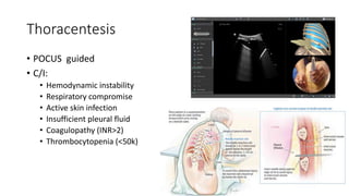
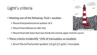
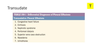
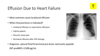
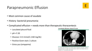
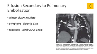
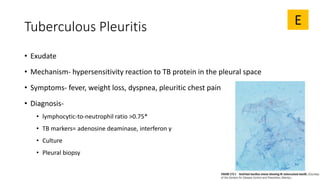
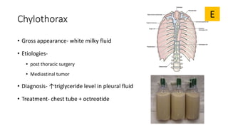
Pleural Effusion
Itamar Tzadok
Internal medicine department D
Kaplan medical center
18.02.2019
Outline
• Definition
• Signs and symptoms
• Diagnosis
• Transudate and exudate- selected etiologies
• Algorithm
Signs and symptoms
• Asymptomatic
• Dyspnea
• Cough (non-productive)
• Pleuritic chest pain
• In the effected side:
• Dullness to percussion
• Reduced chest expansion
• Reduced breath sounds
Signs and symptoms cont
Wong CL, et al. Does this patient have a pleural effusion? JAMA. 2009;301(3):309-317
Signs and symptoms – clues to etiology
Diagnosis
• Radiological verification
• Thoracentesis
• Gross appearance
• Transudate vs exudate
• Complimentary tests
Radiological verification
• Lateral decubitus
• Measurement of effusion
• Small- <1.5 cm
• Moderate- 1.5-4.5 cm
• Large- >4.5 cm
• More than 1cm can be tapped
Thoracentesis
• POCUS guided
• C/I:
• Hemodynamic instability
• Respiratory compromise
• Active skin infection
• Insufficient pleural fluid
• Coagulopathy (INR>2)
• Thrombocytopenia (<50k)
Light’s criteria
• Meeting one of the following- fluid = exudate
• Pleural fluid protein/serum protein >0.5
• Pleural fluid LDH/serum LDH >0.6
• Pleural fluid LDH more than two-thirds the normal upper limit for serum
• These criteria misidentify ~25% of transudates as exudates
• Serum-Pleural fluid protein gradient >31 g/L (3.1 g/dL) = transudate
Pleural effusion gross appearance
Pleural effusion complimentary tests
• Total protein + LDH
• Glucose levels
• Differential cell count
• pH
• Gram stain and culture
• TB markers
• Cytology
Transudate T
Effusion Due to Heart Failure
• Most common cause to pleural effusion
• When thoracentesis is indicated?
• Unilateral effusion or asymmetric effusions
• Febrile patient
• Pleuritic chest pain
• Persistent effusion after CHF therapy
• Diagnosis- pleural fluid N-terminal pro-brain natriuretic peptide
(NT-proBNP) >1500 pg/mL
T
Hepatic Hydrothorax
• 5% of patients with cirrhosis and ascites
• Usually right sided effusions
• Direct movement of peritoneal fluid
through defects in the diaphragm
T
Exudate E
Parapneumonic Effusion
• Most common cause of exudate
• History- bacterial pneumonia
• Complicated effusion = needs more than therapeutic thoracentesis
• Loculated pleural fluid
• pH <7.20
• Glucose <3.3 mmol/L (<60 mg/dL)
• Positive Gram stain / culture
• Gross pus (empyema)
E
Effusion Secondary to Malignancy
• 2nd most common cause of exudate
• 75% of cases:
• Lung CA
• Breast CA
• Lymphoma
• Diagnosis- cytology, thoracoscopy
E
Mesothelioma
• History- asbestos exposure
• Symptoms- pleuritic pain, dyspnea
• CXR- thickening of the pleura
• Diagnosis- biopsy
E
Effusion Secondary to Pulmonary
Embolization
• Almost always exudate
• Symptoms- pleuritic pain
• Diagnosis- spiral CT, CT-angio
E
Tuberculous Pleuritis
• Exudate
• Mechanism- hypersensitivity reaction to TB protein in the pleural space
• Symptoms- fever, weight loss, dyspnea, pleuritic chest pain
• Diagnosis-
• lymphocytic-to-neutrophil ratio >0.75*
• TB markers= adenosine deaminase, interferon γ
• Culture
• Pleural biopsy
E
Chylothorax
• Gross appearance- white milky fluid
• Etiologies-
• post thoracic surgery
• Mediastinal tumor
• Diagnosis- ↑triglyceride level in pleural fluid
• Treatment- chest tube + octreotide
E
Hemothorax
• Gross appearance- white milky fluid
• Etiology-
• Trauma
• Vascular tumor
• Diagnosis- pleural fluid Hct ≥ 0.5 peripheral blood Hct
• Treatment- tube thoracostomy
E
Drug-induced pleural disease
• 91 documented drugs
• Mentioned in Harrison 20th ed:
• Nitrofurantoin
• Dantrolene
• Methysergide
• Bromocriptine
• Procarbazine
• Amiodarone
• Dasatinib
E
Meigs' syndrome
• Ascites
• Pleural effusion
• Benign ovarian tumor
E
Yellow nail syndrome (YNS)
• Rare syndrome
• “Primary lymphedema associated with yellow nails and pleural effusion”
• Adults
• Pleural effusions
• Lymphedema
• Yellow dystrophic nails
E
Recap
• CXR + decubitus = first step in diagnosis
• Light’s criteria
• Unilateral effusion + no history of pneumonia => malignancy
• Bilateral effusion- usually cardiac/renal/hepatic problem => treat
underlying condition
• Exudate without findings in preliminary evaluation=> suspect PE
Recap cont.
• Common causes to transudate:
• CHF
• Cirrhosis
• Common causes to exudate:
• Malignancy
• Parapneumonic effusions
• TB
E
T
Thank you!
Questions?

Pleural effusion 18022019

  • 1.
    Pleural Effusion Itamar Tzadok Internalmedicine department D Kaplan medical center 18.02.2019
  • 2.
    Outline • Definition • Signsand symptoms • Diagnosis • Transudate and exudate- selected etiologies • Algorithm
  • 5.
    Signs and symptoms •Asymptomatic • Dyspnea • Cough (non-productive) • Pleuritic chest pain • In the effected side: • Dullness to percussion • Reduced chest expansion • Reduced breath sounds
  • 6.
    Signs and symptomscont Wong CL, et al. Does this patient have a pleural effusion? JAMA. 2009;301(3):309-317
  • 8.
    Signs and symptoms– clues to etiology
  • 9.
    Diagnosis • Radiological verification •Thoracentesis • Gross appearance • Transudate vs exudate • Complimentary tests
  • 10.
    Radiological verification • Lateraldecubitus • Measurement of effusion • Small- <1.5 cm • Moderate- 1.5-4.5 cm • Large- >4.5 cm • More than 1cm can be tapped
  • 11.
    Thoracentesis • POCUS guided •C/I: • Hemodynamic instability • Respiratory compromise • Active skin infection • Insufficient pleural fluid • Coagulopathy (INR>2) • Thrombocytopenia (<50k)
  • 12.
    Light’s criteria • Meetingone of the following- fluid = exudate • Pleural fluid protein/serum protein >0.5 • Pleural fluid LDH/serum LDH >0.6 • Pleural fluid LDH more than two-thirds the normal upper limit for serum • These criteria misidentify ~25% of transudates as exudates • Serum-Pleural fluid protein gradient >31 g/L (3.1 g/dL) = transudate
  • 13.
  • 14.
    Pleural effusion complimentarytests • Total protein + LDH • Glucose levels • Differential cell count • pH • Gram stain and culture • TB markers • Cytology
  • 15.
  • 16.
    Effusion Due toHeart Failure • Most common cause to pleural effusion • When thoracentesis is indicated? • Unilateral effusion or asymmetric effusions • Febrile patient • Pleuritic chest pain • Persistent effusion after CHF therapy • Diagnosis- pleural fluid N-terminal pro-brain natriuretic peptide (NT-proBNP) >1500 pg/mL T
  • 17.
    Hepatic Hydrothorax • 5%of patients with cirrhosis and ascites • Usually right sided effusions • Direct movement of peritoneal fluid through defects in the diaphragm T
  • 18.
  • 19.
    Parapneumonic Effusion • Mostcommon cause of exudate • History- bacterial pneumonia • Complicated effusion = needs more than therapeutic thoracentesis • Loculated pleural fluid • pH <7.20 • Glucose <3.3 mmol/L (<60 mg/dL) • Positive Gram stain / culture • Gross pus (empyema) E
  • 20.
    Effusion Secondary toMalignancy • 2nd most common cause of exudate • 75% of cases: • Lung CA • Breast CA • Lymphoma • Diagnosis- cytology, thoracoscopy E
  • 21.
    Mesothelioma • History- asbestosexposure • Symptoms- pleuritic pain, dyspnea • CXR- thickening of the pleura • Diagnosis- biopsy E
  • 22.
    Effusion Secondary toPulmonary Embolization • Almost always exudate • Symptoms- pleuritic pain • Diagnosis- spiral CT, CT-angio E
  • 23.
    Tuberculous Pleuritis • Exudate •Mechanism- hypersensitivity reaction to TB protein in the pleural space • Symptoms- fever, weight loss, dyspnea, pleuritic chest pain • Diagnosis- • lymphocytic-to-neutrophil ratio >0.75* • TB markers= adenosine deaminase, interferon γ • Culture • Pleural biopsy E
  • 24.
    Chylothorax • Gross appearance-white milky fluid • Etiologies- • post thoracic surgery • Mediastinal tumor • Diagnosis- ↑triglyceride level in pleural fluid • Treatment- chest tube + octreotide E
  • 25.
    Hemothorax • Gross appearance-white milky fluid • Etiology- • Trauma • Vascular tumor • Diagnosis- pleural fluid Hct ≥ 0.5 peripheral blood Hct • Treatment- tube thoracostomy E
  • 26.
    Drug-induced pleural disease •91 documented drugs • Mentioned in Harrison 20th ed: • Nitrofurantoin • Dantrolene • Methysergide • Bromocriptine • Procarbazine • Amiodarone • Dasatinib E
  • 27.
    Meigs' syndrome • Ascites •Pleural effusion • Benign ovarian tumor E
  • 28.
    Yellow nail syndrome(YNS) • Rare syndrome • “Primary lymphedema associated with yellow nails and pleural effusion” • Adults • Pleural effusions • Lymphedema • Yellow dystrophic nails E
  • 31.
    Recap • CXR +decubitus = first step in diagnosis • Light’s criteria • Unilateral effusion + no history of pneumonia => malignancy • Bilateral effusion- usually cardiac/renal/hepatic problem => treat underlying condition • Exudate without findings in preliminary evaluation=> suspect PE
  • 32.
    Recap cont. • Commoncauses to transudate: • CHF • Cirrhosis • Common causes to exudate: • Malignancy • Parapneumonic effusions • TB E T
  • 33.

Editor's Notes

  • #2 מבוסס על 288 Disorders of the Pleura, מתוך האריסון 20
  • #4 פיזיולוגיה ופתופיזיולוגיה קישור לתמונה- http://library.open.oregonstate.edu/aandp/chapter/22-3-the-process-of-breathing/
  • #5 מצבים שיכולים לגרום להצטברות: הגברת הלחץ הורידי הסיסטמי- יותר הסננה דרך הפלאורה הפריאטלית ירידה ברמות החלבון- הסננת הנוזלים כלפי החלל הפלאורלי פגיעה בניקוז הלימפטי- בגלל גידול למשל במצב רגיל- 0.26 מ"ל/ק"ג נוזל (באדם השוקל 70 ק"ג- 18 מ"ל)
  • #6 לרוב יימצאו 1 מתוך 3 התלונות הנ"ל (שיעול, קוצ"נ, כאב פלאוריטי) בחולים עם תפליט כמו"כ, ישנם ממצאים בבדיקה הגופנית, בצד שבו יש יותר תפליט
  • #7 הרגישות והסגוליות הרגישים ביותר- ניקוש
  • #9 מהי הקליניקה? יותר מדויק לומר שתפליט הוא סימן שמלמד על פתולוגיה סמויה.. למשל, חולה שיש לו סימפטומים של שחמת כבד, אז תפליט פלאורלי הוא עוד ממצא שמהווה חלק מכלל התמונה הקלינית
  • #11 https://www.med-ed.virginia.edu/courses/rad/cxr/technique4chest.html מאמר UTD- Imaging of pleural effusions in adults
  • #12 מתוך UTD- Ultrasound-guided thoracentesis
  • #13 רגישות של 99.5% לזיהוי של אקסודט 93-96% מהמקרים הקריטריונים טובים להבחנה של אקסודט מטרנסודט (ההפך אינו נכון..)
  • #14 הטבלה מתוך מאמר AAFP תמונה מתוך Respiratory: An Integrated Approach to Disease פרק Chapter 19: Evaluating Sputum and Pleural Effusions
  • #15 לפי אלגוריתם- לוקחים רק חלבון וLDH. לפי המציאות- אם מנקרים את המטופל, שולחים כבר את כל הנוזל לבדיקות בדיקות ציטולוגיה- להקפיד ולתאם עם המעבדה, משום שזמני הפעילות משתנים ממקום למקום ולא נרצה להרוס את הדגימה
  • #17 קישור- https://www.med-ed.virginia.edu/courses/rad/cxr/pathology7chest.html
  • #18 קישור https://www.annalsthoracicsurgery.org/article/S0003-4975(13)00993-4/pdf ניתן להבחין בפגמים בסרעפת ע"י הזרקת הצבען ICG במהלך טורקוסקופיה
  • #20 Loculated= אין שינוי בפיזור הנוזל כאשר עושים צילום דקוביטוס
  • #21 במידה והציטולוגיה שלילית והחשד הקליני גדול, נעשה טורקוסקופיה כאשר יש תפליט- הדבר מלמד על מחלה מפושטת. לרוב כימו' כבר לא תעזור
  • #22 מקורות- https://www.bmj.com/content/346/bmj.e8376 https://newsnetwork.mayoclinic.org/discussion/living-with-cancer-the-connection-between-asbestos-and-mesothelioma/
  • #23 ב-75% מהמקרים בהם היה PE עם תפליט, אז היתה קליניקה של כאב פלאוריטי
  • #24 * בתפליט של חולה עם חשד מוגבר לTB נראה יותר לימפוציטים
  • #25 לרוב נגרם מפגיעה בthoracic duct
  • #26 קישור לתמונה- https://medical-dictionary.thefreedictionary.com/hemothorax
  • #27 https://www.pneumotox.com/pattern/view/31/V.a/pleural-effusion – אתר עם רשימה מפורטת של כל הכדורים
  • #28 קישור לתמונה- www.radiologycases.com/index.php/radiologycases/article/viewArticle/3209
  • #29 קישור לתמונה- http://www.cmaj.ca/content/181/9/614 האבחנה- קלינית. תסמונת של משפחות, אבל לא ברור הקשר הגנטי
  • #32 מתוך Thorax 2010;65(Suppl 2):ii4eii17. doi:10.1136/thx.2010.136978
  • #33 מתוך Thorax 2010;65(Suppl 2):ii4eii17. doi:10.1136/thx.2010.136978
  • #34 קישור- https://medcomic.com/medcomic/pleural-effusions-transudate-vs-exudate/