Downloaded 109 times
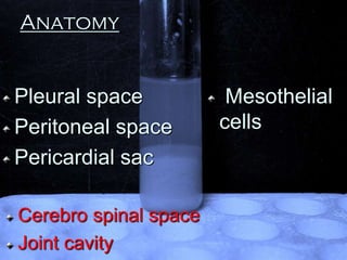
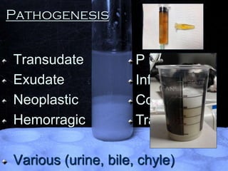


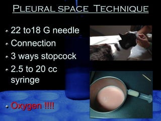


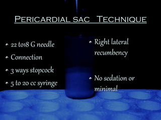


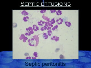


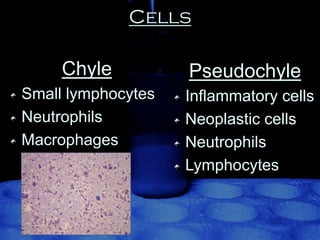

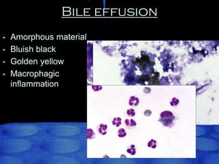


This document discusses cytology techniques and findings for different body cavity fluids. It covers the anatomy of pleural, peritoneal, pericardial, and other spaces. Common etiologies that can cause fluid accumulation include transudates, exudates, and hemorrhage. Techniques are described for safely collecting fluid samples from these spaces using needles and syringes. The cytological appearance of different cell types like mesothelial cells, macrophages, and malignant cells are outlined. Specific conditions that can be identified include infections, chylous effusions, bile effusions, and different types of cancers. The cytological features that can help distinguish between mesothelioma and carcinomas are also summarized.


