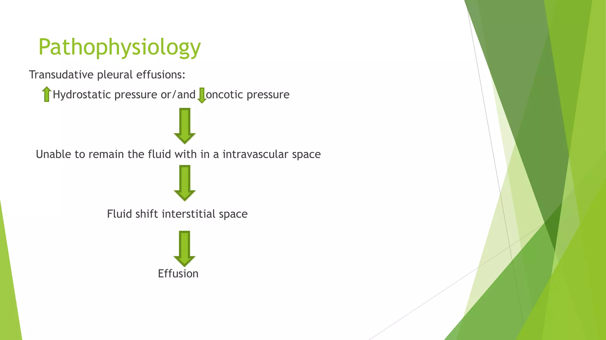
Pleural Effusion
Prepared by:
Dr.Akhilesh Sah
Pleura
Definition and Introduction
 The accumulation of fluid within the pleural space is termed pleural effusion
 The pleural space normally contains only about 10-20ml fluid
 Pleural effusion is a collection of abnormal amount of fluid in the pleural space
 It can occur by itself or can be the result of surrounding parenchymal disease like
infection, malignancy, or inflammatory conditions
 The accumulation of pus is termed empyema , that of blood is haemothorax, and
that of chyle(made of lymph and tiny fat droplets) is a chylothorax
 Pleural fluid accumulates as a result of either increased hydrostatic pressure or
decreased osmotic pressure(transudative effusion, as seen in cardiac, liver or renal
failure), or from increased microvascular pressure due to disease of the pleura or
injury in the adjacent lung (exudative effusion)
Classification
Transudative effusions
Exudative effusions
Transudative effusions
 Also known as hydrothoraces, occurs primarily in non-
inflammatory condtions:
 Is an accumulation of low-protein, low cell count fluid
Cause of transudative effusion
• Increase hydrostatic pressure found in heart failure ( most
common cause of pleural effusion)
• Decrease oncotic pressure (From hypoalbuminemia) found in
cirrhosis of liver or renal disease.
• In this condition, fluid movement is faciliated out of the
capillaries and into the pleural space
Exudative effusions:
 Exudative effusions occur in an area of inflammation; is an
accumulation of high-protein fluid.
 An exudative effusion results from increased capillary
permeability characteristic of inflammatory reaction.
 This types of effusion occurs secondary to conditions such as
pulmonary malignancies, pulmonary infections(TB,
SUBPHRENIC ABSCESS, bacterial or fungal infection) and
pulmonary embolization.
Causes and Etiology
Common causes
• Pneumonia (parapneumonic effusion)
• Tuberculosis
• Pulmonary infarction
• Malignant disease
• Cardiac failure: may cause bilateral effusion
• Subdiaphragmatic disorders (subphrenic abscess,
pancreatitis etc.)
Cont..
Uncommon causes
• Hypoproteinaemia (nephrotic syndrome, liver failure, malnutrition)
• Connective tissue diseases (particularly systemic lupus erythematosus
and rheumatoid arthritis)
• Post-myocardial infarction syndrome
• Acute rheumatic fever
• Meigs’ syndrome (ovarian tumour plus pleural effusion)
• Myxoedema
• Uraemia
• Asbestos-related benign pleural effusion
Pathophysiology
Transudative pleural effusions:
Hydrostatic pressure or/and oncotic pressure
Unable to remain the fluid with in a intravascular space
Fluid shift interstitial space
Effusion
Cont
Exudative Effusions
Clinical Features
Clinical features are those caused by the underlying disease and severity of
effusions
Symptoms
• Fever : chills and rigor
• Chest pain:
• Shortness of breathe
• Cough
Signs
• Decreased breathe Sound
• Dullness on Percussion
Diagnosis and Investigation
• Chest x-ray (around 200ml fluid required to detect in xray)or USG scan to
detect fluid
• Diagnostic Thoracentesis: for Biochemical, bacteriological and cytological
studies
Others : to determine causes and etiology
 CBC: leukocytosis or leukopenia, and others evaluative tests: RFT,LFT, s.LDH
 Pleural aspiration and biopsy
 CT-scan of chest
 Echocardiography: to rule out cardiac disease
 USG A/P: to rule out liver disease
Chest Xray
Cont ..
Cont
Management / Treatment
Objectives of treatment are
 to discover the underlying cause,
 to prevent reaccumulation of fluid, and
 to relieve discomfort, dyspnea, and respiratory compromise
General treatment
•Treatment is aimed at underlying cause (heart disease, infection).
•Thoracentesis(Pleural Aspiration) is done to remove fluid, collect a specimen,
and relieve dyspnea
Therapeutic aspiration:
• Therapeutic aspiration may be required to palliate breathlessness
• removing more than 1.5 L at a time is associated with a small risk of
re-expansion pulmonary oedema
Complication:
 Respiratory failure
 Pneumothorax after drainage

pleural effusion.pptx

  • 1.
  • 2.
  • 3.
    Definition and Introduction The accumulation of fluid within the pleural space is termed pleural effusion  The pleural space normally contains only about 10-20ml fluid  Pleural effusion is a collection of abnormal amount of fluid in the pleural space  It can occur by itself or can be the result of surrounding parenchymal disease like infection, malignancy, or inflammatory conditions  The accumulation of pus is termed empyema , that of blood is haemothorax, and that of chyle(made of lymph and tiny fat droplets) is a chylothorax  Pleural fluid accumulates as a result of either increased hydrostatic pressure or decreased osmotic pressure(transudative effusion, as seen in cardiac, liver or renal failure), or from increased microvascular pressure due to disease of the pleura or injury in the adjacent lung (exudative effusion)
  • 4.
  • 5.
    Transudative effusions  Alsoknown as hydrothoraces, occurs primarily in non- inflammatory condtions:  Is an accumulation of low-protein, low cell count fluid Cause of transudative effusion • Increase hydrostatic pressure found in heart failure ( most common cause of pleural effusion) • Decrease oncotic pressure (From hypoalbuminemia) found in cirrhosis of liver or renal disease. • In this condition, fluid movement is faciliated out of the capillaries and into the pleural space
  • 6.
    Exudative effusions:  Exudativeeffusions occur in an area of inflammation; is an accumulation of high-protein fluid.  An exudative effusion results from increased capillary permeability characteristic of inflammatory reaction.  This types of effusion occurs secondary to conditions such as pulmonary malignancies, pulmonary infections(TB, SUBPHRENIC ABSCESS, bacterial or fungal infection) and pulmonary embolization.
  • 7.
    Causes and Etiology Commoncauses • Pneumonia (parapneumonic effusion) • Tuberculosis • Pulmonary infarction • Malignant disease • Cardiac failure: may cause bilateral effusion • Subdiaphragmatic disorders (subphrenic abscess, pancreatitis etc.)
  • 8.
    Cont.. Uncommon causes • Hypoproteinaemia(nephrotic syndrome, liver failure, malnutrition) • Connective tissue diseases (particularly systemic lupus erythematosus and rheumatoid arthritis) • Post-myocardial infarction syndrome • Acute rheumatic fever • Meigs’ syndrome (ovarian tumour plus pleural effusion) • Myxoedema • Uraemia • Asbestos-related benign pleural effusion
  • 9.
    Pathophysiology Transudative pleural effusions: Hydrostaticpressure or/and oncotic pressure Unable to remain the fluid with in a intravascular space Fluid shift interstitial space Effusion
  • 10.
  • 11.
    Clinical Features Clinical featuresare those caused by the underlying disease and severity of effusions Symptoms • Fever : chills and rigor • Chest pain: • Shortness of breathe • Cough Signs • Decreased breathe Sound • Dullness on Percussion
  • 12.
    Diagnosis and Investigation •Chest x-ray (around 200ml fluid required to detect in xray)or USG scan to detect fluid • Diagnostic Thoracentesis: for Biochemical, bacteriological and cytological studies Others : to determine causes and etiology  CBC: leukocytosis or leukopenia, and others evaluative tests: RFT,LFT, s.LDH  Pleural aspiration and biopsy  CT-scan of chest  Echocardiography: to rule out cardiac disease  USG A/P: to rule out liver disease
  • 14.
  • 16.
  • 17.
  • 18.
    Management / Treatment Objectivesof treatment are  to discover the underlying cause,  to prevent reaccumulation of fluid, and  to relieve discomfort, dyspnea, and respiratory compromise General treatment •Treatment is aimed at underlying cause (heart disease, infection). •Thoracentesis(Pleural Aspiration) is done to remove fluid, collect a specimen, and relieve dyspnea
  • 19.
    Therapeutic aspiration: • Therapeuticaspiration may be required to palliate breathlessness • removing more than 1.5 L at a time is associated with a small risk of re-expansion pulmonary oedema
  • 20.
    Complication:  Respiratory failure Pneumothorax after drainage