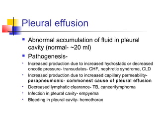
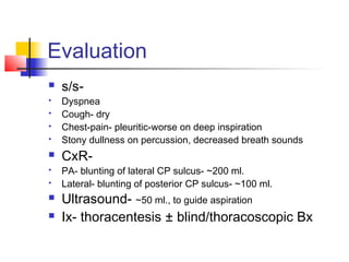
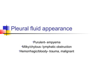
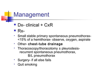
Pleural diseases
Pleural efffusion
Pneumothorax
Mesothelioma
Pleural effusion
 Abnormal accumulation of fluid in pleural
cavity (normal- ~20 ml)
 Pathogenesis-
 Increased production due to increased hydrostatic or decreased
oncotic pressure- transudates- CHF, nephrotic syndrome, CLD
 Increased production due to increased capillary permeability-
parapneumonic- commonest cause of pleural effusion
 Decreased lymphatic clearance- TB, cancer/lymphoma
 Infection in pleural cavity- empyema
 Bleeding in pleural cavity- hemothorax
Evaluation
 s/s-
 Dyspnea
 Cough- dry
 Chest-pain- pleuritic-worse on deep inspiration
 Stony dullness on percussion, decreased breath sounds
 CxR-
 PA- blunting of lateral CP sulcus- ~200 ml.
 Lateral- blunting of posterior CP sulcus- ~100 ml.
 Ultrasound- ~50 ml., to guide aspiration
 Ix- thoracentesis ± blind/thoracoscopic Bx
Pleural fluid appearance
Purulent- empyema
Milky/chylous- lymphatic obstruction
Hemorrhagic/bloody- trauma, malignant
Pleural fluid investigations
 Exudate
 Protein PF/S >0.5
 LDH PF/S >0.6
 PF LDH > 2/3rd
S LDH
 PF glucose < S glucose
 Cells >1000/µL-
neutrophilic- empyema
lymphocytic- TB
 pH <7.30
 Amylase increased
 Gram stain/Z-N stain +ve
 Malignant cells +ve
 Transudate
 No criterion for
exudate fulfilled
Treatment
 Transudative- treat underlying condition
 Exudative-
 Parapneumonic- treat underlying pneumonia,
chest-tube drainage if pH <7.2 or glucose <60
mg/dl
 Empyema & hemothorax- chest-tube drainage
 Malignancy- depends on symptoms-
infrequent- repeated thoracentesis
frequent-
chest-tube drainagepleurodesis
Pleurodesis- doxycycline, talc, bleomycin
Pneumothorax
 Air in pleural space
 Causes-
 Trauma- exogenous or iatrogenic
 Primary- rupture of subpleural blebs-
common in tall, thin, young, smoker males
 Secondary- complication of underlying lung disease
 Presentation-
 Symptom- chest-pain, dyspnea
 Signs- decreased movements, tracheal/mediastinal shift,
increased resonance, decreased breath sounds
Tension pneumothorax
 One-way valve making intra-pleural pressure
more than ambient pressure throughout the
respiratory cycle
 Common causes-
 Penetrating trauma, CPR
 Positive pressure mechanical ventilation
 s/s- marked tachycardia, hypotension in
patient with pneumothorax
 Complication- subcutaneous emphysema
Management
 Dx- clinical + CxR
 Rx-
 Small stable primary spontaneous pneumothorax-
<15% of a hemithorax- observe, oxygen, aspirate
 Other- chest-tube drainage
 Thoracoscopy/thoracotomy ± pleurodesis-
recurrent spontaneous pneumothorax,
B/L pneumothorax
 Surgery- if all else fails
 Quit smoking
Mesothelioma
 Rare, tumor of pleura
 More in elderly, males
 Asbestos exposure- crocidolite most
carcinogenic
 Symptom- dull pain, SOB, pleural effusion
 Ix- CxR, CT scan, thoracoscopy,
IHC-cytokeratin +ve
 Median survival- 9-21 months
 Rx- poor response- Sx/RT/CT-Pemetrexed

Pleural diseases

  • 1.
  • 2.
    Pleural effusion  Abnormalaccumulation of fluid in pleural cavity (normal- ~20 ml)  Pathogenesis-  Increased production due to increased hydrostatic or decreased oncotic pressure- transudates- CHF, nephrotic syndrome, CLD  Increased production due to increased capillary permeability- parapneumonic- commonest cause of pleural effusion  Decreased lymphatic clearance- TB, cancer/lymphoma  Infection in pleural cavity- empyema  Bleeding in pleural cavity- hemothorax
  • 3.
    Evaluation  s/s-  Dyspnea Cough- dry  Chest-pain- pleuritic-worse on deep inspiration  Stony dullness on percussion, decreased breath sounds  CxR-  PA- blunting of lateral CP sulcus- ~200 ml.  Lateral- blunting of posterior CP sulcus- ~100 ml.  Ultrasound- ~50 ml., to guide aspiration  Ix- thoracentesis ± blind/thoracoscopic Bx
  • 4.
    Pleural fluid appearance Purulent-empyema Milky/chylous- lymphatic obstruction Hemorrhagic/bloody- trauma, malignant
  • 5.
    Pleural fluid investigations Exudate  Protein PF/S >0.5  LDH PF/S >0.6  PF LDH > 2/3rd S LDH  PF glucose < S glucose  Cells >1000/µL- neutrophilic- empyema lymphocytic- TB  pH <7.30  Amylase increased  Gram stain/Z-N stain +ve  Malignant cells +ve  Transudate  No criterion for exudate fulfilled
  • 6.
    Treatment  Transudative- treatunderlying condition  Exudative-  Parapneumonic- treat underlying pneumonia, chest-tube drainage if pH <7.2 or glucose <60 mg/dl  Empyema & hemothorax- chest-tube drainage  Malignancy- depends on symptoms- infrequent- repeated thoracentesis frequent- chest-tube drainagepleurodesis Pleurodesis- doxycycline, talc, bleomycin
  • 7.
    Pneumothorax  Air inpleural space  Causes-  Trauma- exogenous or iatrogenic  Primary- rupture of subpleural blebs- common in tall, thin, young, smoker males  Secondary- complication of underlying lung disease  Presentation-  Symptom- chest-pain, dyspnea  Signs- decreased movements, tracheal/mediastinal shift, increased resonance, decreased breath sounds
  • 8.
    Tension pneumothorax  One-wayvalve making intra-pleural pressure more than ambient pressure throughout the respiratory cycle  Common causes-  Penetrating trauma, CPR  Positive pressure mechanical ventilation  s/s- marked tachycardia, hypotension in patient with pneumothorax  Complication- subcutaneous emphysema
  • 9.
    Management  Dx- clinical+ CxR  Rx-  Small stable primary spontaneous pneumothorax- <15% of a hemithorax- observe, oxygen, aspirate  Other- chest-tube drainage  Thoracoscopy/thoracotomy ± pleurodesis- recurrent spontaneous pneumothorax, B/L pneumothorax  Surgery- if all else fails  Quit smoking
  • 10.
    Mesothelioma  Rare, tumorof pleura  More in elderly, males  Asbestos exposure- crocidolite most carcinogenic  Symptom- dull pain, SOB, pleural effusion  Ix- CxR, CT scan, thoracoscopy, IHC-cytokeratin +ve  Median survival- 9-21 months  Rx- poor response- Sx/RT/CT-Pemetrexed