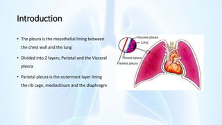
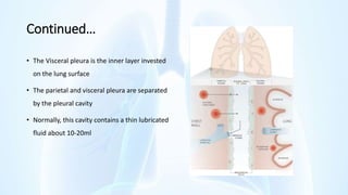
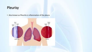
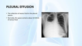
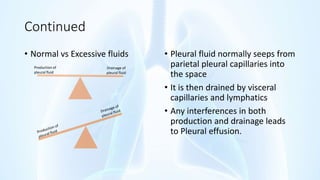
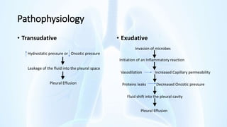
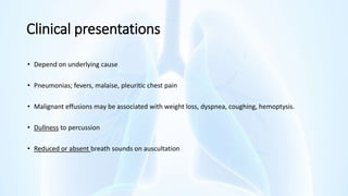
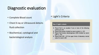
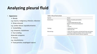
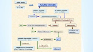
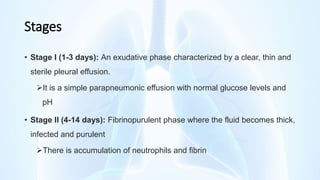
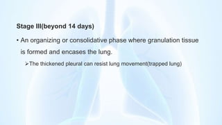
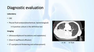
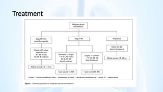
Pleural diseases define a group of diseases that affect the coverings of the lungs. These may be primary or secondary in origin. Relevant references are provided in the slides for further reading.
Note that this information must be used not to replace your lecturer but to supplement and provided a basis for further reading.
Bronchiectasis refers to the congenital/acquired irreversible airway dilation that involves the bronchi/bronchioles in either a focal or a diffuse manner.
It is a pulmonary disease related to chronic infections in the background of inability of respiratory mucosa to clear the infections and impaired ciliary function.
It is chronic disease with high morbidity and mortality
Anatomy & Physiology of The Respiratory System & its DiseasesRaghad AlDuhaylib
This presentation is an overall review of the respiratory system anatomy and physiology. Also, some diseases of the respiratory system are mentioned briefly in the slides.
Help for medical students about topic Suppurative lung diseases - Abscess and gangrene of the lungs, Pneumothorax, Hematorax, Purulent pleurisy. And useful material as required by students. Everything is inserted as per outlines of topics.
Thank you for selecting our 𝐏𝐥𝐞𝐮𝐫𝐚𝐥 𝐞𝐟𝐟𝐮𝐬𝐢𝐨𝐧PPT
This medical PowerPoint template about 𝐏𝐥𝐞𝐮𝐫𝐚𝐥 𝐞𝐟𝐟𝐮𝐬𝐢𝐨𝐧
You can download our template by visiting our website:
https://www.rxslides.com/product/pleural-effusion-powerpoint-template
copy and paste this URL into the browser and download the full editable template.
This 𝐏𝐥𝐞𝐮𝐫𝐚𝐥 𝐞𝐟𝐟𝐮𝐬𝐢𝐨𝐧animated template is designed by RxSlides, a medical professional team covering the following topics about 𝐏𝐥𝐞𝐮𝐫𝐚𝐥 𝐞𝐟𝐟𝐮𝐬𝐢𝐨𝐧
𝐃𝐞𝐟𝐢𝐧𝐢𝐭𝐢𝐨𝐧:
• Respiratory illness caused by the buildup of fluid between lung and chest cavity tissue layers.
𝐀𝐧𝐚𝐭𝐨𝐦𝐲 𝐨𝐟 𝐭𝐡𝐞 𝐋𝐮𝐧𝐠
• The lung is a vital organ responsible for gas exchange.
• It is surrounded by two thin membranes called the pleurae.
• The visceral pleura covers the lung surface.
• The parietal pleura lines the inner chest wall.
𝐩𝐫𝐞𝐯𝐚𝐥𝐞𝐧𝐜𝐞:
• 𝐏𝐥𝐞𝐮𝐫𝐚𝐥 𝐞𝐟𝐟𝐮𝐬𝐢𝐨𝐧 is a common condition, affecting millions of people worldwide.
• The prevalence varies by region, with higher rates in developing countries
𝐏𝐚𝐭𝐡𝐨𝐩𝐡𝐲𝐬𝐢𝐨𝐥𝐨𝐠𝐲:
• Illustrated anatomy of the respiratory system included.
• Animated illustrations demonstrate fluid formation and maintenance.
• Parietal and visceral pleura form the pleural space.
• Fluid produced by filtration from systemic capillaries.
• Fluid accumulation in the pleural cavity causes Pleural Effusion.
𝐑𝐢𝐬𝐤 𝐅𝐚𝐜𝐭𝐨𝐫𝐬
• Smoking
• Heart disease
• Liver disease
• Alcohol
• Lung disease
• Asbestos exposure
𝐜𝐚𝐮𝐬𝐞𝐬:
𝐓𝐫𝐚𝐧𝐬𝐮𝐝𝐚𝐭𝐢𝐯𝐞
o Increased systemic/pulmonary capillary pressure and decreased osmotic pressure.
o Major causes: cirrhosis, heart failure, nephrotic syndrome, protein-losing enteropathy.
𝐄𝐱𝐮𝐝𝐚𝐭𝐢𝐯𝐞:
o Local processes leading to increased capillary permeability.
o Fluid, protein, cells, and serum constituents exude.
o Major causes: inflammation, infection, lung injury, tumors, lung cancer, rheumatoid arthritis, pneumonia, tuberculosis, surgical damage, lymphatic fluid accumulation.
𝐒𝐲𝐦𝐩𝐭𝐨𝐦𝐬:
• Dyspnea (shortness of breath)
• Labored breathing
• Non-productive cough
• Chest tightness
• Orthopnea (difficulty breathing lying down)
𝐃𝐢𝐚𝐠𝐧𝐨𝐬𝐭𝐢𝐜 𝐦𝐞𝐭𝐡𝐨𝐝𝐬
• Physical examination
• Chest radiographs
• Imaging
𝐓𝐫𝐞𝐚𝐭𝐦𝐞𝐧𝐭 𝐎𝐩𝐭𝐢𝐨𝐧𝐬:
Thoracentesis (fluid removal)
Catheter drainage
Pleurodesis (scarring of pleural space)
Pleuroperitoneal shunt (fluid drainage to abdomen)
Pleurectomy (surgical removal of pleura)
Thoracoscopy (visual examination of pleural space)
Thoracostomy (surgical opening of chest cavity)
Visit our site for more animated templates
𝗵𝘁𝘁𝗽𝘀://𝘄𝘄𝘄.𝗿𝘅𝘀𝗹𝗶𝗱𝗲𝘀.𝗰𝗼𝗺
RxSlides PowerPoint icons and illustrations related to 𝐏𝐥𝐞𝐮𝐫𝐚𝐥 𝐞𝐟𝐟𝐮𝐬𝐢𝐨𝐧 will help you customize the content of this editable presentation according to your content and audience interest.
- Video recording of this lecture in English language: https://youtu.be/lK81BzxMqdo
- Video recording of this lecture in Arabic language: https://youtu.be/Ve4P0COk9OI
- Link to download the book free: https://nephrotube.blogspot.com/p/nephrotube-nephrology-books.html
- Link to NephroTube website: www.NephroTube.com
- Link to NephroTube social media accounts: https://nephrotube.blogspot.com/p/join-nephrotube-on-social-media.html
The prostate is an exocrine gland of the male mammalian reproductive system
It is a walnut-sized gland that forms part of the male reproductive system and is located in front of the rectum and just below the urinary bladder
Function is to store and secrete a clear, slightly alkaline fluid that constitutes 10-30% of the volume of the seminal fluid that along with the spermatozoa, constitutes semen
A healthy human prostate measures (4cm-vertical, by 3cm-horizontal, 2cm ant-post ).
It surrounds the urethra just below the urinary bladder. It has anterior, median, posterior and two lateral lobes
It’s work is regulated by androgens which are responsible for male sex characteristics
Generalised disease of the prostate due to hormonal derangement which leads to non malignant enlargement of the gland (increase in the number of epithelial cells and stromal tissue)to cause compression of the urethra leading to symptoms (LUTS
Title: Sense of Taste
Presenter: Dr. Faiza, Assistant Professor of Physiology
Qualifications:
MBBS (Best Graduate, AIMC Lahore)
FCPS Physiology
ICMT, CHPE, DHPE (STMU)
MPH (GC University, Faisalabad)
MBA (Virtual University of Pakistan)
Learning Objectives:
Describe the structure and function of taste buds.
Describe the relationship between the taste threshold and taste index of common substances.
Explain the chemical basis and signal transduction of taste perception for each type of primary taste sensation.
Recognize different abnormalities of taste perception and their causes.
Key Topics:
Significance of Taste Sensation:
Differentiation between pleasant and harmful food
Influence on behavior
Selection of food based on metabolic needs
Receptors of Taste:
Taste buds on the tongue
Influence of sense of smell, texture of food, and pain stimulation (e.g., by pepper)
Primary and Secondary Taste Sensations:
Primary taste sensations: Sweet, Sour, Salty, Bitter, Umami
Chemical basis and signal transduction mechanisms for each taste
Taste Threshold and Index:
Taste threshold values for Sweet (sucrose), Salty (NaCl), Sour (HCl), and Bitter (Quinine)
Taste index relationship: Inversely proportional to taste threshold
Taste Blindness:
Inability to taste certain substances, particularly thiourea compounds
Example: Phenylthiocarbamide
Structure and Function of Taste Buds:
Composition: Epithelial cells, Sustentacular/Supporting cells, Taste cells, Basal cells
Features: Taste pores, Taste hairs/microvilli, and Taste nerve fibers
Location of Taste Buds:
Found in papillae of the tongue (Fungiform, Circumvallate, Foliate)
Also present on the palate, tonsillar pillars, epiglottis, and proximal esophagus
Mechanism of Taste Stimulation:
Interaction of taste substances with receptors on microvilli
Signal transduction pathways for Umami, Sweet, Bitter, Sour, and Salty tastes
Taste Sensitivity and Adaptation:
Decrease in sensitivity with age
Rapid adaptation of taste sensation
Role of Saliva in Taste:
Dissolution of tastants to reach receptors
Washing away the stimulus
Taste Preferences and Aversions:
Mechanisms behind taste preference and aversion
Influence of receptors and neural pathways
Impact of Sensory Nerve Damage:
Degeneration of taste buds if the sensory nerve fiber is cut
Abnormalities of Taste Detection:
Conditions: Ageusia, Hypogeusia, Dysgeusia (parageusia)
Causes: Nerve damage, neurological disorders, infections, poor oral hygiene, adverse drug effects, deficiencies, aging, tobacco use, altered neurotransmitter levels
Neurotransmitters and Taste Threshold:
Effects of serotonin (5-HT) and norepinephrine (NE) on taste sensitivity
Supertasters:
25% of the population with heightened sensitivity to taste, especially bitterness
Increased number of fungiform papillae
These lecture slides, by Dr Sidra Arshad, offer a quick overview of physiological basis of a normal electrocardiogram.
Learning objectives:
1. Define an electrocardiogram (ECG) and electrocardiography
2. Describe how dipoles generated by the heart produce the waveforms of the ECG
3. Describe the components of a normal electrocardiogram of a typical bipolar leads (limb II)
4. Differentiate between intervals and segments
5. Enlist some common indications for obtaining an ECG
Study Resources:
1. Chapter 11, Guyton and Hall Textbook of Medical Physiology, 14th edition
2. Chapter 9, Human Physiology - From Cells to Systems, Lauralee Sherwood, 9th edition
3. Chapter 29, Ganong’s Review of Medical Physiology, 26th edition
4. Electrocardiogram, StatPearls - https://www.ncbi.nlm.nih.gov/books/NBK549803/
5. ECG in Medical Practice by ABM Abdullah, 4th edition
6. ECG Basics, http://www.nataliescasebook.com/tag/e-c-g-basics
MANAGEMENT OF ATRIOVENTRICULAR CONDUCTION BLOCK.pdfJim Jacob Roy
Cardiac conduction defects can occur due to various causes.
Atrioventricular conduction blocks ( AV blocks ) are classified into 3 types.
This document describes the acute management of AV block.
Anti ulcer drugs and their Advance pharmacology ||
Anti-ulcer drugs are medications used to prevent and treat ulcers in the stomach and upper part of the small intestine (duodenal ulcers). These ulcers are often caused by an imbalance between stomach acid and the mucosal lining, which protects the stomach lining.
||Scope: Overview of various classes of anti-ulcer drugs, their mechanisms of action, indications, side effects, and clinical considerations.
Title: Sense of Smell
Presenter: Dr. Faiza, Assistant Professor of Physiology
Qualifications:
MBBS (Best Graduate, AIMC Lahore)
FCPS Physiology
ICMT, CHPE, DHPE (STMU)
MPH (GC University, Faisalabad)
MBA (Virtual University of Pakistan)
Learning Objectives:
Describe the primary categories of smells and the concept of odor blindness.
Explain the structure and location of the olfactory membrane and mucosa, including the types and roles of cells involved in olfaction.
Describe the pathway and mechanisms of olfactory signal transmission from the olfactory receptors to the brain.
Illustrate the biochemical cascade triggered by odorant binding to olfactory receptors, including the role of G-proteins and second messengers in generating an action potential.
Identify different types of olfactory disorders such as anosmia, hyposmia, hyperosmia, and dysosmia, including their potential causes.
Key Topics:
Olfactory Genes:
3% of the human genome accounts for olfactory genes.
400 genes for odorant receptors.
Olfactory Membrane:
Located in the superior part of the nasal cavity.
Medially: Folds downward along the superior septum.
Laterally: Folds over the superior turbinate and upper surface of the middle turbinate.
Total surface area: 5-10 square centimeters.
Olfactory Mucosa:
Olfactory Cells: Bipolar nerve cells derived from the CNS (100 million), with 4-25 olfactory cilia per cell.
Sustentacular Cells: Produce mucus and maintain ionic and molecular environment.
Basal Cells: Replace worn-out olfactory cells with an average lifespan of 1-2 months.
Bowman’s Gland: Secretes mucus.
Stimulation of Olfactory Cells:
Odorant dissolves in mucus and attaches to receptors on olfactory cilia.
Involves a cascade effect through G-proteins and second messengers, leading to depolarization and action potential generation in the olfactory nerve.
Quality of a Good Odorant:
Small (3-20 Carbon atoms), volatile, water-soluble, and lipid-soluble.
Facilitated by odorant-binding proteins in mucus.
Membrane Potential and Action Potential:
Resting membrane potential: -55mV.
Action potential frequency in the olfactory nerve increases with odorant strength.
Adaptation Towards the Sense of Smell:
Rapid adaptation within the first second, with further slow adaptation.
Psychological adaptation greater than receptor adaptation, involving feedback inhibition from the central nervous system.
Primary Sensations of Smell:
Camphoraceous, Musky, Floral, Pepperminty, Ethereal, Pungent, Putrid.
Odor Detection Threshold:
Examples: Hydrogen sulfide (0.0005 ppm), Methyl-mercaptan (0.002 ppm).
Some toxic substances are odorless at lethal concentrations.
Characteristics of Smell:
Odor blindness for single substances due to lack of appropriate receptor protein.
Behavioral and emotional influences of smell.
Transmission of Olfactory Signals:
From olfactory cells to glomeruli in the olfactory bulb, involving lateral inhibition.
Primitive, less old, and new olfactory systems with different path
TEST BANK for Operations Management, 14th Edition by William J. Stevenson, Ve...kevinkariuki227
TEST BANK for Operations Management, 14th Edition by William J. Stevenson, Verified Chapters 1 - 19, Complete Newest Version.pdf
Factory Supply Best Quality Pmk Oil CAS 28578–16–7 PMK Powder in Stockrebeccabio
Factory Supply Best Quality Pmk Oil CAS 28578–16–7 PMK Powder in Stock
Telegram: bmksupplier
signal: +85264872720
threema: TUD4A6YC
You can contact me on Telegram or Threema
Communicate promptly and reply
Free of customs clearance, Double Clearance 100% pass delivery to USA, Canada, Spain, Germany, Netherland, Poland, Italy, Sweden, UK, Czech Republic, Australia, Mexico, Russia, Ukraine, Kazakhstan.Door to door service
Hot Selling Organic intermediates
2. Introduction
• The pleura is the mesothelial lining between
the chest wall and the lung
• Divided into 2 layers; Parietal and the Visceral
pleura
• Parietal pleura is the outermost layer lining
the rib cage, mediastinum and the diaphragm
3. Continued…
• The Visceral pleura is the inner layer invested
on the lung surface
• The parietal and visceral pleura are separated
by the pleural cavity
• Normally, this cavity contains a thin lubricated
fluid about 10-20ml
6. Etiology
A. Primary pleural disease:
1. Tuberculosis;
2. Rheumatic fever;
3. Viral disease: Coxsackie B virus may cause a recurrent pleuromyositis, named “Pleurodynia” or “Bernholm
disease”;
4. Malignant (mesothelioma).
B. Secondary to:
1. Lung disease: pneumonia, tuberculosis, lung abscess, pulmonary Embolism;
2. Mediastinal disease: pericarditis, mediastinitis or malignancy;
3. Subdiaphragmatic disease: amoebic or subphrenic abscess.
7. Clinical presentation
• Chest Pain. It is characteristically sharp, localized and worsened by deep inspiration or coughing.
• Evidence of infection; fever, malaise, cough, dyspnea
• Intercostal tenderness on palpation
• Pleural rub on auscultation
8. Diagnosis
• Lab investigations: CBC, D-dimer test, arterial blood gases and culture
• Imaging: Chest x-ray, ECG, CT scan depending on your differentials
• Findings on chest x-ray may include; infiltrations (i.e., pneumonia), effusions (i.e., pulmonary
embolism, malignancy), or lack of identifiable lung markings (i.e., pneumothorax)
9. Management
• The goal of initial management is to find the underlying etiology and symptomatic treatment
• Prescribe appropriate analgesics(WHO analgesic ladder)
• Supplemental oxygen may be necessary
• Patient monitoring
• Inflammation resolves once the underlying cause is treated
11. Pneumothorax
• Pneumothorax is the presence of air outside the lung, within the pleural space which can impair
oxygenation or ventilation.
• Air can enter the intrapleural space through a communication from the chest wall (ie, trauma) or
through the lung parenchyma across the visceral pleura(i.e spontaneous pneumothorax)
• Spontaneous pneumothorax occurs when the visceral pleura leaks as part of an underlying lung
disease e.g. tuberculosis, any degenerative or cavitating lung disease and necrotizing tumors
12. Clinical features
Spontaneous pneumothorax
• May be asymptomatic. Symptomatic cases are associated with acute onset of chest pain and
shortness of breath
Iatrogenic pneumothorax
• Symptoms similar to those of spontaneous pneumothorax
Tension pneumothorax
• Hypotension, hypoxia, chest pain, dyspnea
Pneumomediastinum
• May or may not have symptoms; chest pain, persistent cough, sore throat, dysphagia, shortness of
breath, or nausea/vomiting
13. Diagnostic evaluation
• Diagnosis is majorly clinical
• Imaging provides additional information and
should be performed;
Extent of pneumothorax, potential
causes, and assistance with the
therapeutic plan.
14. PLEURAL EFFUSION
• The collection of excess fluid in the pleural
spaces.
• Normally, this space contains about 10-20mls
of serous fluid
15. Continued
• Normal vs Excessive fluids • Pleural fluid normally seeps from
parietal pleural capillaries into
the space
• It is then drained by visceral
capillaries and lymphatics
• Any interferences in both
production and drainage leads
to Pleural effusion.
Production of
pleural fluid
Drainage of
pleural fluid
16. Transudative effusion
• Also known as hydrothoraces occurs in non-inflammatory conditions.
• There is no associated increase in capillary permeability and therefore low protein and Cell counts
• May be due to increased hydrostatic pressure, as in cardiac failure
• OR: A decrease in oncotic pressure as happens in hypoalbuminemia(Liver Cirrhosis or renal
disease)
17. Exudative effusion
• It occurs in inflammatory conditions and results in a protein rich fluid
• Common conditions include;
• Pulmonary infections(Pneumonia, TB, etc)
• Pulmonary Embolism
• Malignancies
18. Pathophysiology
• Transudative • Exudative
Hydrostatic pressure or Oncotic pressure
Leakage of the fluid into the pleural space
Pleural Effusion
Initiation of an Inflammatory reaction
Vasodilation Increased Capillary permeability
Invasion of microbes
Proteins leaks Decreased Oncotic pressure
Fluid shift into the pleural cavity
Pleural Effusion
19. Clinical presentations
• Depend on underlying cause
• Pneumonias; fevers, malaise, pleuritic chest pain
• Malignant effusions may be associated with weight loss, dyspnea, coughing, hemoptysis.
• Dullness to percussion
• Reduced or absent breath sounds on auscultation
22. Analyzing pleural fluid
• Appearance
Bloody
e.g. trauma, malignancy, infection, infarction
Straw-coloured
e.g. cardiac failure, hypoalbuminemia
Turbid/Milky
e.g. empyema, chylothorax
Foul smelling
Anaerobic empyema
Viscous
e.g. mesothelioma
Food particles; esophageal rupture
23.
24. Management
• Identify and treat the cause
• Relive discomfort, dyspnea and respiratory compromise
• Thoracentesis to remove the excess fluid
• A chest drain may be necessary in malignant effusions
25.
26. Empyema
• Collection of purulent material (pus) from a lung infection in the pleural space
• It is commonly a consequence of pneumonia, injury, or chest surgery
• 20% to 57% of people with pneumonia develop a parapneumonic effusion, of
whom some can progress to pleural empyema
• Commonly S. aureus, S. pneumoniae, S. pyogens
27. Stages
• Stage I (1-3 days): An exudative phase characterized by a clear, thin and
sterile pleural effusion.
It is a simple parapneumonic effusion with normal glucose levels and
pH
• Stage II (4-14 days): Fibrinopurulent phase where the fluid becomes thick,
infected and purulent
There is accumulation of neutrophils and fibrin
28. Stage III(beyond 14 days)
• An organizing or consolidative phase where granulation tissue
is formed and encases the lung.
The thickened pleural can resist lung movement(trapped lung)
29. Clinical features
• Features of pneumonia
Fever, dyspnea, cough, chest pain
• Abdominal pain, vomiting
• Splinting of the affected side
30. Diagnostic evaluation
Laboratory
• CBC
• Pleural fluid analysis(biochemical, bacteriological)
A positive culture is the definitive test
Imaging
• Ultrasound(pleural loculations and septations)
• Chest X ray(Pleural effusion)
• CT scan(pleural thickening and enhancement)
A: Air B: Fluid
31. Management
• Antibiotic use alone is curative in stage I
• In Stage II &III, drainage of the effusion is necessary to supplement antibiotics
• Drainage techniques may be surgical or non-surgical
• Non-surgical interventions
Thoracentesis(through a needle)
Thoracostomy(through a chest tube)
32. Management continued…
Surgical interventions include
• Video‐assisted thoracoscopic surgery (VATS)
Enables visualization of the pleural cavity for drainage of pus and disruption of septations
A temporary chest tube is left in place for postoperative drainage of any re‐accumulated
effusions
• Open thoracotomy
Involves surgical exploration of the pleural space and drainage of the empyema
33. Other forms of pleural effusions
• Hemothorax
Accumulation of blood in the pleural cavities
• Chylothorax
Accumulation of chile in the pleural cavities due to rupture of the thoracic duct
• Urinothorax
Rare condition in which there's accumulation of urine in the pleural cavities. It can be
obstructive or traumatic
34. Mesothelioma
• Malignancy involving mesothelial cells that normally line the body
cavities
• It affects pleura in 87% of cases but may also involve peritoneum,
pericardium, and testis
• 3 major histologic types are sarcomatous, epithelial, and mixed
35. Malignant pleural mesothelioma
• Tumor growth usually starts at the lower part of the chest
• The tumor may invade the diaphragm and encase the surface of the
lung and interlobar fissures
36. Etiology
• Asbestos is the principal carcinogen implicated
• Others
• Age: Risk increases with age; rare under 45 years
• Gender: More common in males
• Smoking
• Radiatiion
• Genetic mutation: BAP1 gene
37. Clinical presentation
Classical symptoms ;-
• Non pleuritic chest pain .
• Dyspnoea
• Systemic symptoms; Fatigue, Weight loss, Sweating & fever
Physical Examination
• Finger clubbing .
• Signs of pleural effusion or sold pleural tumor .
Signs of advanced disease ;-
• Palpable chest mass
• Hoarse voice , vocal cord palsy .
• SVC Obstruction
• Horner's syndrome
• Ascites due to involvement of the peritoneum
41. References
1. Frank W. Sellke, Pedro J. del Nido, and Scott J. Swanson. Sabiston and Spencer Surgery of the
Chest; 9th Edition(2016). Chapters 27-31 pages 462-518
2. F. Charles Brunicardi, Katie S. Nason, Rose B. Ganim, and James D. Luketich. Schwartz’s Principles
of Surgery; 11th edition volume 1 (2019). Chapter 19 pages 736-744
3. Sriram Bhat M; SRB’s Manual of Surgery 5th Edition. Chapter 28 Pages 1116-1120
The Parietal pleura has somatic innervation via the Phrenic nerve. Irritation of this layers therefore, results in sharp and localized pain.
The Visceral pleural has no somatic innervation and derives its innervation from autonomic nerves. Pain if any, is dull, slowly aching and not localized.
The pleural fluid is normally produced by parietal vessels due to an increase in the negative pressure created by inspiration. It then exits through parietal lymphatic vessels and continuously absorbed by the visceral pleura.
Other symptoms may include; reduced breath sounds, wheezing, productive cough, or rapid, shallow breathing.
Spontaneous pneumothorax: Occurs when the visceral pleura ruptures without an external traumatic event. Primary; no identifiable cause. In secondary, there is an underlying lung disease such as COPD.
Tension pneumothorax: Is a life-threatening condition that develops when air is trapped in the pleural cavity under positive pressure, displacing mediastinal structures and compromising cardiopulmonary function.
Pneumomediastinum: air is present in the mediastinum which may be from trauma of the lungs, airways or bowel.