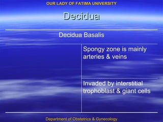
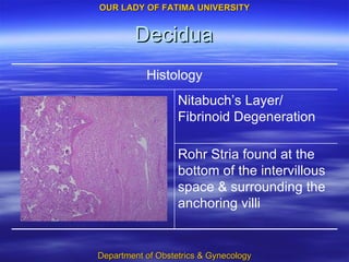
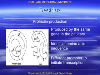
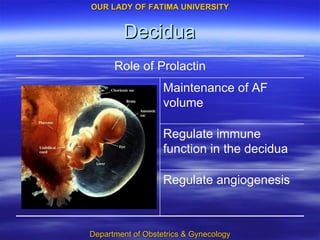
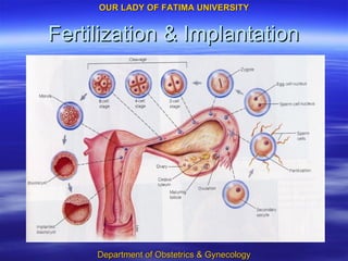
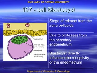
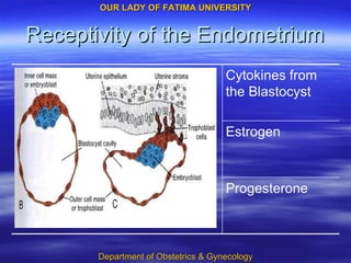
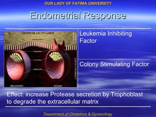
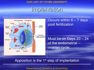
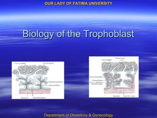
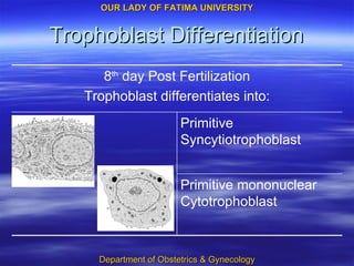
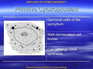
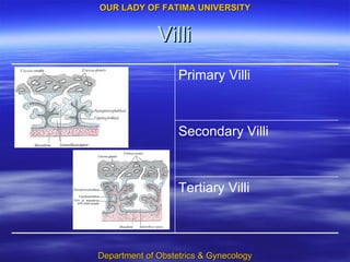
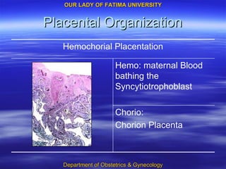
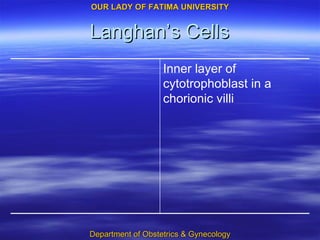
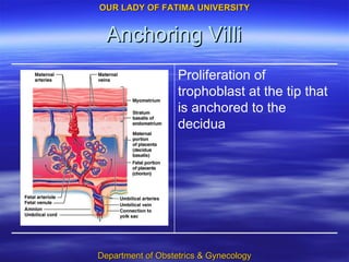
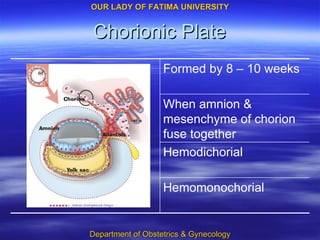
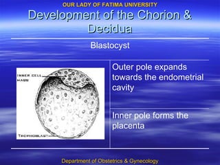
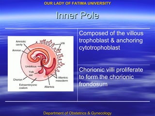
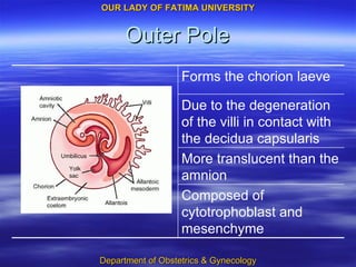
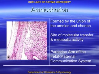
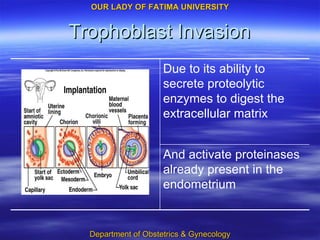
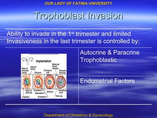
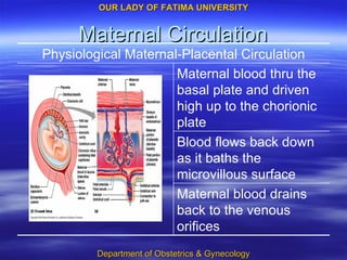
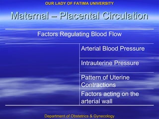
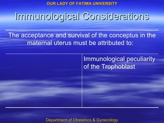
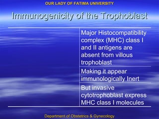
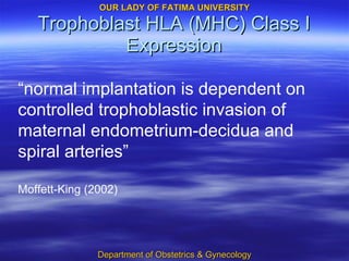
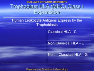
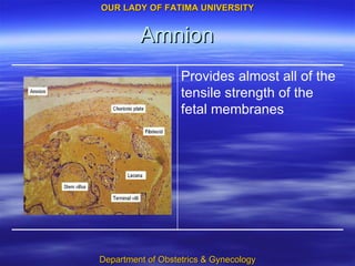
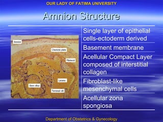
Placenta & Fetal Membranes Crisostomo S. O. Ordoño Jr. MD
Decidua Highly modified Specialized
Decidua Dependent on: Blastocyst Implantation Progesterone Estrogen
Decidua of Pregnancy Decidua Parietalis Decidua Capsularis Decidua Basalis Structure based on anatomical location:
Decidua 14 – 16 weeks AOG Decidua Vera
Decidua Zona Basalis Zona Spongiosa Zona Compacta Layers of the Decidua Parietalis & Basalis
Decidua Reaction Completed with Blastocyst Implantation Commences mid-luteal phase
Decidual Endometrial Stromal Cells Enlarge, polygonal Round & vesicular nuclei Clear & basophilic cytoplasm Pericellular membrane Decidual Reaction
Decidua Protection against proteases Attachment for the Trophoblast Pericellular Membrane provides:
Decidua Decidua Basalis Decidua Capsularis Decidua Parietalis Blood Supply:
Decidual Histology Bone Marrow Derived Cells Endometrial large granular lymphocytes Endometrial Stroma True Decidual Cells
Decidua Becoming cuboidal to flat in later pregnancy Large distended glands with hyperplasia minimal stroma Zona Spongiosa
Decidua Invaded by interstitial trophoblast & giant cells Spongy zone is mainly arteries & veins Decidua Basalis
Decidua Rohr Stria found at the bottom of the intervillous space & surrounding the anchoring villi Nitabuch’s Layer/ Fibrinoid Degeneration Histology
Decidua Different promoter to initiate transcription Identical amino acid sequence Produced by the same gene in the pituitary Prolactin production
Decidua Regulate angiogenesis Regulate immune function in the decidua Maintenance of AF volume Role of Prolactin
Fertilization & Implantation
107 - cell Blastocyst Blastocyst directly influence the receptivity of the endometrium Due to proteases from the secretory endometrium Stage of release from the zona pellucida
Receptivity of the Endometrium Progesterone Estrogen Cytokines from the Blastocyst
Endometrial Response Effect: increase Protease secretion by Trophoblast to degrade the extracellular matrix Colony Stimulating Factor Leukemia Inhibiting Factor
Implantation Must be on Days 20 – 24 of the endometrial – ovarian cycle Apposition is the 1 st  step of implantation Occurs within 6 – 7 days post fertilization
Biology of the Trophoblast
Trophoblast Differentiation Primitive mononuclear Cytotrophoblast Primitive Syncytiotrophoblast 8 th  day Post Fertilization Trophoblast differentiates into:
Primitive Cytotrophoblast Can undergo DNA synthesis Well-demarcated cell border Germinal cells of the syncytium
Primitive Syncytiotrophoblast Lacks the ability for DNA synthesis No individual cells Multi-nucleated
Trophoblast Differentiation Extravillous Trophoblast Villous Trophoblast
Villous Trophoblast Function: Transport of oxygen & nutrients Gives rise to the chorionic villi
Extravillous Trophoblast Migrates to the decidua & myometrium Penetrates the maternal vasculature Interstitial trophoblast Endovascular trophoblast Gives rise to the primary villous stalk
Interstitial Trophoblast Surrounds the maternal spiral arteries Penetrates the maternal decidua & myometrium
Endovascular Trophoblast Penetrates the lumen of the spiral arteries
Trophoblast Invasion Similar invasion characteristic of metastatic cancer cells 10 th  day the blastocyst is totally encased within the decidua
Chorion Trophoblast with an underlying mesenchyme
Villi Tertiary Villi Secondary Villi Primary Villi
Placental Organization Chorio:  Chorion Placenta Hemo: maternal Blood bathing the Syncytiotrophoblast Hemochorial Placentation
Langhan’s Cells Inner layer of cytotrophoblast in a chorionic villi
Anchoring Villi Proliferation of trophoblast at the tip that is anchored to the decidua
Chorionic Plate Hemomonochorial Hemodichorial When amnion & mesenchyme of chorion fuse together Formed by 8 – 10 weeks
Placental Development
Development of the Chorion & Decidua Inner pole forms the placenta Outer pole expands towards the endometrial cavity Blastocyst
Inner Pole Chorionic villi proliferate to form the chorionic frondosum Composed of the villous trophoblast & anchoring cytotrophoblast
Outer Pole Composed of cytotrophoblast and mesenchyme More translucent than the amnion Due to the degeneration of the villi in contact with the decidua capsularis Forms the chorion laeve
Amniochorion Paracrine Arm of the Fetal-Maternal Communication System Site of molecular transfer & metabolic activity Formed by the union of the amnion and chorion
Trophoblast Invasion And activate proteinases already present in the endometrium Due to its ability to secrete proteolytic enzymes to digest the extracellular matrix
Trophoblast Invasion Endometrial Factors Autocrine & Paracrine Trophoblastic  Ability to invade in the 1 st  trimester and limited Invasiveness in the last trimester is controlled by:
Trophoblast Invasion Promotes invasion into the endometrium Acts in an autocrine manner Insulin – like growth Factor Trophoblast
Trophoblast Invasion HENCE: controlled invasiveness Blocks the autocrine loop Insulin like growth factor binding protein type 4 Decidual Cells
Trophoblast Invasion A Trophoblastic Glue Oncofetal Fibronectin or Fetal-Specific Fibronectin Secured by Fetal Fibronectin
Decidual Spiral Artery Invasion Endovascular Trophoblast Interstitial Trophoblast Carried out by two populations of Extravillous Trophoblast:
Decidual Spiral Artery Invasion Prepares the arteries to facilitate invasion Aggregates around the maternal spiral arteries Interstitial Trophoblast
Decidual Spiral Artery Invasion Destroys the endothelium Fibrinoid material replaces muscle and connective tissue Initially forming cellular plugs Enters the lumen of the spiral arteries Endovascular Trophoblast
Decidual Spiral Artery Invasion Decidual Veins are not invaded Does not involve the Decidua Parietalis
Decidual Spiral Artery Invasion 1 st  Stage Before 12 weeks AOG Invasion & Modification of Spiral Arteries Stages of Uteroplacental Vessel Development
Decidual Spiral Artery Invasion 2 nd  Stage Between 12 – 16 weeks Invasion of intramyometrial parts of the spiral arteries Producing low-resistance vessels Stages of Uteroplacental Vessel Development
Establishment of Maternal Blood Flow Occurs approximately 4 weeks post fertilization
Villus Branching Each lobule has one artery and vein Each truncal villi and ramification is one lobule/ cotyledon Majority would arborize and end freely in the intervillous space Some become anchoring villi
Placental Growth & Maturation
Placental Growth At Term, placenta is 1/6 of the fetal weight  17 th  week AOG placenta & fetal weights are approximately equal
Placental Dimensions Weight 508 grams Volume 497 Thickness 23 mm Diameter 185 mm
Placental Maturation Infiltration of Hofbauer cells Later becomes dense and cells more spindly and closely packed Abundant and loose intracellular matrix early in gestation Stroma of the Villi
Placental Maturation Capable of paracrine regulation of Trophoblast Function Increase in number and maturation as pregnancy progress Fetal Macrophages Hofbauer Cells
Placental Maturation Increase Vascularity Decrease Stroma Decrease in thickness of the syncytium Histological Changes that provides an increased efficiency of transport & exchange
Fetal & Maternal Blood Circulation in Mature Placenta
Fetal Circulation Blood with a higher O2 content passes thru the umbilical vein Umbilical vessels branch repeatedly beneath the amnion and diving villi Deoxygenated Blood flows through two umbilical arteries
Fetal Circulation Arteries always cross over the veins Responsive to vasoactive substances Umbilical vessels along the fetal surface of the chorionic plate Placental Surface / Chorionic vessels
Fetal Circulation Decrease amount of smooth muscle but has increase in caliber Each supply one cotyledon Perforating branches of the surface arteries Truncal Arteries
Maternal Circulation Maternal blood drains back to the venous orifices Blood flows back down as it baths the microvillous surface Maternal blood thru the basal plate and driven high up to the chorionic plate Physiological Maternal-Placental Circulation
Maternal – Placental Circulation Arterial Blood Pressure Factors acting on the arterial wall Pattern of Uterine Contractions Intrauterine Pressure Factors Regulating Blood Flow
Immunological Considerations Immunological peculiarity of the Trophoblast The acceptance and survival of the conceptus in the maternal uterus must be attributed to:
Immunogenicity of the Trophoblast But invasive cytotrophoblast express MHC class I molecules Making it appear immunologically Inert Major Histocompatibility complex (MHC) class I and II antigens are absent from villous trophoblast
Trophoblast HLA (MHC) Class I Expression “ normal implantation is dependent on controlled trophoblastic invasion of maternal endometrium-decidua and spiral arteries” Moffett-King (2002)
Trophoblast HLA (MHC) Class I Expression Non – Classical HLA - G Non Classical HLA - E Classical HLA - C Human Leukocyte Antigens Express by the Trophoblasts
Amnion Provides almost all of the tensile strength of the fetal membranes
Amnion Structure Single layer of epithelial cells-ectoderm derived Acellular zona spongiosa Fibroblast-like mesenchymal cells Acellular Compact Layer composed of interstitial collagen Basement membrane
Amnion Development End of the 1 st  trimester – apposition of the mesoblast of the chorion and amnion Growth of the amnion eventually engulfs the embryo and prolapses into the cavity Amniogenic cells – precursor of amnionic epithelium
Amnion Epithelial Cells Metabolically Active Major site of transfer between AF and amnion
Metabolic Activity of the Amnion Synthesis of vasoactive peptides Site of Prostaglandin production Produce PGE2 & Fetal Fibronectin Inhibits metalloprotinase-1 synthesis
Vasoactive peptides produced by the Amnion Corticotrophin – Releasing Hormone –  smooth muscle relaxant Brain natriuretic peptide –  smooth muscle relaxant Parathyroid – related protein -  vasodilator   Endothelin -  vasoconstrictor
Importance of the Metabolic Activity of the Amnion Availability to the fetus of the bioactive agents Modulates chorionic vessel tone and blood flow
Amnion Anatomy Umbilical Cord Amnion Placental Amnion
Amnion Tensile Strength Composed of x-linked collagens I & III Exclusively resides in the compact layer
Metabolic Function Sensitive to mechanical stretching: Triggers autocrine & paracrine responses Production of bioactive agents Maintain AF homeostasis
Amnionic Fluid Average Volume : 1,000 ml at term Continuously increase amount until 34 weeks Clear fluid
Umbilical Cord & Related Structures
Cord Structure & Function Covered by amnion Moist Dull white Extends from the fetal umbilicus to the fetal surface of the placenta
Cord Structure & Function Contains : 2 arteries 1 vein Wharton jelly :  extracellular matrix Average Length: 55 cm Diameter: 0.8 – 2.0 cm
Thank You!

Placenta and fetal membranes

  • 1.
    Placenta & FetalMembranes Crisostomo S. O. Ordoño Jr. MD
  • 2.
  • 3.
    Decidua Dependent on:Blastocyst Implantation Progesterone Estrogen
  • 4.
    Decidua of PregnancyDecidua Parietalis Decidua Capsularis Decidua Basalis Structure based on anatomical location:
  • 5.
    Decidua 14 –16 weeks AOG Decidua Vera
  • 6.
    Decidua Zona BasalisZona Spongiosa Zona Compacta Layers of the Decidua Parietalis & Basalis
  • 7.
    Decidua Reaction Completedwith Blastocyst Implantation Commences mid-luteal phase
  • 8.
    Decidual Endometrial StromalCells Enlarge, polygonal Round & vesicular nuclei Clear & basophilic cytoplasm Pericellular membrane Decidual Reaction
  • 9.
    Decidua Protection againstproteases Attachment for the Trophoblast Pericellular Membrane provides:
  • 10.
    Decidua Decidua BasalisDecidua Capsularis Decidua Parietalis Blood Supply:
  • 11.
    Decidual Histology BoneMarrow Derived Cells Endometrial large granular lymphocytes Endometrial Stroma True Decidual Cells
  • 12.
    Decidua Becoming cuboidalto flat in later pregnancy Large distended glands with hyperplasia minimal stroma Zona Spongiosa
  • 13.
    Decidua Invaded byinterstitial trophoblast & giant cells Spongy zone is mainly arteries & veins Decidua Basalis
  • 14.
    Decidua Rohr Striafound at the bottom of the intervillous space & surrounding the anchoring villi Nitabuch’s Layer/ Fibrinoid Degeneration Histology
  • 15.
    Decidua Different promoterto initiate transcription Identical amino acid sequence Produced by the same gene in the pituitary Prolactin production
  • 16.
    Decidua Regulate angiogenesisRegulate immune function in the decidua Maintenance of AF volume Role of Prolactin
  • 17.
  • 18.
    107 - cellBlastocyst Blastocyst directly influence the receptivity of the endometrium Due to proteases from the secretory endometrium Stage of release from the zona pellucida
  • 19.
    Receptivity of theEndometrium Progesterone Estrogen Cytokines from the Blastocyst
  • 20.
    Endometrial Response Effect:increase Protease secretion by Trophoblast to degrade the extracellular matrix Colony Stimulating Factor Leukemia Inhibiting Factor
  • 21.
    Implantation Must beon Days 20 – 24 of the endometrial – ovarian cycle Apposition is the 1 st step of implantation Occurs within 6 – 7 days post fertilization
  • 22.
    Biology of theTrophoblast
  • 23.
    Trophoblast Differentiation Primitivemononuclear Cytotrophoblast Primitive Syncytiotrophoblast 8 th day Post Fertilization Trophoblast differentiates into:
  • 24.
    Primitive Cytotrophoblast Canundergo DNA synthesis Well-demarcated cell border Germinal cells of the syncytium
  • 25.
    Primitive Syncytiotrophoblast Lacksthe ability for DNA synthesis No individual cells Multi-nucleated
  • 26.
    Trophoblast Differentiation ExtravillousTrophoblast Villous Trophoblast
  • 27.
    Villous Trophoblast Function:Transport of oxygen & nutrients Gives rise to the chorionic villi
  • 28.
    Extravillous Trophoblast Migratesto the decidua & myometrium Penetrates the maternal vasculature Interstitial trophoblast Endovascular trophoblast Gives rise to the primary villous stalk
  • 29.
    Interstitial Trophoblast Surroundsthe maternal spiral arteries Penetrates the maternal decidua & myometrium
  • 30.
    Endovascular Trophoblast Penetratesthe lumen of the spiral arteries
  • 31.
    Trophoblast Invasion Similarinvasion characteristic of metastatic cancer cells 10 th day the blastocyst is totally encased within the decidua
  • 32.
    Chorion Trophoblast withan underlying mesenchyme
  • 33.
    Villi Tertiary VilliSecondary Villi Primary Villi
  • 34.
    Placental Organization Chorio: Chorion Placenta Hemo: maternal Blood bathing the Syncytiotrophoblast Hemochorial Placentation
  • 35.
    Langhan’s Cells Innerlayer of cytotrophoblast in a chorionic villi
  • 36.
    Anchoring Villi Proliferationof trophoblast at the tip that is anchored to the decidua
  • 37.
    Chorionic Plate HemomonochorialHemodichorial When amnion & mesenchyme of chorion fuse together Formed by 8 – 10 weeks
  • 38.
  • 39.
    Development of theChorion & Decidua Inner pole forms the placenta Outer pole expands towards the endometrial cavity Blastocyst
  • 40.
    Inner Pole Chorionicvilli proliferate to form the chorionic frondosum Composed of the villous trophoblast & anchoring cytotrophoblast
  • 41.
    Outer Pole Composedof cytotrophoblast and mesenchyme More translucent than the amnion Due to the degeneration of the villi in contact with the decidua capsularis Forms the chorion laeve
  • 42.
    Amniochorion Paracrine Armof the Fetal-Maternal Communication System Site of molecular transfer & metabolic activity Formed by the union of the amnion and chorion
  • 43.
    Trophoblast Invasion Andactivate proteinases already present in the endometrium Due to its ability to secrete proteolytic enzymes to digest the extracellular matrix
  • 44.
    Trophoblast Invasion EndometrialFactors Autocrine & Paracrine Trophoblastic Ability to invade in the 1 st trimester and limited Invasiveness in the last trimester is controlled by:
  • 45.
    Trophoblast Invasion Promotesinvasion into the endometrium Acts in an autocrine manner Insulin – like growth Factor Trophoblast
  • 46.
    Trophoblast Invasion HENCE:controlled invasiveness Blocks the autocrine loop Insulin like growth factor binding protein type 4 Decidual Cells
  • 47.
    Trophoblast Invasion ATrophoblastic Glue Oncofetal Fibronectin or Fetal-Specific Fibronectin Secured by Fetal Fibronectin
  • 48.
    Decidual Spiral ArteryInvasion Endovascular Trophoblast Interstitial Trophoblast Carried out by two populations of Extravillous Trophoblast:
  • 49.
    Decidual Spiral ArteryInvasion Prepares the arteries to facilitate invasion Aggregates around the maternal spiral arteries Interstitial Trophoblast
  • 50.
    Decidual Spiral ArteryInvasion Destroys the endothelium Fibrinoid material replaces muscle and connective tissue Initially forming cellular plugs Enters the lumen of the spiral arteries Endovascular Trophoblast
  • 51.
    Decidual Spiral ArteryInvasion Decidual Veins are not invaded Does not involve the Decidua Parietalis
  • 52.
    Decidual Spiral ArteryInvasion 1 st Stage Before 12 weeks AOG Invasion & Modification of Spiral Arteries Stages of Uteroplacental Vessel Development
  • 53.
    Decidual Spiral ArteryInvasion 2 nd Stage Between 12 – 16 weeks Invasion of intramyometrial parts of the spiral arteries Producing low-resistance vessels Stages of Uteroplacental Vessel Development
  • 54.
    Establishment of MaternalBlood Flow Occurs approximately 4 weeks post fertilization
  • 55.
    Villus Branching Eachlobule has one artery and vein Each truncal villi and ramification is one lobule/ cotyledon Majority would arborize and end freely in the intervillous space Some become anchoring villi
  • 56.
  • 57.
    Placental Growth AtTerm, placenta is 1/6 of the fetal weight 17 th week AOG placenta & fetal weights are approximately equal
  • 58.
    Placental Dimensions Weight508 grams Volume 497 Thickness 23 mm Diameter 185 mm
  • 59.
    Placental Maturation Infiltrationof Hofbauer cells Later becomes dense and cells more spindly and closely packed Abundant and loose intracellular matrix early in gestation Stroma of the Villi
  • 60.
    Placental Maturation Capableof paracrine regulation of Trophoblast Function Increase in number and maturation as pregnancy progress Fetal Macrophages Hofbauer Cells
  • 61.
    Placental Maturation IncreaseVascularity Decrease Stroma Decrease in thickness of the syncytium Histological Changes that provides an increased efficiency of transport & exchange
  • 62.
    Fetal & MaternalBlood Circulation in Mature Placenta
  • 63.
    Fetal Circulation Bloodwith a higher O2 content passes thru the umbilical vein Umbilical vessels branch repeatedly beneath the amnion and diving villi Deoxygenated Blood flows through two umbilical arteries
  • 64.
    Fetal Circulation Arteriesalways cross over the veins Responsive to vasoactive substances Umbilical vessels along the fetal surface of the chorionic plate Placental Surface / Chorionic vessels
  • 65.
    Fetal Circulation Decreaseamount of smooth muscle but has increase in caliber Each supply one cotyledon Perforating branches of the surface arteries Truncal Arteries
  • 66.
    Maternal Circulation Maternalblood drains back to the venous orifices Blood flows back down as it baths the microvillous surface Maternal blood thru the basal plate and driven high up to the chorionic plate Physiological Maternal-Placental Circulation
  • 67.
    Maternal – PlacentalCirculation Arterial Blood Pressure Factors acting on the arterial wall Pattern of Uterine Contractions Intrauterine Pressure Factors Regulating Blood Flow
  • 68.
    Immunological Considerations Immunologicalpeculiarity of the Trophoblast The acceptance and survival of the conceptus in the maternal uterus must be attributed to:
  • 69.
    Immunogenicity of theTrophoblast But invasive cytotrophoblast express MHC class I molecules Making it appear immunologically Inert Major Histocompatibility complex (MHC) class I and II antigens are absent from villous trophoblast
  • 70.
    Trophoblast HLA (MHC)Class I Expression “ normal implantation is dependent on controlled trophoblastic invasion of maternal endometrium-decidua and spiral arteries” Moffett-King (2002)
  • 71.
    Trophoblast HLA (MHC)Class I Expression Non – Classical HLA - G Non Classical HLA - E Classical HLA - C Human Leukocyte Antigens Express by the Trophoblasts
  • 72.
    Amnion Provides almostall of the tensile strength of the fetal membranes
  • 73.
    Amnion Structure Singlelayer of epithelial cells-ectoderm derived Acellular zona spongiosa Fibroblast-like mesenchymal cells Acellular Compact Layer composed of interstitial collagen Basement membrane
  • 74.
    Amnion Development Endof the 1 st trimester – apposition of the mesoblast of the chorion and amnion Growth of the amnion eventually engulfs the embryo and prolapses into the cavity Amniogenic cells – precursor of amnionic epithelium
  • 75.
    Amnion Epithelial CellsMetabolically Active Major site of transfer between AF and amnion
  • 76.
    Metabolic Activity ofthe Amnion Synthesis of vasoactive peptides Site of Prostaglandin production Produce PGE2 & Fetal Fibronectin Inhibits metalloprotinase-1 synthesis
  • 77.
    Vasoactive peptides producedby the Amnion Corticotrophin – Releasing Hormone – smooth muscle relaxant Brain natriuretic peptide – smooth muscle relaxant Parathyroid – related protein - vasodilator Endothelin - vasoconstrictor
  • 78.
    Importance of theMetabolic Activity of the Amnion Availability to the fetus of the bioactive agents Modulates chorionic vessel tone and blood flow
  • 79.
    Amnion Anatomy UmbilicalCord Amnion Placental Amnion
  • 80.
    Amnion Tensile StrengthComposed of x-linked collagens I & III Exclusively resides in the compact layer
  • 81.
    Metabolic Function Sensitiveto mechanical stretching: Triggers autocrine & paracrine responses Production of bioactive agents Maintain AF homeostasis
  • 82.
    Amnionic Fluid AverageVolume : 1,000 ml at term Continuously increase amount until 34 weeks Clear fluid
  • 83.
    Umbilical Cord &Related Structures
  • 84.
    Cord Structure &Function Covered by amnion Moist Dull white Extends from the fetal umbilicus to the fetal surface of the placenta
  • 85.
    Cord Structure &Function Contains : 2 arteries 1 vein Wharton jelly : extracellular matrix Average Length: 55 cm Diameter: 0.8 – 2.0 cm
  • 86.

Editor's Notes

  • #3 Decidua is a specialized, highly modified endometrium of pregnancy and is a function of the hemochorial placentation
  • #4 Transformation is dependent on: Estrogen, Progesterone & factors secreted by the blastocyst during trophoblast invasion
  • #5 The decidua of pregnancy is anatomically composed of three parts: Decidua Basalis, Decidua Capsularis, Decidua Parietalis
  • #6 Decidua vera is the point where decidua parietalis and capsularis are joined
  • #7 The decidua basalis & parietalis are composed of three layers
  • #8 Predecidual reaction starts during the mid luteal phase of the endometrial cycle
  • #10 Protection against proteases of the cytotrophoblast
  • #11 Decidua Parietalis = retain a smooth muscle wall and endothelium and remain responsive to vasoactive agents Decidua Capsularis = blood supply is lost as the embryo grows Decidua Basalis = spiral arteries are invaded by the trophoblast and a vascular shell is left behind, not responsive to vasoactive agents
  • #12 The primary cell components of the true decidual cells are from endometrial stromal cells & numerous bone marrow derived Cells
  • #13 Initially with large distended glands with abundant secreatory activity for nourishment of the bastocyst
  • #14 Decidua Basalis contributes to the formation of the basal plate of the placenta It differs from the decidua parietalis in two important respects: D. Basalis consist mainly of arteries and veins D. Basalis s invaded by large numbers of interstitial trophoblast cells & giant cells
  • #15 Where the invading trophoblast meet the decidua there is a zone of fibrinoid degeneration/ Nitabuch’s layer
  • #16 Decidua is the source of the prolactin that is present in enormous amounts in amnionic fluid during human pregnancy
  • #17 Role in solute and water transport across the amniochorion Act on human T cells in an autocrine & paracrine manner Regulate angiogenesis during implantation
  • #18 Fertilization must occur within a few hours Almost all pregnancies occur during the 2 days preceding or on the day of ovulation Zygote undergoes slow cleavage for 3 days while still in the fallopian tube Morula enters the uterine cavity about 3 days after fertilization 58 cell blastula differentiates into 5 cell inner cell mass & 53 outer cell mass 107 cell blastocyst = 8 inner cell mass & 99 trophoblastic cells.
  • #20 Cytokines secreted Interleukin 1 α (IL- 1 α ) and Interleukin 1Ăź(IL-1Ăź)
  • #75 Amniogenic cells line the inner surface of the trophoblast
  • #76 Apical surface of the amnion if full of developed microvilli