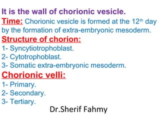
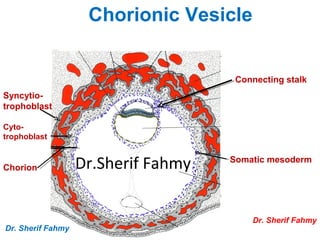
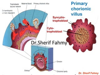
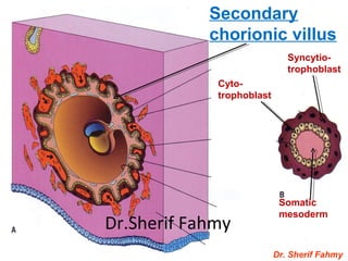
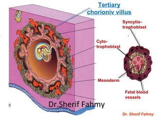
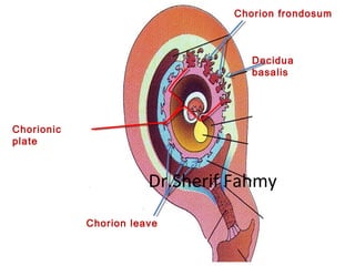
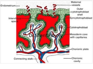
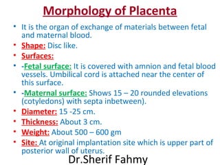
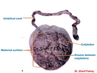
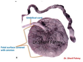
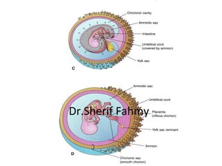
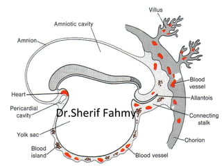
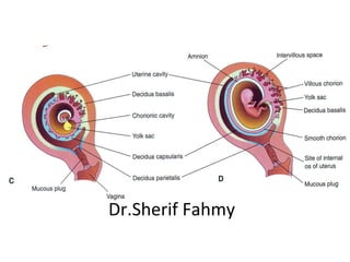
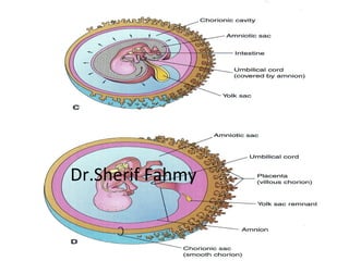
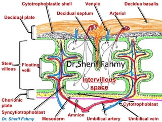
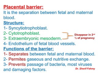
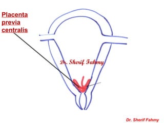
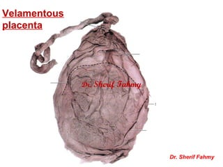
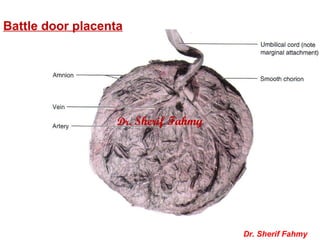
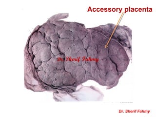
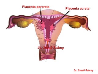
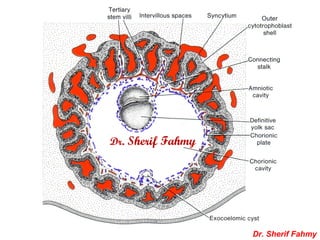
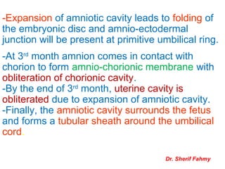
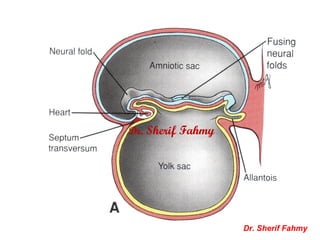
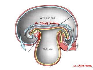
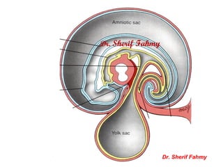
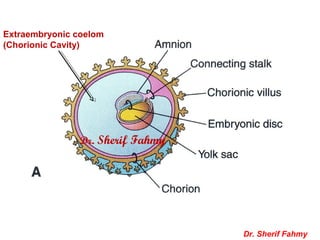
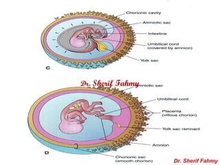
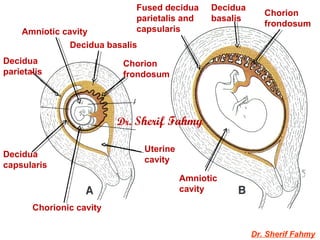
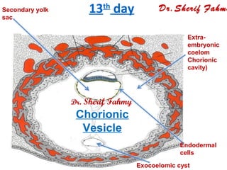
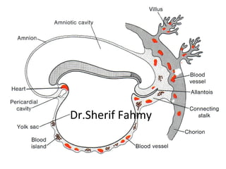
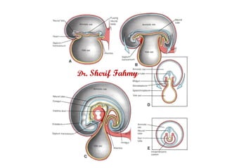
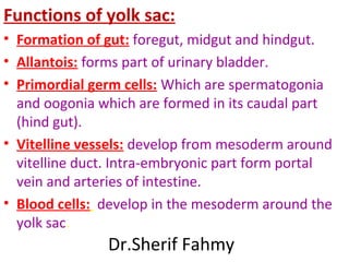
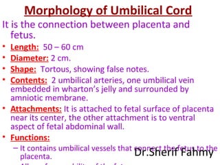
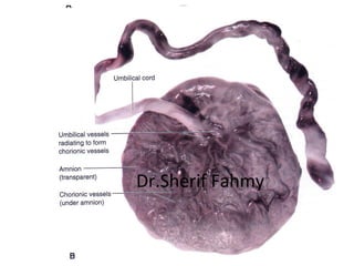
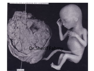
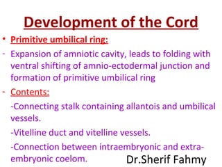
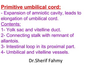
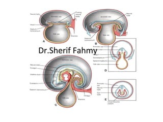
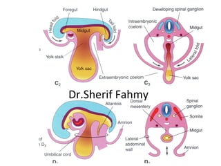
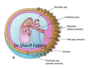
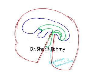
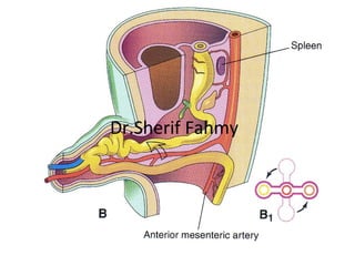
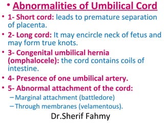
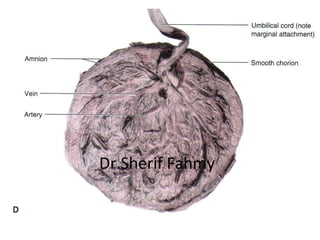
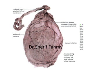
Fetal Membranes
Dr.Sherif Fahmy
Fetal membranes:
1- Chorion
2- Placenta.
2- Amnion.
3- Umbilical cord.
4- Yolk sac.
Dr.Sherif Fahmy
Chorion
Dr.Sherif Fahmy
It is the wall of chorionic vesicle.
Time: Chorionic vesicle is formed at the 12th
day
by the formation of extra-embryonic mesoderm.
Structure of chorion:
1- Syncytiotrophoblast.
2- Cytotrophoblast.
3- Somatic extra-embryonic mesoderm.
Chorionic velli:
1- Primary.
2- Secondary.
3- Tertiary.
Dr.Sherif Fahmy
Connecting stalk
Somatic mesoderm
Syncytio-
trophoblast
Cyto-
trophoblast
Chorion
Chorionic Vesicle
Dr. Sherif Fahmy
Dr. Sherif Fahmy
Dr.Sherif Fahmy
Dr. Sherif Fahmy
Primary
chorionic
villus
Cyto-
trophoblast
Syncytio-
trophoblast
Dr.Sherif Fahmy
Dr. Sherif Fahmy
Syncytio-
trophoblast
Cyto-
trophoblast
Somatic
mesoderm
Secondary
chorionic villus
Dr.Sherif Fahmy
Dr. Sherif Fahmy
Syncytio-
trophoblast
Cyto-
trophoblast
Mesoderm
Fetal blood
vessels
Tertiary
chorioniv villus
Dr.Sherif Fahmy
Decidua
basalis
Chorion frondosum
Chorionic
plate
Chorion leave
Dr.Sherif Fahmy
Dr.Sherif Fahmy
PLACENTA
(Page 38)
Dr.Sherif Fahmy
Morphology of Placenta
• It is the organ of exchange of materials between fetal
and maternal blood.
• Shape: Disc like.
• Surfaces:
• -Fetal surface: It is covered with amnion and fetal blood
vessels. Umbilical cord is attached near the center of
this surface.
• -Maternal surface: Shows 15 – 20 rounded elevations
(cotyledons) with septa inbetween).
• Diameter: 15 -25 cm.
• Thickness: About 3 cm.
• Weight: About 500 – 600 gm
• Site: At original implantation site which is upper part of
posterior wall of uterus.
Dr.Sherif Fahmy
Cotyledon
Groove between
cotyledons
Umbilical cord
Maternal surface
Dr. Sherif Fahmy
Dr.Sherif Fahmy
Fetal surface covered
with amnion
Umbilical cord
Dr. Sherif Fahmy
Dr.Sherif Fahmy
Formation of Placenta
Dr.Sherif Fahmy
Dr.Sherif Fahmy
Dr.Sherif Fahmy
Dr.Sherif Fahmy
Structure of Placenta
Dr.Sherif Fahmy
Dr.Sherif Fahmy
Decidua basalis
Arteriol
Venule
Decidual septum
Cytotrophoblastic shell
Decidual plate
Stem
villous
Floating
velli
Intervillous
space
Chorionic
plate
Umbilical veinUmbilical artery
Amnion
Mesoderm
Syncytiotrophoblast
Cytotrophoblast
Dr. Sherif Fahmy
Dr.Sherif Fahmy
Placental barrier:
It is the separation between fetal and maternal
blood.
Structure:
1- Syncytiotrophoblast.
2- Cytotrophoblast.
3- Extraembryonic mesoderm.
4- Endothelium of fetal blood vessels.
Functions of the barrier:
1- Separates between fetal and maternal blood.
2- Permites gaseous and nutritive exchange.
3- Prevents passage of bacteria, most viruses
and damaging factors.
Disappear in 2nd
½ of pregnancy
Dr. Sherif Fahmy
Placental circulation:
1- Maternal part: Maternal blood flow from
endometrial arterioles to the intervillous
spaces where floating velli are bathed in
maternal blood. Exchange of gases and
nutritive materials occurs. Then blood flows
back from chorionic plate to endometrial
veins.
2- Fetal part: umbilical arteries carry
venous blood of the fetus to placenta while
umbilical veins carry blood loaded with
nutritive material and oxygen. Dr. Sherif Fahmy
Functions of placenta
1- Exchange of gases and metabolites.
2- Transmission of maternal antibodies
starting from 14th
week.
3- Production of hormones as progesterone,
estrogen, HCG and somatomammotropin
3- Barrier against bacteria and most of viruses.
4- Excretory function as it excretes urea and
creatinine.
Dr. Sherif Fahmy
Anomalies of Placenta
1- Abnormalities in position:
A- Placenta previa parietalis.
B- Placenta brevia marginalis.
C- Placenta brevia centralis.
2- Abnormality in shape:
A- biloped placenta.
B- Triloped placenta.
3- Abnormality in number:
A- Twin placenta.
B- Accessory placenta.
4- Abnormality in attachement of umbilical cord:
A- Velamentous.
B- Battle door. Dr. Sherif Fahmy
Dr. Sherif Fahmy
Placenta
previa
Marginalis &
parietalis
Dr. Sherif Fahmy
Dr. Sherif Fahmy
Placenta
previa
centralis
Dr. Sherif Fahmy
Dr. Sherif Fahmy
Velamentous
placenta
Dr. Sherif Fahmy
Dr. Sherif Fahmy
Battle door placenta
Dr. Sherif Fahmy
Dr. Sherif Fahmy
Accessory placenta
Dr. Sherif Fahmy
Dr. Sherif Fahmy
Placenta acreta
Placenta percreta
Dr. Sherif Fahmy
Dr. Sherif Fahmy
AMNION
Dr.Sherif Fahmy
AMNION
-It is a membrane that enclose amniotic cavity.
-Formation:
-It is formed at the 8th
day as a small cavity in
epiblast cells with formation of amnioblasts.
-So, floor of the cavity is epiblast while the
roof is formed from amnioblasts.
-By the 12th
day it becomes separated from
cytotrophoblasts by primary mesoderm
(Extraembryonic).
-Amnio-ectodermal junction is at the margin
of oval embryonic disc at the 3rd
week.
8th
day of pregnancy
Dr. Sherif Fahmy
Amnioblast
Amniotic cavity
Epiblast Hypoblast
Dr. Sherif Fahmy
9th
& 10th
days
Dr. Sherif Fahmy
Cyto-
trophoblast
Amnioblast
Amniotic
cavity
Epiblast
Dr. Sherif Fahmy
Dr. Sherif Fahmy
Dr. Sherif Fahmy
-At 3rd
month amnion comes in contact with
chorion to form amnio-chorionic membrane with
obliteration of chorionic cavity.
-By the end of 3rd
month, uterine cavity is
obliterated due to expansion of amniotic cavity.
-Finally, the amniotic cavity surrounds the fetus
and forms a tubular sheath around the umbilical
cord.
-Expansion of amniotic cavity leads to folding of
the embryonic disc and amnio-ectodermal
junction will be present at primitive umbilical ring.
Dr. Sherif Fahmy
Dr. Sherif Fahmy
Dr. Sherif Fahmy
Dr. Sherif Fahmy
Dr. Sherif Fahmy
Dr. Sherif Fahmy
Dr. Sherif Fahmy
Dr. Sherif Fahmy
Extraembryonic coelom
(Chorionic Cavity)
Dr. Sherif Fahmy
Dr. Sherif Fahmy
Dr. Sherif Fahmy
Dr. Sherif Fahmy
Decidua basalis
Decidua
parietalis
Decidua
capsularis
Chorion
frondosum
Uterine
cavity
Amniotic cavity
Chorion
frondosum
Fused decidua
parietalis and
capsularis
Amniotic
cavity
Decidua
basalis
Chorionic cavity
Dr. Sherif Fahmy
Amniotic fluid
- Normal volume is 1000 – 1500 cc clear
watery fluid.
- Source: 1st
from amnioblast then from
kidney.
- If the volume is less than 500 cc it is
called oligohydramnios.
-If the volume is more than 2000 cc is
called polyhydramnios. Dr. Sherif Fahmy
Functions of amniotic fluid:
1- At early pregnancy:
1- Acts as water cushion that absorbs external
shocks.
2- Acts as heat insulator.
3- Prevents adhesion of embryo to wall of uterus.
4- Prevents adhesion of fetal parts.
2- At late pregnancy:
1- A space for accumulated urine.
2- Allows fetal movements to help body muscles
to develop.
3- Help suckling training and development of gut
muscles. Dr. Sherif Fahmy
3- During labor:
1- Protects against uterine contractions.
2- Formation of bag of water that gradually
dilate the cervix.
3- Sterile amniotic washes vagina before
passage of baby.
4- Rupture of amniotic sac is a sign of start
of delivery.
Dr. Sherif Fahmy
Abnormalities of amniotic fluid:
1- Polyhydramnios.
Causes:
1- No cause (35 %).
2- Maternal diabetes.
3- Congenital malformation e.g.
anencephaly and esophageal atresia.
2- Oligohydramnios.
Cause:
-Renal agenesis.
3- Premature rupture of amnion.
Dr. Sherif Fahmy
YOLK SAC
Dr.Sherif Fahmy
Fate & development of yolk sac
• Primary yolk sac: It replaces cavity of blastocyst
after the formation of Heuser’s membrane which is
formed of flat cells that originate from hypoblast
cells at 9th
& 10th
day.
• Secondary yolk sac: additional endodermal cells
from hypoblast cells will line the Heuser’s
membrane, reduction of size of yolk sac and
formation of allantois. This occurs in the 13th
day.
• Defenitive yolk sac: During 3rd
week, hypoblast
become replaced by endoderm. After folding, it
shares in formation of gut and the part remains
outside the embryo is called defenitive yolk sac. It is
connected to yolk sac by vitello-intestinal duct.
7th
day:
Dr. Sherif Fahmy
Dr. Sherif Fahmy
8TH
Day of Pregnancy
Endometrium
Cytotrophoblasts Hypoblasts
Amniotic cavity
Epiblast
Dr.Sherif Fahmy
Dr. Sherif Fahmy
9th
& 10th
days
Primary yolk sac
Heuser’s
membrane
Hypoblast
Amniotic
cavity
Epiblast
Dr. Sherif Fahmy
13th
day
Endodermal
cells
Secondary yolk
sac
Exocoelomic cyst
Extra-
embryonic
coelom
Chorionic
cavity)
Dr.Sherif Fahmy
Chorionic
Vesicle
Dr. Sherif Fahmy
Dr.Sherif Fahmy
Dr. Sherif Fahmy
Functions of yolk sac:
• Formation of gut: foregut, midgut and hindgut.
• Allantois: forms part of urinary bladder.
• Primordial germ cells: Which are spermatogonia
and oogonia which are formed in its caudal part
(hind gut).
• Vitelline vessels: develop from mesoderm around
vitelline duct. Intra-embryonic part form portal
vein and arteries of intestine.
• Blood cells: develop in the mesoderm around the
yolk sac.
Dr.Sherif Fahmy
Abnormalities of Yolk
Sac
• 1- Vitelline cyst and fistula
due to persistence of vitelline
duct.
• 2- Urachal cyst and fistula due
to persistence of urachus
from allantois.
Dr.Sherif Fahmy
Dr. Sherif Fahmy
Dr. Sherif Fahmy
UMBILICAL
CORD
Dr.Sherif Fahmy
Morphology of Umbilical Cord
It is the connection between placenta and
fetus.
• Length: 50 – 60 cm
• Diameter: 2 cm.
• Shape: Tortous, showing false notes.
• Contents: 2 umbilical arteries, one umbilical vein
embedded in wharton’s jelly and surrounded by
amniotic membrane.
• Attachments: It is attached to fetal surface of placenta
near its center, the other attachment is to ventral
aspect of fetal abdominal wall.
• Functions:
– It contains umbilical vessels that connect the fetus to the
placenta. Dr.Sherif Fahmy
Dr.Sherif Fahmy
Dr.Sherif Fahmy
Development of the Cord
• Primitive umbilical ring:
- Expansion of amniotic cavity, leads to folding with
ventral shifting of amnio-ectodermal junction and
formation of primitive umbilical ring
- Contents:
-Connecting stalk containing allantois and umbilical
vessels.
-Vitelline duct and vitelline vessels.
-Connection between intraembryonic and extra-
embryonic coelom. Dr.Sherif Fahmy
Primitive umbilical cord:
- Expansion of amniotic cavity, leads to
elongation of umbilical cord.
Contents:
1- Yolk sac and vitelline duct.
2- Connecting stalk with remnant of
allantois.
3- Intestinal loop in its proximal part.
4- Umbilical and vitelline vessels.
Dr.Sherif Fahmy
Definitive umbilical cord:
- Return of intestinal loop to abdominal cavity at
3rd
month.
-Obliteration of vitelline duct, allantois, extra-
embryonic part of vitelline vessels.
-Degeneration of one umbilical vein with
persistence of other vein and 2 umbilical
arteries.
-Transformation of mesoderm of connecting
stalk into wharton’s jelly.
Dr.Sherif Fahmy
Development
Dr.Sherif Fahmy
Embryonic disc with removed
ectoderm
Intra-
embryonic
ceolom
Dr.Sherif Fahmy
Dr.Sherif Fahmy
Dr.Sherif Fahmy
Dr.Sherif Fahmy
Dr.Sherif Fahmy
Dr.Sherif Fahmy
Dr.Sherif Fahmy
Dr.Sherif Fahmy
• Abnormalities of Umbilical Cord
• 1- Short cord: leads to premature separation
of placenta.
• 2- Long cord: It may encircle neck of fetus and
may form true knots.
• 3- Congenital umbilical hernia
(omphalocele): the cord contains coils of
intestine.
• 4- Presence of one umbilical artery.
• 5- Abnormal attachment of the cord:
–Marginal attachment (battledore)
–Through membranes (velamentous).
Dr.Sherif Fahmy
Dr.Sherif Fahmy
Dr.Sherif Fahmy
Dr.Sherif Fahmy
Twins
Dr.Sherif Fahmy
Types of TWINS
Dizygotic (Fraternal) twins:
- It is the commonest type as it represent 2/3 of
twins and 7 – 11 / 1000 births.
- Fertilization of 2 separate ova.
- Each embryo has its own placenta, chorion
and amniotic cavity.
- Twins are non-identical and may of same sex
or different.
Dr.Sherif Fahmy
Monozygotic (Identical) twins:
Developed from division of a fertilized ovum. Twins of this type are
identical and of same sex. Its incidence is 0.3 – 0.4 %
Division may occure at 3 different stages:
1- At morula stage: Twins has separate amnion,
chorion and placentae (as in dizygotic).
2- At blastocyst stage: due to division of inner cell
mass. Twins has separate amniotic cavity but single
chorion and placenta.
3- At embryonic disc: Midline division of the embryonic
disc. Twins has common amniotic cavity, common
chorion and common placenta .
Dr.Sherif Fahmy
Morula
stage
EmbryonicDisc StageEarly
blastcyste
Dr.Sherif Fahmy
SIAMESE (CONJOINED) TWINS
• Fused monozygotic twins that occurs
due to incomplete separation of
emberyonic disc. They could be
either:
Craniopagus: Fusion between 2 heads.
Thoracopagus: Fusion at thoracic region.
Pygopagus: Fusion at the pelvic region.
Dr.Sherif Fahmy
Conjoined Twines
Dr.Sherif Fahmy
Birth Defects
Dr.Sherif Fahmy
Down Syndrome
Dr.Sherif Fahmy
Dr.Sherif Fahmy
Cri du Chat Syndrome
Dr.Sherif Fahmy
Turner Syndrome
Dr.Sherif Fahmy
Achondroplasia
Dr.Sherif Fahmy
A Child of A Mother Treated by
Antiepileptic drug
A Child of A Mother Treated with
Antithyroid drug
A Child of A mother Exposed to
Rubella Infection
External Appearance of the Embryo
(4th
– 8th
week)
At the embryonic period (4th
– 8th
week),
human shape becomes easily identified.
-Head, body and limb buds are easily
identified.
-Eyes, nose and ears are seen.
Dr.Sherif Fahmy
C-R length in mms Age of embryo in weeks
5 – 8 5
10 – 14 6
17 – 22 7
28 – 30 8
Dr.Sherif Fahmy
Fetal Period
Dr.Sherif Fahmy
C-R length in cm Age of embryo in months
5 – 8 cm 3rd
month
18 cm 5th
month
36 cm Full term fetus at birth
Dr.Sherif Fahmy
Relative size of head to body:
-At the beginning of the 3rd
month, the head is ½
the CR length.
-At the beginning of the 5th
month, the head is 1/3
the CH length.
-At birth, the head is ¼ of CH length.
Weight growth:
-At the end of 5th
month, the weight is ½ kg.
-At the 7th
month, the weight is 1.75 kg.
-At full term, the weight is 3.5 kg.
Dr.Sherif Fahmy
Changes in external features:
-Face becomes human looking.
-Limbs become longer.
-External genitalia are differentiated at 12th
week.
-Lanugo hair covers the fetus since the 4th
month.
-The skin is wrinkled till the end of 6th
month.
-Testes descend to scrotum just before birth.
-Skin is covered by fatty substance called vernix
caesosa.
Fetal movement:
It is clearly recognized since the 5th
month.
Dr.Sherif Fahmy
The End &
Beginning
With my best wishes
Dr. Sherif Fahmy

Revision on General Embryology 2

  • 1.
  • 2.
    Fetal membranes: 1- Chorion 2-Placenta. 2- Amnion. 3- Umbilical cord. 4- Yolk sac. Dr.Sherif Fahmy
  • 3.
  • 4.
    It is thewall of chorionic vesicle. Time: Chorionic vesicle is formed at the 12th day by the formation of extra-embryonic mesoderm. Structure of chorion: 1- Syncytiotrophoblast. 2- Cytotrophoblast. 3- Somatic extra-embryonic mesoderm. Chorionic velli: 1- Primary. 2- Secondary. 3- Tertiary. Dr.Sherif Fahmy
  • 5.
  • 6.
  • 7.
  • 8.
    Dr. Sherif Fahmy Syncytio- trophoblast Cyto- trophoblast Mesoderm Fetalblood vessels Tertiary chorioniv villus Dr.Sherif Fahmy
  • 9.
  • 10.
  • 11.
  • 12.
    Morphology of Placenta •It is the organ of exchange of materials between fetal and maternal blood. • Shape: Disc like. • Surfaces: • -Fetal surface: It is covered with amnion and fetal blood vessels. Umbilical cord is attached near the center of this surface. • -Maternal surface: Shows 15 – 20 rounded elevations (cotyledons) with septa inbetween). • Diameter: 15 -25 cm. • Thickness: About 3 cm. • Weight: About 500 – 600 gm • Site: At original implantation site which is upper part of posterior wall of uterus. Dr.Sherif Fahmy
  • 13.
    Cotyledon Groove between cotyledons Umbilical cord Maternalsurface Dr. Sherif Fahmy Dr.Sherif Fahmy
  • 14.
    Fetal surface covered withamnion Umbilical cord Dr. Sherif Fahmy Dr.Sherif Fahmy
  • 15.
  • 16.
  • 17.
  • 18.
  • 19.
  • 20.
  • 21.
    Decidua basalis Arteriol Venule Decidual septum Cytotrophoblasticshell Decidual plate Stem villous Floating velli Intervillous space Chorionic plate Umbilical veinUmbilical artery Amnion Mesoderm Syncytiotrophoblast Cytotrophoblast Dr. Sherif Fahmy Dr.Sherif Fahmy
  • 22.
    Placental barrier: It isthe separation between fetal and maternal blood. Structure: 1- Syncytiotrophoblast. 2- Cytotrophoblast. 3- Extraembryonic mesoderm. 4- Endothelium of fetal blood vessels. Functions of the barrier: 1- Separates between fetal and maternal blood. 2- Permites gaseous and nutritive exchange. 3- Prevents passage of bacteria, most viruses and damaging factors. Disappear in 2nd ½ of pregnancy Dr. Sherif Fahmy
  • 23.
    Placental circulation: 1- Maternalpart: Maternal blood flow from endometrial arterioles to the intervillous spaces where floating velli are bathed in maternal blood. Exchange of gases and nutritive materials occurs. Then blood flows back from chorionic plate to endometrial veins. 2- Fetal part: umbilical arteries carry venous blood of the fetus to placenta while umbilical veins carry blood loaded with nutritive material and oxygen. Dr. Sherif Fahmy
  • 24.
    Functions of placenta 1-Exchange of gases and metabolites. 2- Transmission of maternal antibodies starting from 14th week. 3- Production of hormones as progesterone, estrogen, HCG and somatomammotropin 3- Barrier against bacteria and most of viruses. 4- Excretory function as it excretes urea and creatinine. Dr. Sherif Fahmy
  • 25.
    Anomalies of Placenta 1-Abnormalities in position: A- Placenta previa parietalis. B- Placenta brevia marginalis. C- Placenta brevia centralis. 2- Abnormality in shape: A- biloped placenta. B- Triloped placenta. 3- Abnormality in number: A- Twin placenta. B- Accessory placenta. 4- Abnormality in attachement of umbilical cord: A- Velamentous. B- Battle door. Dr. Sherif Fahmy Dr. Sherif Fahmy
  • 26.
  • 27.
  • 28.
  • 29.
    Battle door placenta Dr.Sherif Fahmy Dr. Sherif Fahmy
  • 30.
    Accessory placenta Dr. SherifFahmy Dr. Sherif Fahmy
  • 31.
    Placenta acreta Placenta percreta Dr.Sherif Fahmy Dr. Sherif Fahmy
  • 32.
  • 33.
    AMNION -It is amembrane that enclose amniotic cavity. -Formation: -It is formed at the 8th day as a small cavity in epiblast cells with formation of amnioblasts. -So, floor of the cavity is epiblast while the roof is formed from amnioblasts. -By the 12th day it becomes separated from cytotrophoblasts by primary mesoderm (Extraembryonic). -Amnio-ectodermal junction is at the margin of oval embryonic disc at the 3rd week.
  • 34.
    8th day of pregnancy Dr.Sherif Fahmy Amnioblast Amniotic cavity Epiblast Hypoblast Dr. Sherif Fahmy
  • 35.
    9th & 10th days Dr. SherifFahmy Cyto- trophoblast Amnioblast Amniotic cavity Epiblast Dr. Sherif Fahmy
  • 36.
  • 37.
    -At 3rd month amnioncomes in contact with chorion to form amnio-chorionic membrane with obliteration of chorionic cavity. -By the end of 3rd month, uterine cavity is obliterated due to expansion of amniotic cavity. -Finally, the amniotic cavity surrounds the fetus and forms a tubular sheath around the umbilical cord. -Expansion of amniotic cavity leads to folding of the embryonic disc and amnio-ectodermal junction will be present at primitive umbilical ring. Dr. Sherif Fahmy
  • 38.
  • 39.
  • 40.
  • 41.
    Dr. Sherif Fahmy Extraembryoniccoelom (Chorionic Cavity) Dr. Sherif Fahmy
  • 42.
  • 43.
    Dr. Sherif Fahmy Deciduabasalis Decidua parietalis Decidua capsularis Chorion frondosum Uterine cavity Amniotic cavity Chorion frondosum Fused decidua parietalis and capsularis Amniotic cavity Decidua basalis Chorionic cavity Dr. Sherif Fahmy
  • 44.
    Amniotic fluid - Normalvolume is 1000 – 1500 cc clear watery fluid. - Source: 1st from amnioblast then from kidney. - If the volume is less than 500 cc it is called oligohydramnios. -If the volume is more than 2000 cc is called polyhydramnios. Dr. Sherif Fahmy
  • 45.
    Functions of amnioticfluid: 1- At early pregnancy: 1- Acts as water cushion that absorbs external shocks. 2- Acts as heat insulator. 3- Prevents adhesion of embryo to wall of uterus. 4- Prevents adhesion of fetal parts. 2- At late pregnancy: 1- A space for accumulated urine. 2- Allows fetal movements to help body muscles to develop. 3- Help suckling training and development of gut muscles. Dr. Sherif Fahmy
  • 46.
    3- During labor: 1-Protects against uterine contractions. 2- Formation of bag of water that gradually dilate the cervix. 3- Sterile amniotic washes vagina before passage of baby. 4- Rupture of amniotic sac is a sign of start of delivery. Dr. Sherif Fahmy
  • 47.
    Abnormalities of amnioticfluid: 1- Polyhydramnios. Causes: 1- No cause (35 %). 2- Maternal diabetes. 3- Congenital malformation e.g. anencephaly and esophageal atresia. 2- Oligohydramnios. Cause: -Renal agenesis. 3- Premature rupture of amnion. Dr. Sherif Fahmy
  • 48.
  • 49.
    Fate & developmentof yolk sac • Primary yolk sac: It replaces cavity of blastocyst after the formation of Heuser’s membrane which is formed of flat cells that originate from hypoblast cells at 9th & 10th day. • Secondary yolk sac: additional endodermal cells from hypoblast cells will line the Heuser’s membrane, reduction of size of yolk sac and formation of allantois. This occurs in the 13th day. • Defenitive yolk sac: During 3rd week, hypoblast become replaced by endoderm. After folding, it shares in formation of gut and the part remains outside the embryo is called defenitive yolk sac. It is connected to yolk sac by vitello-intestinal duct.
  • 50.
  • 51.
    8TH Day of Pregnancy Endometrium CytotrophoblastsHypoblasts Amniotic cavity Epiblast Dr.Sherif Fahmy Dr. Sherif Fahmy
  • 52.
    9th & 10th days Primary yolksac Heuser’s membrane Hypoblast Amniotic cavity Epiblast Dr. Sherif Fahmy
  • 53.
  • 54.
  • 55.
  • 56.
    Functions of yolksac: • Formation of gut: foregut, midgut and hindgut. • Allantois: forms part of urinary bladder. • Primordial germ cells: Which are spermatogonia and oogonia which are formed in its caudal part (hind gut). • Vitelline vessels: develop from mesoderm around vitelline duct. Intra-embryonic part form portal vein and arteries of intestine. • Blood cells: develop in the mesoderm around the yolk sac. Dr.Sherif Fahmy
  • 57.
    Abnormalities of Yolk Sac •1- Vitelline cyst and fistula due to persistence of vitelline duct. • 2- Urachal cyst and fistula due to persistence of urachus from allantois. Dr.Sherif Fahmy
  • 58.
  • 59.
  • 60.
  • 61.
    Morphology of UmbilicalCord It is the connection between placenta and fetus. • Length: 50 – 60 cm • Diameter: 2 cm. • Shape: Tortous, showing false notes. • Contents: 2 umbilical arteries, one umbilical vein embedded in wharton’s jelly and surrounded by amniotic membrane. • Attachments: It is attached to fetal surface of placenta near its center, the other attachment is to ventral aspect of fetal abdominal wall. • Functions: – It contains umbilical vessels that connect the fetus to the placenta. Dr.Sherif Fahmy
  • 62.
  • 63.
  • 64.
    Development of theCord • Primitive umbilical ring: - Expansion of amniotic cavity, leads to folding with ventral shifting of amnio-ectodermal junction and formation of primitive umbilical ring - Contents: -Connecting stalk containing allantois and umbilical vessels. -Vitelline duct and vitelline vessels. -Connection between intraembryonic and extra- embryonic coelom. Dr.Sherif Fahmy
  • 65.
    Primitive umbilical cord: -Expansion of amniotic cavity, leads to elongation of umbilical cord. Contents: 1- Yolk sac and vitelline duct. 2- Connecting stalk with remnant of allantois. 3- Intestinal loop in its proximal part. 4- Umbilical and vitelline vessels. Dr.Sherif Fahmy
  • 66.
    Definitive umbilical cord: -Return of intestinal loop to abdominal cavity at 3rd month. -Obliteration of vitelline duct, allantois, extra- embryonic part of vitelline vessels. -Degeneration of one umbilical vein with persistence of other vein and 2 umbilical arteries. -Transformation of mesoderm of connecting stalk into wharton’s jelly. Dr.Sherif Fahmy
  • 67.
  • 68.
    Embryonic disc withremoved ectoderm Intra- embryonic ceolom Dr.Sherif Fahmy
  • 69.
  • 70.
  • 71.
  • 72.
  • 73.
  • 74.
  • 75.
  • 76.
    • Abnormalities ofUmbilical Cord • 1- Short cord: leads to premature separation of placenta. • 2- Long cord: It may encircle neck of fetus and may form true knots. • 3- Congenital umbilical hernia (omphalocele): the cord contains coils of intestine. • 4- Presence of one umbilical artery. • 5- Abnormal attachment of the cord: –Marginal attachment (battledore) –Through membranes (velamentous). Dr.Sherif Fahmy
  • 77.
  • 78.
  • 79.
  • 80.
  • 81.
    Types of TWINS Dizygotic(Fraternal) twins: - It is the commonest type as it represent 2/3 of twins and 7 – 11 / 1000 births. - Fertilization of 2 separate ova. - Each embryo has its own placenta, chorion and amniotic cavity. - Twins are non-identical and may of same sex or different. Dr.Sherif Fahmy
  • 82.
    Monozygotic (Identical) twins: Developedfrom division of a fertilized ovum. Twins of this type are identical and of same sex. Its incidence is 0.3 – 0.4 % Division may occure at 3 different stages: 1- At morula stage: Twins has separate amnion, chorion and placentae (as in dizygotic). 2- At blastocyst stage: due to division of inner cell mass. Twins has separate amniotic cavity but single chorion and placenta. 3- At embryonic disc: Midline division of the embryonic disc. Twins has common amniotic cavity, common chorion and common placenta . Dr.Sherif Fahmy
  • 83.
  • 84.
    SIAMESE (CONJOINED) TWINS •Fused monozygotic twins that occurs due to incomplete separation of emberyonic disc. They could be either: Craniopagus: Fusion between 2 heads. Thoracopagus: Fusion at thoracic region. Pygopagus: Fusion at the pelvic region. Dr.Sherif Fahmy
  • 85.
  • 87.
  • 88.
  • 89.
  • 90.
    Cri du ChatSyndrome Dr.Sherif Fahmy
  • 91.
  • 92.
  • 93.
    A Child ofA Mother Treated by Antiepileptic drug
  • 94.
    A Child ofA Mother Treated with Antithyroid drug
  • 95.
    A Child ofA mother Exposed to Rubella Infection
  • 96.
    External Appearance ofthe Embryo (4th – 8th week) At the embryonic period (4th – 8th week), human shape becomes easily identified. -Head, body and limb buds are easily identified. -Eyes, nose and ears are seen. Dr.Sherif Fahmy
  • 97.
    C-R length inmms Age of embryo in weeks 5 – 8 5 10 – 14 6 17 – 22 7 28 – 30 8 Dr.Sherif Fahmy
  • 98.
  • 99.
    C-R length incm Age of embryo in months 5 – 8 cm 3rd month 18 cm 5th month 36 cm Full term fetus at birth Dr.Sherif Fahmy
  • 100.
    Relative size ofhead to body: -At the beginning of the 3rd month, the head is ½ the CR length. -At the beginning of the 5th month, the head is 1/3 the CH length. -At birth, the head is ¼ of CH length. Weight growth: -At the end of 5th month, the weight is ½ kg. -At the 7th month, the weight is 1.75 kg. -At full term, the weight is 3.5 kg. Dr.Sherif Fahmy
  • 101.
    Changes in externalfeatures: -Face becomes human looking. -Limbs become longer. -External genitalia are differentiated at 12th week. -Lanugo hair covers the fetus since the 4th month. -The skin is wrinkled till the end of 6th month. -Testes descend to scrotum just before birth. -Skin is covered by fatty substance called vernix caesosa. Fetal movement: It is clearly recognized since the 5th month. Dr.Sherif Fahmy
  • 102.
    The End & Beginning Withmy best wishes Dr. Sherif Fahmy