SlideShare a Scribd company logo
1 of 56
Download to read offline
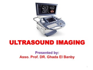
ULTRASOUND IMAGING
Presented by:
Asso. Prof. DR. Ghada El Banby
1
Echo Ranging
• The basis for ultrasound imaging.
• First successful application SONAR in world war 1 (Sound
Navigation And Ranging)
• To display each echo in a position corresponding to that of
the interface or feature (known as a target) that caused it.
• 1880- Jacques and Pierre, discoverers of the
piezoelectric effect.
History
Definitions
• sound is a vibration that propagates as an
acoustic wave, through a transmission medium
such as a gas, liquid or solid.
• Ultrasound: inaudible sound waves with a
frequency of 2–15 megahertz (MHz) when used
for medical imaging
• Ultrasound imaging: the use of ultrasound to
generate anatomical images.
6
Sound
• Mechanical & Longitudinal
wave
• Travels in a straight Line
• Human ear range is 20Hz -
20kHz
• Cannot travel through
Vacuum
Ultrasound
• Mechanical & Longitudinal
wave
• Exceeding the upper limit of
human hearing
• > 20,000H or 20kHz.
7
Ultrasound Physics
Characteristics of Ultrasounds
8
Ultrasound Physics
– The velocity of a sound wave in a medium, c, is related to its
wavelength f and frequency λ by :
* For 5 MHz ultrasound beam has a wavelength of 0.308 mm in soft
tissue with a velocity of 1540 m/sec.
– Image forming depends on acoustic impedance between tissues
• Reflections at boundaries between two different media occur because of
differences in a characteristic known as the acoustic impedance Z of each
substance.
• The acoustic impedance is defined as Z = ρc, ρ is the density of a medium
through which the sound travels and c is the speed of sound through that
medium.
• Acoustic impedance Z
Ultrasound Physics
Acoustic impedance is the resistance of a tissue to the passage of
ultrasound. The higher the degree of impedance mismatch, the greater the
amount of reflection.
The amount of reflection depends on differences in acoustic impedance
(z) between media.
 The intensity reflection coefficient R is defined as the ratio of the intensity of
the reflected wave relative to the incident (transmitted) wave. This statement
can be written mathematically as :
 where Z1 and Z2 are the acoustic impedances of the two media making up the
boundary.
 A reflection coefficient of zero (corresponding to total transmission and no
reflection) occurs when the acoustic impedances of the two media are the
same.
 The image formed in an ultrasound is made by tracking reflections (as shown in
Figure ) and mapping the intensity of the reflected sound waves in a two-
dimensional plane.
15
Interactions of Ultrasound with tissue
Reflection
Transmission
Attenuation
Scattering
16
Interactions of Ultrasound with tissue
Reflection
 Occurs at a boundary between 2
adjacent tissues or Media.
 Acoustic impedance (z)
 The ultrasound image is formed
from reflected echoes.
 Not all the sound wave is
reflected, some continues
deeper into the body.
 These waves will reflect from
deeper tissue structures.
Transmission
17
 Redirection of sound in several
directions
 Caused by interaction with small
reflector or rough surface
 Beam angle-independent appearance
in the image unlike reflections.
Scattering
 The deeper the wave travels in
the body, the weaker it becomes
 The amplitude of the wave
decreases with increasing depth
Attenuation
18
Interactions of Ultrasound with tissue
size less than wavelength
Using known
information about
the speed of sound in
the tissues.
• Measured time for the echo to be
received
• Distance from the transmitter to
organ
• can be calculated, and used to
create an image.
Principle of Ultrasound
By measuring these echo waves it
is possible to determine
How far away the object is and its
size, shape and consistency
19
• Dynamic range of echoes: function in number of
bits for A/D converter….To change Contrast
(dynamic range of gray levels)
26
27
• Frame time / Frame rate
– Time to scan a complete image
– Example: time to scan 1 cm= 2x1cm/c= 2 cm/(1540 m/s) = 13 s
Then, frame time to scan a 20 cm depth with 128 lines=13 s x20 x128
Frame rate = 1/ frame time = 30 frames/s
Smaller D
Smaller N
Higher Frame Rate
• Temporal resolution:
- The time it takes to create one image.
- The more images can be produced and presented per unit of
time, the greater the temporal resolution.
- Frame rate : is the number of images(frames) displayed per
second. High frame rate (many /sec.) is desirable to provide
better temporal resolution.
Depth
width
Advantages
• Lack of radiation
• Quick, adaptable
• Looking at different layers/planes
• Portable
• Less expensive
Parts of Ultrasound Machine
Transducer
Probe
Pulse
control
CPU (Central
Processing
Unit)
Key Board Display
Storage
device
Printer
35
Doppler
Freeze
Depth
Gain
The Ultrasound Machine
• Transducer probe - probe that sends and receives the sound waves
• Central processing unit (CPU) - computer that does all of the calculations
and contains the electrical power supplies for itself and the transducer
probe
• Transducer pulse controls - changes the amplitude, frequency and
duration of the pulses emitted from the transducer probe
• Display - displays the image from the ultrasound data processed by the
CPU
• Keyboard/cursor - inputs data and takes measurements from the display
• Disk storage device (hard, floppy, CD) - stores the acquired images
• Printer - prints the image from the displayed data
Transducer probe
• Is the main part of the ultrasound machine.
• The transducer probe makes the sound waves and receives the echoes.
• The mouth and ears of the ultrasound machine. The transducer probe
generates and receives sound waves using a principle called the
piezoelectric (pressure electricity) effect, which was discovered by Pierre
and Jacques Curie in 1880.
• In the probe, there are one or more quartz crystals called piezoelectric
crystals.
• When an electric current is applied to these crystals, they change shape
rapidly. The rapid shape changes, or vibrations, of the crystals produce
sound waves that travel outward. Conversely, when sound or pressure
waves hit the crystals, they emit electrical currents.
• Therefore, the same crystals can be used to send and receive sound
waves. The probe also has a sound absorbing substance to eliminate
back reflections from the probe itself, and an acoustic lens to help focus
the emitted sound waves.
Transducer
44
eliminate back reflections from the probe itself
46
Ensures no other sound waves affect
the transducer.
Suppresses the vibrations of crystals to
allow shorter pulses to be sent to increase
resolution.
To reduce the difference in
impedance bet. Tissues
&crystals.
Reduce reflection of input
pulses.
Transducer
Matching Layer
 Has acoustic impedance
between that of tissue and the
Piezoelectric elements
 Reduces the reflection of
ultrasound at the scan head
surface
Piezoelectric Elements
 Produce a voltage when
deformed by an applied
pressure
 Quartz, ceramics, man-made
material
Damping Material
 Reduces “ringing” of the element
 Helps to produce very short pulses
47
Piezoelectric Crystals and Frequency
Higher frequency (10MHz)
Shorter Wavelength
Better Resolution
Less Penetrate
Lower frequency (3MHz)
Longer Wavelength
Poorer resolution
Deeper penetration
48
Transducer probe…
• Transducer probes come in many shapes and sizes.
• The shape of the probe determines its field of view, and the frequency
of emitted sound waves determines how deep the sound waves
penetrate and the resolution of the image.
• Transducer probes may contain one or more crystal elements; in
multiple-element probes, each crystal has its own circuit.
• Multiple-element probes have the advantage that the ultrasounic beam
can be "steered" by changing the timing in which each element gets
pulsed; steering the beam is especially important for cardiac ultrasound .
• In addition to probes that can be moved across the surface of the body,
some probes are designed to be inserted through various openings of
the body (e.x, esophagus) so that they can get closer to the organ being
examined.
Types of Ultrasound Transducer
50
51
Types of Ultrasound Transducer
Sector Transducer Linear Transducer Convex Transducer
Crystal Arrangement phased array linear Curvilinear
Footprint Size small Big ( small for hockey
transducers )
Big(small for the micro
convex
Frequency 1-5 MHz 3-12 MHz 1-5 MHz
Beam Shape sector, almost triangular rectangular Convex
Applications gynecological ultrasound,
upper body
ultrasound
obstetrics ultrasound ,
breast, thyroid, vascular
ultrasound
typically abdominal
,pelvic and lung ( micro
convex transducer
52
Selection of Transducer
Superficial vessels and organs (1 to 3cm depth )
 7.5 to 15 MHz
Deeper structures in abdomen and pelvis (12 to 15cm)
 2.25 to 3.5 MHz
53
Central processing unit (CPU)
• The CPU is the brain of the ultrasound machine.
• The CPU is basically a computer that contains the
microprocessor, memory, amplifiers and power supplies for
the microprocessor and transducer probe.
• The CPU sends electrical currents to the transducer probe to
emit sound waves, and also receives the electrical pulses
from the probes that were created from the returning
echoes.
• The CPU does all of the calculations involved in processing
the data. Once the raw data are processed, the CPU forms
the image on the monitor. The CPU can also store the
processed data and/or image on disk.
Transducer pulse controls
• The transducer pulse controls allow the
operator, called the ultrasonographer, to set
and change the frequency and duration of
the ultrasound pulses, as well as the scan
mode of the machine.
• The commands from the operator are
translated into changing electric currents that
are applied to the piezoelectric crystals in the
transducer probe.
Modes Ultrasound
• A-mode- amplitude mode.
• B-mode- brightness mode.
• M-mode- motion mode.
• Doppler-mode.
A-mode
• A-mode (Amplitude-mode) ultrasound is
used to judge the depth of an organ, or
otherwise assess an organ's dimensions.
• Display of echo amplitude (Y-axis) versus
distance (X-axis) into the tissue.
Cornea- ant.Lens- pos. lens- retina- fat
B-Mode
• B-mode ultrasound (Brightness-mode) is the display of a 2D-map of
B-mode data, currently the most common form of ultrasound
imaging.
• This form of display (solid areas appear white and fluid areas
appear black) is also called gray scale.
• The B-mode scan is the basis of 2D scanning. The transducer is
moved about to view the body from a variety of angles. The probe can
be moved in a line (linear scan), or rotated from a particular
position (sector scan).
How is an image formed on the monitor?
 The amplitude of each reflected wave is represented by a dot
 The position of the dot represents the depth from which the echo is
received
 The brightness of the dot represents the strength of the returning
echo
 These dots are combined to form a complete image
61
How is an image formed on the monitor?
Strong Reflections = White dots
As : calcified structures, diaphragm
Weaker Reflections = Grey dots
Myocardium, valve tissue, vessel walls, liver
No Reflections = Black dots
Intra-cardiac cavities, gall bladder
62
M-mode
• The M-mode (Motion-mode) ultrasound is used for analyzing
moving body parts (also called time-motion or TM-mode)
commonly in cardiac and fetal cardiac imaging.
• Used for studying the motion of interface, particularly in cardiac
structures such as the various valves and the chamber walls.
• Analyses a single ultrasound line.
Doppler-Mode
64
• Ultrasound (US) can measure blood velocity based on
Doppler phenomenon.
f_received > f_transmitted
up_shift
Towards each other
f_received < f_transmitted
down_shift
Opposite to each other
65
• US signal from blood originates due to the scattering from red blood cells (RBCs)
(diameter 7-10 mm).
• Blood flow , either towards or away from transducer, alters the frequency of
backscattered echoes compared to the transmitted US frequency from transducer.
• If the blood flows towards the transducer the detected frequency is higher than
transmitted frequency and vice versa.
Doppler-Mode
Doppler frequency
• Doppler shift or Doppler effect
is defined as the change in
frequency of sound wave due
to a reflector moving towards
or away from an object, which
in the case of ultrasound is the
transducer.
Color Flow Mapping Mode (CFM)
• Spatial map overlaid on a B-mode gray-scale
image that depicts an estimate of blood flow
mean velocity
– Direction of flow encoded in colors (blue away
from the transducer and red toward it)
Power Doppler Mode
• This color-coded image of blood flow is based
on intensity rather than on direction of flow,
with a paler color representing higher
intensity.
– It is also known as ‘‘angio’’
Secondary Modes
• Duplex
– Presentation of two modes simultaneously:
usually 2D and pulsed (wave) Doppler
• Triplex
– Presentation of three modes simultaneously:
usually 2D, color flow, and pulsed Doppler
• 3D
– Display or Surface/volume rendering used to
visualize volume composed of multiple 2D slices.
• 4D
– A 3D image moving in time
Major Uses of Ultrasound
 Obstetrics and Gynecology
• measuring the size of the fetus to determine the due date
• determining the position of the fetus to see if it is in the normal .
• seeing the number of fetuses in the uterus.
• checking the sex of the baby .
• checking the fetus's growth rate by making many measurements over time
• seeing tumors of the ovary and breast
 Cardiology
• seeing the inside of the heart to identify abnormal structures or functions
• measuring blood flow through the heart and major blood vessels
 Urology
• measuring blood flow through the kidney
• seeing kidney stones
• detecting prostate cancer early
Kidneys – parenchyma hypoechoic
to liver
Image from UCMC
Liver
Kidney
Liver-
kidney
interface
Cortex
Fat
https://www.instructables.com/Body-
ultrasound-Sonography-With-Arduino/
102
Quick review questions
1. What is ultrasound? What types of tissues does it
travel well through? Poorly through?
2. How does the frequency of the ultrasound affect
resolution and penetration?
3. What are some pros using ultrasound?
4. What shapes do the probes come in? Can you
describe a few of the differences of the probe
types?

More Related Content

What's hot

Gradient echo pulse sequence swastik
Gradient echo pulse sequence  swastikGradient echo pulse sequence  swastik
Gradient echo pulse sequence swastikBipul Poudel
 
Ultrasound physcics, techniques &amp; knobology
Ultrasound physcics, techniques &amp; knobologyUltrasound physcics, techniques &amp; knobology
Ultrasound physcics, techniques &amp; knobologySuzanneCain2
 
ULTRASOUND IMAGING PRINCIPLES
ULTRASOUND IMAGING PRINCIPLESULTRASOUND IMAGING PRINCIPLES
ULTRASOUND IMAGING PRINCIPLESINDIA ULTRASOUND
 
Ultrasound Physics Made easy - By Dr Chandni Wadhwani
Ultrasound Physics Made easy - By Dr Chandni WadhwaniUltrasound Physics Made easy - By Dr Chandni Wadhwani
Ultrasound Physics Made easy - By Dr Chandni WadhwaniChandni Wadhwani
 
Ultrasound physics and image optimization1 (1)
Ultrasound physics and image optimization1 (1)Ultrasound physics and image optimization1 (1)
Ultrasound physics and image optimization1 (1)Prajwith Rai
 
Encoding and image formation
Encoding and image formationEncoding and image formation
Encoding and image formationAnkit Mishra
 
Physical Principles Of Ultrasound
Physical Principles Of UltrasoundPhysical Principles Of Ultrasound
Physical Principles Of Ultrasoundu.surgery
 
Basic physics of Ultrasound
Basic physics of UltrasoundBasic physics of Ultrasound
Basic physics of UltrasoundNouman Ahmed
 
Understanding ultrasound
Understanding ultrasoundUnderstanding ultrasound
Understanding ultrasoundMohammad Amir
 
Ultrasound Basic Knobology & Controls
Ultrasound Basic Knobology & ControlsUltrasound Basic Knobology & Controls
Ultrasound Basic Knobology & ControlsMadhuka Perera
 
Types of Ultrasound Probes
Types of Ultrasound ProbesTypes of Ultrasound Probes
Types of Ultrasound Probeshafsatalat1
 
Types of transducers in ultrasound diagnostic
Types of transducers in ultrasound diagnosticTypes of transducers in ultrasound diagnostic
Types of transducers in ultrasound diagnosticMohamed M. Elsaied
 
Basic Pulse Sequences In MRI
Basic Pulse Sequences In MRIBasic Pulse Sequences In MRI
Basic Pulse Sequences In MRIUpakar Paudel
 
MRI spin echo pulse sequences
MRI spin echo pulse sequencesMRI spin echo pulse sequences
MRI spin echo pulse sequencesShiva Prakash
 
PRINCIPLE OF ULTRAOUND
PRINCIPLE OF ULTRAOUNDPRINCIPLE OF ULTRAOUND
PRINCIPLE OF ULTRAOUNDhanisahwarrior
 
ultrasound transducers and resolution
ultrasound transducers and resolutionultrasound transducers and resolution
ultrasound transducers and resolutionVallabhaneni Bhupal
 
Ultrasound Physics
Ultrasound PhysicsUltrasound Physics
Ultrasound Physicsu.surgery
 
MRI Parameters for Imaging
MRI Parameters for ImagingMRI Parameters for Imaging
MRI Parameters for ImagingAnjan Dangal
 

What's hot (20)

Gradient echo pulse sequence swastik
Gradient echo pulse sequence  swastikGradient echo pulse sequence  swastik
Gradient echo pulse sequence swastik
 
Basics Physics of ultrasound
Basics Physics of ultrasoundBasics Physics of ultrasound
Basics Physics of ultrasound
 
Ultrasound physcics, techniques &amp; knobology
Ultrasound physcics, techniques &amp; knobologyUltrasound physcics, techniques &amp; knobology
Ultrasound physcics, techniques &amp; knobology
 
ULTRASOUND IMAGING PRINCIPLES
ULTRASOUND IMAGING PRINCIPLESULTRASOUND IMAGING PRINCIPLES
ULTRASOUND IMAGING PRINCIPLES
 
Ultrasound Physics Made easy - By Dr Chandni Wadhwani
Ultrasound Physics Made easy - By Dr Chandni WadhwaniUltrasound Physics Made easy - By Dr Chandni Wadhwani
Ultrasound Physics Made easy - By Dr Chandni Wadhwani
 
Ultrasound physics and image optimization1 (1)
Ultrasound physics and image optimization1 (1)Ultrasound physics and image optimization1 (1)
Ultrasound physics and image optimization1 (1)
 
Encoding and image formation
Encoding and image formationEncoding and image formation
Encoding and image formation
 
Physical Principles Of Ultrasound
Physical Principles Of UltrasoundPhysical Principles Of Ultrasound
Physical Principles Of Ultrasound
 
Basic physics of Ultrasound
Basic physics of UltrasoundBasic physics of Ultrasound
Basic physics of Ultrasound
 
Understanding ultrasound
Understanding ultrasoundUnderstanding ultrasound
Understanding ultrasound
 
Ultrasound Basic Knobology & Controls
Ultrasound Basic Knobology & ControlsUltrasound Basic Knobology & Controls
Ultrasound Basic Knobology & Controls
 
Types of Ultrasound Probes
Types of Ultrasound ProbesTypes of Ultrasound Probes
Types of Ultrasound Probes
 
Types of transducers in ultrasound diagnostic
Types of transducers in ultrasound diagnosticTypes of transducers in ultrasound diagnostic
Types of transducers in ultrasound diagnostic
 
Basic Pulse Sequences In MRI
Basic Pulse Sequences In MRIBasic Pulse Sequences In MRI
Basic Pulse Sequences In MRI
 
MRI spin echo pulse sequences
MRI spin echo pulse sequencesMRI spin echo pulse sequences
MRI spin echo pulse sequences
 
Basics in ct
Basics in ctBasics in ct
Basics in ct
 
PRINCIPLE OF ULTRAOUND
PRINCIPLE OF ULTRAOUNDPRINCIPLE OF ULTRAOUND
PRINCIPLE OF ULTRAOUND
 
ultrasound transducers and resolution
ultrasound transducers and resolutionultrasound transducers and resolution
ultrasound transducers and resolution
 
Ultrasound Physics
Ultrasound PhysicsUltrasound Physics
Ultrasound Physics
 
MRI Parameters for Imaging
MRI Parameters for ImagingMRI Parameters for Imaging
MRI Parameters for Imaging
 

Similar to Ultrasound Imaging_2023.pdf

Physics of ultrasound and echocardiography
Physics of ultrasound and echocardiographyPhysics of ultrasound and echocardiography
Physics of ultrasound and echocardiographyjeetshitole
 
Ultrasonography final
Ultrasonography finalUltrasonography final
Ultrasonography finalravikumarbme
 
Principles of Ultrasound Physics.pptx
Principles of Ultrasound Physics.pptxPrinciples of Ultrasound Physics.pptx
Principles of Ultrasound Physics.pptxGemechisAsefa1
 
Sonography
SonographySonography
SonographyFLI
 
Ultrasound and Doppler physics
Ultrasound and Doppler physicsUltrasound and Doppler physics
Ultrasound and Doppler physicsDr Anuradha R
 
Physic Of Ultrasound
Physic Of UltrasoundPhysic Of Ultrasound
Physic Of UltrasoundKhalis Karim
 
ultra sound.pptx
ultra sound.pptxultra sound.pptx
ultra sound.pptxAliMRiyath
 
ultrasound physics
ultrasound physicsultrasound physics
ultrasound physicsRakesh Ca
 
Ultrasound &amp; properties
Ultrasound &amp;  propertiesUltrasound &amp;  properties
Ultrasound &amp; propertiesMahesh Kumar
 
BIS CHAPTER ONE.pptx
BIS CHAPTER ONE.pptxBIS CHAPTER ONE.pptx
BIS CHAPTER ONE.pptxTirusew1
 
BASICS OF US & DUPLEX (NEW).ppt
BASICS OF US & DUPLEX (NEW).pptBASICS OF US & DUPLEX (NEW).ppt
BASICS OF US & DUPLEX (NEW).pptmohamedabushanab1
 
What is Ultrasound.ppt
What is Ultrasound.pptWhat is Ultrasound.ppt
What is Ultrasound.pptNguyenDragon1
 
8159968.ppt
8159968.ppt8159968.ppt
8159968.pptYtchechy
 
Basic physics of ultrasound.JH
Basic physics of ultrasound.JHBasic physics of ultrasound.JH
Basic physics of ultrasound.JHhari baskar
 
basicsofultrasound-170807183128 (1).pdf
basicsofultrasound-170807183128 (1).pdfbasicsofultrasound-170807183128 (1).pdf
basicsofultrasound-170807183128 (1).pdflittlealphonsa
 

Similar to Ultrasound Imaging_2023.pdf (20)

Physics of ultrasound and echocardiography
Physics of ultrasound and echocardiographyPhysics of ultrasound and echocardiography
Physics of ultrasound and echocardiography
 
Ultrasonography final
Ultrasonography finalUltrasonography final
Ultrasonography final
 
Ultrasound Physics Jr.pptx
Ultrasound Physics  Jr.pptxUltrasound Physics  Jr.pptx
Ultrasound Physics Jr.pptx
 
Principles of Ultrasound Physics.pptx
Principles of Ultrasound Physics.pptxPrinciples of Ultrasound Physics.pptx
Principles of Ultrasound Physics.pptx
 
Sonography
SonographySonography
Sonography
 
Ultrasound and Doppler physics
Ultrasound and Doppler physicsUltrasound and Doppler physics
Ultrasound and Doppler physics
 
Physic Of Ultrasound
Physic Of UltrasoundPhysic Of Ultrasound
Physic Of Ultrasound
 
ultra sound.pptx
ultra sound.pptxultra sound.pptx
ultra sound.pptx
 
PRINCIPLES OF ULTRASONOGRAPHY
PRINCIPLES OF ULTRASONOGRAPHYPRINCIPLES OF ULTRASONOGRAPHY
PRINCIPLES OF ULTRASONOGRAPHY
 
Us imaging
Us imagingUs imaging
Us imaging
 
ultrasound physics
ultrasound physicsultrasound physics
ultrasound physics
 
Ultrasound &amp; properties
Ultrasound &amp;  propertiesUltrasound &amp;  properties
Ultrasound &amp; properties
 
BIS CHAPTER ONE.pptx
BIS CHAPTER ONE.pptxBIS CHAPTER ONE.pptx
BIS CHAPTER ONE.pptx
 
USphysics.pptx
USphysics.pptxUSphysics.pptx
USphysics.pptx
 
Us physics (4)
Us physics (4)Us physics (4)
Us physics (4)
 
BASICS OF US & DUPLEX (NEW).ppt
BASICS OF US & DUPLEX (NEW).pptBASICS OF US & DUPLEX (NEW).ppt
BASICS OF US & DUPLEX (NEW).ppt
 
What is Ultrasound.ppt
What is Ultrasound.pptWhat is Ultrasound.ppt
What is Ultrasound.ppt
 
8159968.ppt
8159968.ppt8159968.ppt
8159968.ppt
 
Basic physics of ultrasound.JH
Basic physics of ultrasound.JHBasic physics of ultrasound.JH
Basic physics of ultrasound.JH
 
basicsofultrasound-170807183128 (1).pdf
basicsofultrasound-170807183128 (1).pdfbasicsofultrasound-170807183128 (1).pdf
basicsofultrasound-170807183128 (1).pdf
 

Recently uploaded

Presentation by Andreas Schleicher Tackling the School Absenteeism Crisis 30 ...
Presentation by Andreas Schleicher Tackling the School Absenteeism Crisis 30 ...Presentation by Andreas Schleicher Tackling the School Absenteeism Crisis 30 ...
Presentation by Andreas Schleicher Tackling the School Absenteeism Crisis 30 ...EduSkills OECD
 
POINT- BIOCHEMISTRY SEM 2 ENZYMES UNIT 5.pptx
POINT- BIOCHEMISTRY SEM 2 ENZYMES UNIT 5.pptxPOINT- BIOCHEMISTRY SEM 2 ENZYMES UNIT 5.pptx
POINT- BIOCHEMISTRY SEM 2 ENZYMES UNIT 5.pptxSayali Powar
 
Accessible design: Minimum effort, maximum impact
Accessible design: Minimum effort, maximum impactAccessible design: Minimum effort, maximum impact
Accessible design: Minimum effort, maximum impactdawncurless
 
Industrial Policy - 1948, 1956, 1973, 1977, 1980, 1991
Industrial Policy - 1948, 1956, 1973, 1977, 1980, 1991Industrial Policy - 1948, 1956, 1973, 1977, 1980, 1991
Industrial Policy - 1948, 1956, 1973, 1977, 1980, 1991RKavithamani
 
Incoming and Outgoing Shipments in 1 STEP Using Odoo 17
Incoming and Outgoing Shipments in 1 STEP Using Odoo 17Incoming and Outgoing Shipments in 1 STEP Using Odoo 17
Incoming and Outgoing Shipments in 1 STEP Using Odoo 17Celine George
 
SOCIAL AND HISTORICAL CONTEXT - LFTVD.pptx
SOCIAL AND HISTORICAL CONTEXT - LFTVD.pptxSOCIAL AND HISTORICAL CONTEXT - LFTVD.pptx
SOCIAL AND HISTORICAL CONTEXT - LFTVD.pptxiammrhaywood
 
URLs and Routing in the Odoo 17 Website App
URLs and Routing in the Odoo 17 Website AppURLs and Routing in the Odoo 17 Website App
URLs and Routing in the Odoo 17 Website AppCeline George
 
Alper Gobel In Media Res Media Component
Alper Gobel In Media Res Media ComponentAlper Gobel In Media Res Media Component
Alper Gobel In Media Res Media ComponentInMediaRes1
 
Solving Puzzles Benefits Everyone (English).pptx
Solving Puzzles Benefits Everyone (English).pptxSolving Puzzles Benefits Everyone (English).pptx
Solving Puzzles Benefits Everyone (English).pptxOH TEIK BIN
 
Software Engineering Methodologies (overview)
Software Engineering Methodologies (overview)Software Engineering Methodologies (overview)
Software Engineering Methodologies (overview)eniolaolutunde
 
BASLIQ CURRENT LOOKBOOK LOOKBOOK(1) (1).pdf
BASLIQ CURRENT LOOKBOOK  LOOKBOOK(1) (1).pdfBASLIQ CURRENT LOOKBOOK  LOOKBOOK(1) (1).pdf
BASLIQ CURRENT LOOKBOOK LOOKBOOK(1) (1).pdfSoniaTolstoy
 
Separation of Lanthanides/ Lanthanides and Actinides
Separation of Lanthanides/ Lanthanides and ActinidesSeparation of Lanthanides/ Lanthanides and Actinides
Separation of Lanthanides/ Lanthanides and ActinidesFatimaKhan178732
 
Arihant handbook biology for class 11 .pdf
Arihant handbook biology for class 11 .pdfArihant handbook biology for class 11 .pdf
Arihant handbook biology for class 11 .pdfchloefrazer622
 
Presiding Officer Training module 2024 lok sabha elections
Presiding Officer Training module 2024 lok sabha electionsPresiding Officer Training module 2024 lok sabha elections
Presiding Officer Training module 2024 lok sabha electionsanshu789521
 
Kisan Call Centre - To harness potential of ICT in Agriculture by answer farm...
Kisan Call Centre - To harness potential of ICT in Agriculture by answer farm...Kisan Call Centre - To harness potential of ICT in Agriculture by answer farm...
Kisan Call Centre - To harness potential of ICT in Agriculture by answer farm...Krashi Coaching
 
Contemporary philippine arts from the regions_PPT_Module_12 [Autosaved] (1).pptx
Contemporary philippine arts from the regions_PPT_Module_12 [Autosaved] (1).pptxContemporary philippine arts from the regions_PPT_Module_12 [Autosaved] (1).pptx
Contemporary philippine arts from the regions_PPT_Module_12 [Autosaved] (1).pptxRoyAbrique
 
Hybridoma Technology ( Production , Purification , and Application )
Hybridoma Technology  ( Production , Purification , and Application  ) Hybridoma Technology  ( Production , Purification , and Application  )
Hybridoma Technology ( Production , Purification , and Application ) Sakshi Ghasle
 
Employee wellbeing at the workplace.pptx
Employee wellbeing at the workplace.pptxEmployee wellbeing at the workplace.pptx
Employee wellbeing at the workplace.pptxNirmalaLoungPoorunde1
 

Recently uploaded (20)

Presentation by Andreas Schleicher Tackling the School Absenteeism Crisis 30 ...
Presentation by Andreas Schleicher Tackling the School Absenteeism Crisis 30 ...Presentation by Andreas Schleicher Tackling the School Absenteeism Crisis 30 ...
Presentation by Andreas Schleicher Tackling the School Absenteeism Crisis 30 ...
 
POINT- BIOCHEMISTRY SEM 2 ENZYMES UNIT 5.pptx
POINT- BIOCHEMISTRY SEM 2 ENZYMES UNIT 5.pptxPOINT- BIOCHEMISTRY SEM 2 ENZYMES UNIT 5.pptx
POINT- BIOCHEMISTRY SEM 2 ENZYMES UNIT 5.pptx
 
Accessible design: Minimum effort, maximum impact
Accessible design: Minimum effort, maximum impactAccessible design: Minimum effort, maximum impact
Accessible design: Minimum effort, maximum impact
 
Industrial Policy - 1948, 1956, 1973, 1977, 1980, 1991
Industrial Policy - 1948, 1956, 1973, 1977, 1980, 1991Industrial Policy - 1948, 1956, 1973, 1977, 1980, 1991
Industrial Policy - 1948, 1956, 1973, 1977, 1980, 1991
 
Incoming and Outgoing Shipments in 1 STEP Using Odoo 17
Incoming and Outgoing Shipments in 1 STEP Using Odoo 17Incoming and Outgoing Shipments in 1 STEP Using Odoo 17
Incoming and Outgoing Shipments in 1 STEP Using Odoo 17
 
SOCIAL AND HISTORICAL CONTEXT - LFTVD.pptx
SOCIAL AND HISTORICAL CONTEXT - LFTVD.pptxSOCIAL AND HISTORICAL CONTEXT - LFTVD.pptx
SOCIAL AND HISTORICAL CONTEXT - LFTVD.pptx
 
URLs and Routing in the Odoo 17 Website App
URLs and Routing in the Odoo 17 Website AppURLs and Routing in the Odoo 17 Website App
URLs and Routing in the Odoo 17 Website App
 
Alper Gobel In Media Res Media Component
Alper Gobel In Media Res Media ComponentAlper Gobel In Media Res Media Component
Alper Gobel In Media Res Media Component
 
Solving Puzzles Benefits Everyone (English).pptx
Solving Puzzles Benefits Everyone (English).pptxSolving Puzzles Benefits Everyone (English).pptx
Solving Puzzles Benefits Everyone (English).pptx
 
Software Engineering Methodologies (overview)
Software Engineering Methodologies (overview)Software Engineering Methodologies (overview)
Software Engineering Methodologies (overview)
 
BASLIQ CURRENT LOOKBOOK LOOKBOOK(1) (1).pdf
BASLIQ CURRENT LOOKBOOK  LOOKBOOK(1) (1).pdfBASLIQ CURRENT LOOKBOOK  LOOKBOOK(1) (1).pdf
BASLIQ CURRENT LOOKBOOK LOOKBOOK(1) (1).pdf
 
Separation of Lanthanides/ Lanthanides and Actinides
Separation of Lanthanides/ Lanthanides and ActinidesSeparation of Lanthanides/ Lanthanides and Actinides
Separation of Lanthanides/ Lanthanides and Actinides
 
Staff of Color (SOC) Retention Efforts DDSD
Staff of Color (SOC) Retention Efforts DDSDStaff of Color (SOC) Retention Efforts DDSD
Staff of Color (SOC) Retention Efforts DDSD
 
Arihant handbook biology for class 11 .pdf
Arihant handbook biology for class 11 .pdfArihant handbook biology for class 11 .pdf
Arihant handbook biology for class 11 .pdf
 
Presiding Officer Training module 2024 lok sabha elections
Presiding Officer Training module 2024 lok sabha electionsPresiding Officer Training module 2024 lok sabha elections
Presiding Officer Training module 2024 lok sabha elections
 
Kisan Call Centre - To harness potential of ICT in Agriculture by answer farm...
Kisan Call Centre - To harness potential of ICT in Agriculture by answer farm...Kisan Call Centre - To harness potential of ICT in Agriculture by answer farm...
Kisan Call Centre - To harness potential of ICT in Agriculture by answer farm...
 
Contemporary philippine arts from the regions_PPT_Module_12 [Autosaved] (1).pptx
Contemporary philippine arts from the regions_PPT_Module_12 [Autosaved] (1).pptxContemporary philippine arts from the regions_PPT_Module_12 [Autosaved] (1).pptx
Contemporary philippine arts from the regions_PPT_Module_12 [Autosaved] (1).pptx
 
Hybridoma Technology ( Production , Purification , and Application )
Hybridoma Technology  ( Production , Purification , and Application  ) Hybridoma Technology  ( Production , Purification , and Application  )
Hybridoma Technology ( Production , Purification , and Application )
 
Employee wellbeing at the workplace.pptx
Employee wellbeing at the workplace.pptxEmployee wellbeing at the workplace.pptx
Employee wellbeing at the workplace.pptx
 
TataKelola dan KamSiber Kecerdasan Buatan v022.pdf
TataKelola dan KamSiber Kecerdasan Buatan v022.pdfTataKelola dan KamSiber Kecerdasan Buatan v022.pdf
TataKelola dan KamSiber Kecerdasan Buatan v022.pdf
 

Ultrasound Imaging_2023.pdf

  • 1. ULTRASOUND IMAGING Presented by: Asso. Prof. DR. Ghada El Banby 1
  • 2. Echo Ranging • The basis for ultrasound imaging. • First successful application SONAR in world war 1 (Sound Navigation And Ranging) • To display each echo in a position corresponding to that of the interface or feature (known as a target) that caused it.
  • 3. • 1880- Jacques and Pierre, discoverers of the piezoelectric effect. History
  • 4. Definitions • sound is a vibration that propagates as an acoustic wave, through a transmission medium such as a gas, liquid or solid. • Ultrasound: inaudible sound waves with a frequency of 2–15 megahertz (MHz) when used for medical imaging • Ultrasound imaging: the use of ultrasound to generate anatomical images. 6
  • 5. Sound • Mechanical & Longitudinal wave • Travels in a straight Line • Human ear range is 20Hz - 20kHz • Cannot travel through Vacuum Ultrasound • Mechanical & Longitudinal wave • Exceeding the upper limit of human hearing • > 20,000H or 20kHz. 7 Ultrasound Physics
  • 7. Ultrasound Physics – The velocity of a sound wave in a medium, c, is related to its wavelength f and frequency λ by : * For 5 MHz ultrasound beam has a wavelength of 0.308 mm in soft tissue with a velocity of 1540 m/sec.
  • 8. – Image forming depends on acoustic impedance between tissues • Reflections at boundaries between two different media occur because of differences in a characteristic known as the acoustic impedance Z of each substance. • The acoustic impedance is defined as Z = ρc, ρ is the density of a medium through which the sound travels and c is the speed of sound through that medium. • Acoustic impedance Z Ultrasound Physics Acoustic impedance is the resistance of a tissue to the passage of ultrasound. The higher the degree of impedance mismatch, the greater the amount of reflection. The amount of reflection depends on differences in acoustic impedance (z) between media.
  • 9.
  • 10.  The intensity reflection coefficient R is defined as the ratio of the intensity of the reflected wave relative to the incident (transmitted) wave. This statement can be written mathematically as :  where Z1 and Z2 are the acoustic impedances of the two media making up the boundary.  A reflection coefficient of zero (corresponding to total transmission and no reflection) occurs when the acoustic impedances of the two media are the same.  The image formed in an ultrasound is made by tracking reflections (as shown in Figure ) and mapping the intensity of the reflected sound waves in a two- dimensional plane.
  • 11. 15
  • 12. Interactions of Ultrasound with tissue Reflection Transmission Attenuation Scattering 16
  • 13. Interactions of Ultrasound with tissue Reflection  Occurs at a boundary between 2 adjacent tissues or Media.  Acoustic impedance (z)  The ultrasound image is formed from reflected echoes.  Not all the sound wave is reflected, some continues deeper into the body.  These waves will reflect from deeper tissue structures. Transmission 17
  • 14.  Redirection of sound in several directions  Caused by interaction with small reflector or rough surface  Beam angle-independent appearance in the image unlike reflections. Scattering  The deeper the wave travels in the body, the weaker it becomes  The amplitude of the wave decreases with increasing depth Attenuation 18 Interactions of Ultrasound with tissue size less than wavelength
  • 15. Using known information about the speed of sound in the tissues. • Measured time for the echo to be received • Distance from the transmitter to organ • can be calculated, and used to create an image. Principle of Ultrasound By measuring these echo waves it is possible to determine How far away the object is and its size, shape and consistency 19
  • 16. • Dynamic range of echoes: function in number of bits for A/D converter….To change Contrast (dynamic range of gray levels)
  • 17. 26
  • 18. 27 • Frame time / Frame rate – Time to scan a complete image – Example: time to scan 1 cm= 2x1cm/c= 2 cm/(1540 m/s) = 13 s Then, frame time to scan a 20 cm depth with 128 lines=13 s x20 x128 Frame rate = 1/ frame time = 30 frames/s Smaller D Smaller N Higher Frame Rate • Temporal resolution: - The time it takes to create one image. - The more images can be produced and presented per unit of time, the greater the temporal resolution. - Frame rate : is the number of images(frames) displayed per second. High frame rate (many /sec.) is desirable to provide better temporal resolution. Depth width
  • 19. Advantages • Lack of radiation • Quick, adaptable • Looking at different layers/planes • Portable • Less expensive
  • 20. Parts of Ultrasound Machine Transducer Probe Pulse control CPU (Central Processing Unit) Key Board Display Storage device Printer 35
  • 21.
  • 22.
  • 24. The Ultrasound Machine • Transducer probe - probe that sends and receives the sound waves • Central processing unit (CPU) - computer that does all of the calculations and contains the electrical power supplies for itself and the transducer probe • Transducer pulse controls - changes the amplitude, frequency and duration of the pulses emitted from the transducer probe • Display - displays the image from the ultrasound data processed by the CPU • Keyboard/cursor - inputs data and takes measurements from the display • Disk storage device (hard, floppy, CD) - stores the acquired images • Printer - prints the image from the displayed data
  • 25.
  • 26. Transducer probe • Is the main part of the ultrasound machine. • The transducer probe makes the sound waves and receives the echoes. • The mouth and ears of the ultrasound machine. The transducer probe generates and receives sound waves using a principle called the piezoelectric (pressure electricity) effect, which was discovered by Pierre and Jacques Curie in 1880. • In the probe, there are one or more quartz crystals called piezoelectric crystals. • When an electric current is applied to these crystals, they change shape rapidly. The rapid shape changes, or vibrations, of the crystals produce sound waves that travel outward. Conversely, when sound or pressure waves hit the crystals, they emit electrical currents. • Therefore, the same crystals can be used to send and receive sound waves. The probe also has a sound absorbing substance to eliminate back reflections from the probe itself, and an acoustic lens to help focus the emitted sound waves.
  • 27.
  • 29.
  • 30. 46 Ensures no other sound waves affect the transducer. Suppresses the vibrations of crystals to allow shorter pulses to be sent to increase resolution. To reduce the difference in impedance bet. Tissues &crystals. Reduce reflection of input pulses.
  • 31. Transducer Matching Layer  Has acoustic impedance between that of tissue and the Piezoelectric elements  Reduces the reflection of ultrasound at the scan head surface Piezoelectric Elements  Produce a voltage when deformed by an applied pressure  Quartz, ceramics, man-made material Damping Material  Reduces “ringing” of the element  Helps to produce very short pulses 47
  • 32. Piezoelectric Crystals and Frequency Higher frequency (10MHz) Shorter Wavelength Better Resolution Less Penetrate Lower frequency (3MHz) Longer Wavelength Poorer resolution Deeper penetration 48
  • 33. Transducer probe… • Transducer probes come in many shapes and sizes. • The shape of the probe determines its field of view, and the frequency of emitted sound waves determines how deep the sound waves penetrate and the resolution of the image. • Transducer probes may contain one or more crystal elements; in multiple-element probes, each crystal has its own circuit. • Multiple-element probes have the advantage that the ultrasounic beam can be "steered" by changing the timing in which each element gets pulsed; steering the beam is especially important for cardiac ultrasound . • In addition to probes that can be moved across the surface of the body, some probes are designed to be inserted through various openings of the body (e.x, esophagus) so that they can get closer to the organ being examined.
  • 34. Types of Ultrasound Transducer 50
  • 35. 51
  • 36. Types of Ultrasound Transducer Sector Transducer Linear Transducer Convex Transducer Crystal Arrangement phased array linear Curvilinear Footprint Size small Big ( small for hockey transducers ) Big(small for the micro convex Frequency 1-5 MHz 3-12 MHz 1-5 MHz Beam Shape sector, almost triangular rectangular Convex Applications gynecological ultrasound, upper body ultrasound obstetrics ultrasound , breast, thyroid, vascular ultrasound typically abdominal ,pelvic and lung ( micro convex transducer 52
  • 37. Selection of Transducer Superficial vessels and organs (1 to 3cm depth )  7.5 to 15 MHz Deeper structures in abdomen and pelvis (12 to 15cm)  2.25 to 3.5 MHz 53
  • 38. Central processing unit (CPU) • The CPU is the brain of the ultrasound machine. • The CPU is basically a computer that contains the microprocessor, memory, amplifiers and power supplies for the microprocessor and transducer probe. • The CPU sends electrical currents to the transducer probe to emit sound waves, and also receives the electrical pulses from the probes that were created from the returning echoes. • The CPU does all of the calculations involved in processing the data. Once the raw data are processed, the CPU forms the image on the monitor. The CPU can also store the processed data and/or image on disk.
  • 39. Transducer pulse controls • The transducer pulse controls allow the operator, called the ultrasonographer, to set and change the frequency and duration of the ultrasound pulses, as well as the scan mode of the machine. • The commands from the operator are translated into changing electric currents that are applied to the piezoelectric crystals in the transducer probe.
  • 40. Modes Ultrasound • A-mode- amplitude mode. • B-mode- brightness mode. • M-mode- motion mode. • Doppler-mode.
  • 41. A-mode • A-mode (Amplitude-mode) ultrasound is used to judge the depth of an organ, or otherwise assess an organ's dimensions. • Display of echo amplitude (Y-axis) versus distance (X-axis) into the tissue. Cornea- ant.Lens- pos. lens- retina- fat
  • 42. B-Mode • B-mode ultrasound (Brightness-mode) is the display of a 2D-map of B-mode data, currently the most common form of ultrasound imaging. • This form of display (solid areas appear white and fluid areas appear black) is also called gray scale. • The B-mode scan is the basis of 2D scanning. The transducer is moved about to view the body from a variety of angles. The probe can be moved in a line (linear scan), or rotated from a particular position (sector scan).
  • 43. How is an image formed on the monitor?  The amplitude of each reflected wave is represented by a dot  The position of the dot represents the depth from which the echo is received  The brightness of the dot represents the strength of the returning echo  These dots are combined to form a complete image 61
  • 44. How is an image formed on the monitor? Strong Reflections = White dots As : calcified structures, diaphragm Weaker Reflections = Grey dots Myocardium, valve tissue, vessel walls, liver No Reflections = Black dots Intra-cardiac cavities, gall bladder 62
  • 45. M-mode • The M-mode (Motion-mode) ultrasound is used for analyzing moving body parts (also called time-motion or TM-mode) commonly in cardiac and fetal cardiac imaging. • Used for studying the motion of interface, particularly in cardiac structures such as the various valves and the chamber walls. • Analyses a single ultrasound line.
  • 46. Doppler-Mode 64 • Ultrasound (US) can measure blood velocity based on Doppler phenomenon. f_received > f_transmitted up_shift Towards each other f_received < f_transmitted down_shift Opposite to each other
  • 47. 65 • US signal from blood originates due to the scattering from red blood cells (RBCs) (diameter 7-10 mm). • Blood flow , either towards or away from transducer, alters the frequency of backscattered echoes compared to the transmitted US frequency from transducer. • If the blood flows towards the transducer the detected frequency is higher than transmitted frequency and vice versa.
  • 49. • Doppler shift or Doppler effect is defined as the change in frequency of sound wave due to a reflector moving towards or away from an object, which in the case of ultrasound is the transducer.
  • 50. Color Flow Mapping Mode (CFM) • Spatial map overlaid on a B-mode gray-scale image that depicts an estimate of blood flow mean velocity – Direction of flow encoded in colors (blue away from the transducer and red toward it)
  • 51. Power Doppler Mode • This color-coded image of blood flow is based on intensity rather than on direction of flow, with a paler color representing higher intensity. – It is also known as ‘‘angio’’
  • 52. Secondary Modes • Duplex – Presentation of two modes simultaneously: usually 2D and pulsed (wave) Doppler • Triplex – Presentation of three modes simultaneously: usually 2D, color flow, and pulsed Doppler • 3D – Display or Surface/volume rendering used to visualize volume composed of multiple 2D slices. • 4D – A 3D image moving in time
  • 53. Major Uses of Ultrasound  Obstetrics and Gynecology • measuring the size of the fetus to determine the due date • determining the position of the fetus to see if it is in the normal . • seeing the number of fetuses in the uterus. • checking the sex of the baby . • checking the fetus's growth rate by making many measurements over time • seeing tumors of the ovary and breast  Cardiology • seeing the inside of the heart to identify abnormal structures or functions • measuring blood flow through the heart and major blood vessels  Urology • measuring blood flow through the kidney • seeing kidney stones • detecting prostate cancer early
  • 54. Kidneys – parenchyma hypoechoic to liver Image from UCMC Liver Kidney Liver- kidney interface Cortex Fat
  • 56. Quick review questions 1. What is ultrasound? What types of tissues does it travel well through? Poorly through? 2. How does the frequency of the ultrasound affect resolution and penetration? 3. What are some pros using ultrasound? 4. What shapes do the probes come in? Can you describe a few of the differences of the probe types?