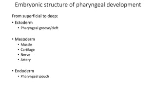
The document discusses the pharyngeal apparatus, which develops in the human embryo between the head and thorax. It has 6 arches that facilitate respiration and feeding. Each arch contributes to the development of specific anatomic structures of the head, face, palate and neck. The arches give rise to cartilages, muscles, nerves, arteries, and endodermal pouches that develop into important structures like the ears, tongue, tonsils, and thyroid gland. Understanding the development and derivatives of the pharyngeal arches explains the anatomy of many craniofacial and neck regions.


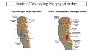


![Endodermal Derivatives of Pharyngeal Pouches
Pharyngeal Pouches Derivatives
1 Tympanic (middle ear) cavity; Auditory (Eustachian) tube
2 Palatine tonsils; Tonsillar fossa
3 Inferior parathyroid gland; Thymus
4 Superior parathyroid gland; ultimobranchial body (parafollicular [C] cells of the thyroid
gland)](https://image.slidesharecdn.com/pharyngealapparatus2021-230124044248-4a5eb515/85/Pharyngeal-Apparatus-pptx-17-320.jpg)


