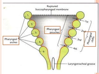
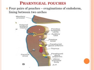
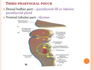
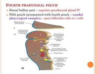
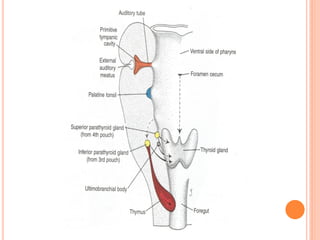
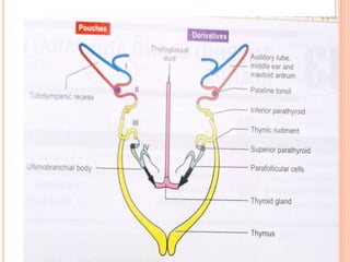
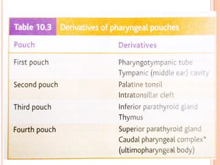
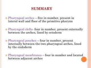
1. The pharyngeal arches form pouches and clefts that give rise to important structures. The parathyroid glands arise from the third and fourth pharyngeal pouches.
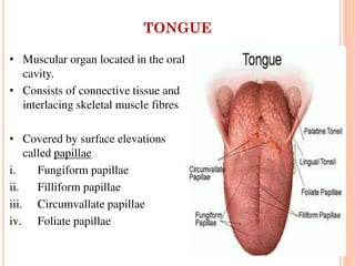
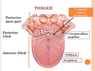
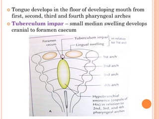
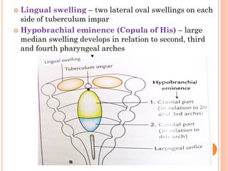
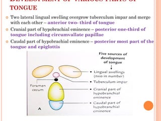
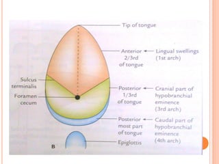
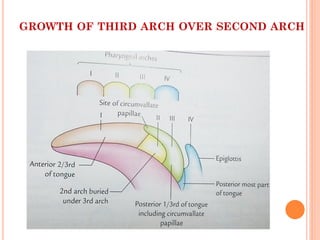
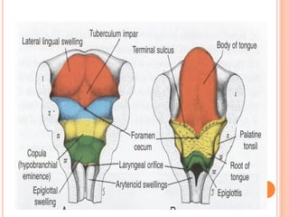
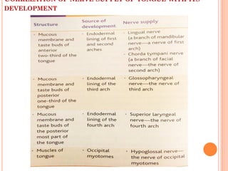
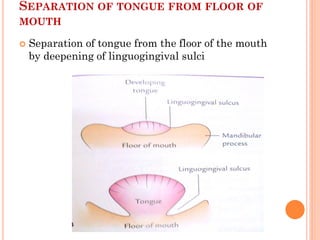
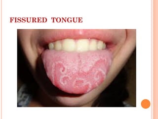
2. The tongue develops from the first, second, third and fourth pharyngeal arches. A median tuberculum impar later merges with two lateral lingual swellings to form the anterior two-thirds of the tongue.
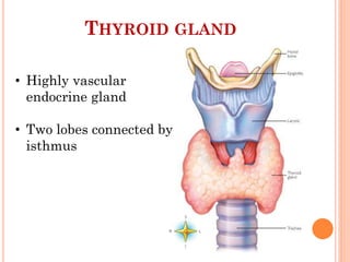
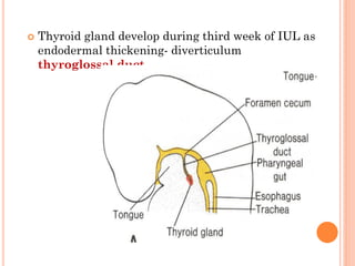
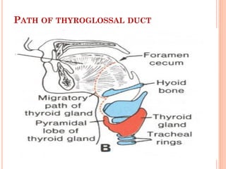
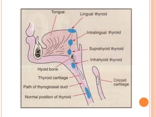
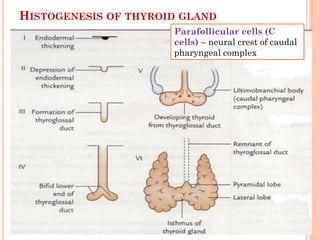
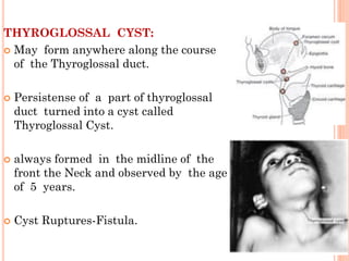
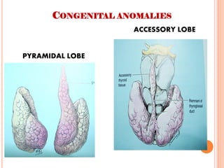
3. The thyroid gland develops from an endodermal thickening that descends as the thyroglossal duct. The duct normally disappears but sometimes leaves remnants that can form cyst
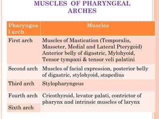


![ Enumerate the Derivatives of second pharyngeal
arch
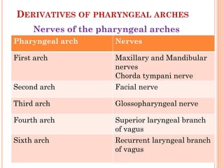
Name the Derivatives of first pharyngeal arch
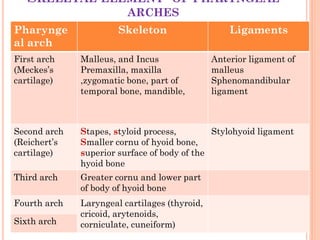
Development oy hyoid bone [apr 2002]
Explain Cervical sinus
Development of parathyroid [dec 2010]
Thyroglossal duct [dec 2011]
Explain the Development of thyroid gland
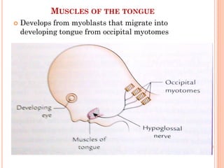
Brief account of Development of tongue](https://image.slidesharecdn.com/pharyngealapparatusii-tonguethyroid-160318040421/85/Pharyngeal-apparatus-ii-tongue-thyroid-50-320.jpg)