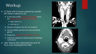
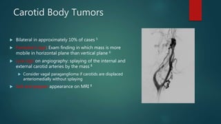
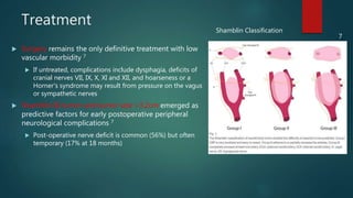
Ms. L
JASON LEPSE, MS4
UNIVERSITY OF KANSAS SCHOOL OF MEDICINE
H&P
 Background: 47 yo female who was referred to an outside ENT in August,
2014
 CC: Right neck mass
 HPI: Patient reported presence of neck mass for 9 years
 Initially fluctuated in size but recently began growing steadily
 Intermittent dull pain over right lateral neck sometimes extending to right year
 No fluctuation of mass with eating; no facial weakness, numbness, dysphagia,
odynophagia or voice change
 No tachycardia, diaphoresis, flushing or tachypnea
*Modified from documentation by Drs. Michael Baker, Kiran Kakarala and Paul Camarata
H&P
 PMH: Anemia, headaches and seasonal allergies
 PSH: Tonsillectomy and adenoidectomy
 Meds/Allergies: Aspirin and Tylenol; allergic to sulfa
 FH: Breast and uterine cancer in mother and sister
 SH: ½ ppd smoker for 35 years; occasional alcohol use
 ROS: + fatigue, hearing loss, tinnitus, palpitations, confusion, light-
headedness, headaches
*Modified from documentation by Drs. Michael Baker, Kiran Kakarala and Paul Camarata
H&P
 Exam
 Vitals: WNL
 General: Well-developed, well-nourished
 Ears: canals normal, TM intact and mobile; no evidence of fluid or infection
 Nose: Normal nasal mucosa, no masses/lesions
 Nasopharynx: No mass, lesion or drainage
 Oral Cavity: Clear and without signs of infection
 Pharynx: No mass or ulceration
 Larynx: Vocal cords mobile, no visible lesions
 Neck: Midline trachea without mass or lesion, large right side mass mobile (more horizontal than vertical) ,
firm, 4x5cm extending to the angle of mandible superiorly inferiorly to the level of cricoid, no TTP, non-
pulsatile
DDX: lymphadenopathy, branchial cleft cysts, salivary gland tumors, neurogenic
tumors, and aneurysms of the carotid artery.
*Modified from documentation by Drs. Michael Baker, Kiran Kakarala and Paul Camarata
Workup
 CT Neck with Contrast ordered by outside
ENT before referral to KU
 In the area of the carotid bifurcations there
are large enhancing masses bilaterally
 Right: 8x4x3 cm
 Left: 6x4 cm
 Masses compress but do not invade IJ
 Unremarkable parotid and submandibular
glands
 Diagnosis: Bilateral Carotid Body Tumors
 Definitive diagnosis would require histologic
verification
 Labs: Plasma-free metanephrines and 24-
hour urine metanephrines WNL
Carotid Body Tumors
 Type of paraganglioma, rare neuroendocrine tumors arising from extra-adrenal autonomic
paraganglia
 Parasympathetic derived
 Head and neck paragangliomas are less likely to be secretory (5%)1
 Most are sporadic but hereditary paragangliomas have been linked to mutations in the
succinate dehydrogenase (SDH) enzyme complex 2
 Sporadic tumors more common in women; hereditary tumors are equally distributed 3
 Mean age at diagnosis was 47 in a study of 236 patients 4
 Vast majority are benign; malignancy is only confirmed by presence of paraganglioma in
lymph nodes 1
Carotid Body Tumors
 Bilateral in approximately 10% of cases 5
 Fontaine’s sign: Exam finding in which mass is more
mobile in horizontal plane than vertical plane 6
 Lyre sign on angiography: splaying of the internal and
external carotid arteries by the mass 8
 Consider vagal paraganglioma if carotids are displaced
anteriomedially without splaying
 Salt and pepper appearance on MRI 8
Treatment
 Surgery remains the only definitive treatment with low
vascular morbidity 7
 If untreated, complications include dysphagia, deficits of
cranial nerves VII, IX, X, XI and XII, and hoarseness or a
Horner's syndrome may result from pressure on the vagus
or sympathetic nerves
 Shamblin III tumor and tumor size >3.2cm emerged as
predictive factors for early postoperative peripheral
neurological complications 7
 Post-operative nerve deficit is common (56%) but often
temporary (17% at 18 months)
7
Shamblin Classification
Ms. L’s Course
 Ms. L saw Dr. Kakarala, Dr. Vamanan and Dr. Camarata prior to operation
 Underwent preoperative right carotid body tumor embolization
 Controversial with some studies reporting no benefit and others reporting reduction
in intraoperative blood loss and associated morbidity
 Right pre-auricular transparotid and transcervical approach to parapharyngeal
space tumor
 Failed swallow with evidence of right vocal cord paralysis
 PEG tube placed
 Underwent Vocal Cord Injection three days after resection
 Discharged with nectar-thick liquids and post-op swallow appointment
References
 1: Johnson, Jonas T., Clark A. Rosen, and Byron J. Bailey. Bailey's Head and Neck Surgery--otolaryngology.
Philadelphia: Wolters Kluwer Health /Lippincott Williams & Wilkins, 2014
 2: Dannenberg H, Dinjens WN, Abbou M, et al. Frequent germ-line succinate dehydrogenase subunit D gene
mutations in patients with apparently sporadic parasympathetic paraganglioma. Clin Cancer Res 2002; 8:2061.
 3: Lin D, Carty S, Young WF Jr. Paragangliomas: Epidemiology, clinical presentation, diagnosis, and histology.
UpToDate. 2015.
 4: Erickson D, Kudva YC, Ebersold MJ, Thompson GB, Grant CS, van Heerden JA, Young WF Jr. Benign
paragangliomas: clinical presentation and treatment outcomes in 236 patients. J Clin Endocrinol Metab.
2001;86(11):5210.
 5: Davidovic LB, Djukic VB, Vasic DM et-al. Diagnosis and treatment of carotid body paraganglioma: 21 years of
experience at a clinical center of Serbia. World J Surg Oncol. 2005;3 (1): 10
 6 :Boedeker CC, Ridder GJ, Schipper J. Paragangliomas of the head and neck: diagnosis and treatment. Fam Cancer.
2005;4(1):55.
 7: Lamblin, E., I. Atallah, E. Reyt, S. Schmerber, J.-L. Magne, and C.a. Righini. "Neurovascular Complications following
Carotid Body Paraganglioma Resection." European Annals of Otorhinolaryngology, Head and Neck Diseases (2016)
 8: Lee, K. J. Essential Otolaryngology: Head and Neck Surgery. Norwalk, CT: Appleton & Lange, 2012.

Carotid body tumors

  • 1.
    Ms. L JASON LEPSE,MS4 UNIVERSITY OF KANSAS SCHOOL OF MEDICINE
  • 2.
    H&P  Background: 47yo female who was referred to an outside ENT in August, 2014  CC: Right neck mass  HPI: Patient reported presence of neck mass for 9 years  Initially fluctuated in size but recently began growing steadily  Intermittent dull pain over right lateral neck sometimes extending to right year  No fluctuation of mass with eating; no facial weakness, numbness, dysphagia, odynophagia or voice change  No tachycardia, diaphoresis, flushing or tachypnea *Modified from documentation by Drs. Michael Baker, Kiran Kakarala and Paul Camarata
  • 3.
    H&P  PMH: Anemia,headaches and seasonal allergies  PSH: Tonsillectomy and adenoidectomy  Meds/Allergies: Aspirin and Tylenol; allergic to sulfa  FH: Breast and uterine cancer in mother and sister  SH: ½ ppd smoker for 35 years; occasional alcohol use  ROS: + fatigue, hearing loss, tinnitus, palpitations, confusion, light- headedness, headaches *Modified from documentation by Drs. Michael Baker, Kiran Kakarala and Paul Camarata
  • 4.
    H&P  Exam  Vitals:WNL  General: Well-developed, well-nourished  Ears: canals normal, TM intact and mobile; no evidence of fluid or infection  Nose: Normal nasal mucosa, no masses/lesions  Nasopharynx: No mass, lesion or drainage  Oral Cavity: Clear and without signs of infection  Pharynx: No mass or ulceration  Larynx: Vocal cords mobile, no visible lesions  Neck: Midline trachea without mass or lesion, large right side mass mobile (more horizontal than vertical) , firm, 4x5cm extending to the angle of mandible superiorly inferiorly to the level of cricoid, no TTP, non- pulsatile DDX: lymphadenopathy, branchial cleft cysts, salivary gland tumors, neurogenic tumors, and aneurysms of the carotid artery. *Modified from documentation by Drs. Michael Baker, Kiran Kakarala and Paul Camarata
  • 5.
    Workup  CT Neckwith Contrast ordered by outside ENT before referral to KU  In the area of the carotid bifurcations there are large enhancing masses bilaterally  Right: 8x4x3 cm  Left: 6x4 cm  Masses compress but do not invade IJ  Unremarkable parotid and submandibular glands  Diagnosis: Bilateral Carotid Body Tumors  Definitive diagnosis would require histologic verification  Labs: Plasma-free metanephrines and 24- hour urine metanephrines WNL
  • 6.
    Carotid Body Tumors Type of paraganglioma, rare neuroendocrine tumors arising from extra-adrenal autonomic paraganglia  Parasympathetic derived  Head and neck paragangliomas are less likely to be secretory (5%)1  Most are sporadic but hereditary paragangliomas have been linked to mutations in the succinate dehydrogenase (SDH) enzyme complex 2  Sporadic tumors more common in women; hereditary tumors are equally distributed 3  Mean age at diagnosis was 47 in a study of 236 patients 4  Vast majority are benign; malignancy is only confirmed by presence of paraganglioma in lymph nodes 1
  • 7.
    Carotid Body Tumors Bilateral in approximately 10% of cases 5  Fontaine’s sign: Exam finding in which mass is more mobile in horizontal plane than vertical plane 6  Lyre sign on angiography: splaying of the internal and external carotid arteries by the mass 8  Consider vagal paraganglioma if carotids are displaced anteriomedially without splaying  Salt and pepper appearance on MRI 8
  • 8.
    Treatment  Surgery remainsthe only definitive treatment with low vascular morbidity 7  If untreated, complications include dysphagia, deficits of cranial nerves VII, IX, X, XI and XII, and hoarseness or a Horner's syndrome may result from pressure on the vagus or sympathetic nerves  Shamblin III tumor and tumor size >3.2cm emerged as predictive factors for early postoperative peripheral neurological complications 7  Post-operative nerve deficit is common (56%) but often temporary (17% at 18 months) 7 Shamblin Classification
  • 9.
    Ms. L’s Course Ms. L saw Dr. Kakarala, Dr. Vamanan and Dr. Camarata prior to operation  Underwent preoperative right carotid body tumor embolization  Controversial with some studies reporting no benefit and others reporting reduction in intraoperative blood loss and associated morbidity  Right pre-auricular transparotid and transcervical approach to parapharyngeal space tumor  Failed swallow with evidence of right vocal cord paralysis  PEG tube placed  Underwent Vocal Cord Injection three days after resection  Discharged with nectar-thick liquids and post-op swallow appointment
  • 10.
    References  1: Johnson,Jonas T., Clark A. Rosen, and Byron J. Bailey. Bailey's Head and Neck Surgery--otolaryngology. Philadelphia: Wolters Kluwer Health /Lippincott Williams & Wilkins, 2014  2: Dannenberg H, Dinjens WN, Abbou M, et al. Frequent germ-line succinate dehydrogenase subunit D gene mutations in patients with apparently sporadic parasympathetic paraganglioma. Clin Cancer Res 2002; 8:2061.  3: Lin D, Carty S, Young WF Jr. Paragangliomas: Epidemiology, clinical presentation, diagnosis, and histology. UpToDate. 2015.  4: Erickson D, Kudva YC, Ebersold MJ, Thompson GB, Grant CS, van Heerden JA, Young WF Jr. Benign paragangliomas: clinical presentation and treatment outcomes in 236 patients. J Clin Endocrinol Metab. 2001;86(11):5210.  5: Davidovic LB, Djukic VB, Vasic DM et-al. Diagnosis and treatment of carotid body paraganglioma: 21 years of experience at a clinical center of Serbia. World J Surg Oncol. 2005;3 (1): 10  6 :Boedeker CC, Ridder GJ, Schipper J. Paragangliomas of the head and neck: diagnosis and treatment. Fam Cancer. 2005;4(1):55.  7: Lamblin, E., I. Atallah, E. Reyt, S. Schmerber, J.-L. Magne, and C.a. Righini. "Neurovascular Complications following Carotid Body Paraganglioma Resection." European Annals of Otorhinolaryngology, Head and Neck Diseases (2016)  8: Lee, K. J. Essential Otolaryngology: Head and Neck Surgery. Norwalk, CT: Appleton & Lange, 2012.