At theend of the lecture, the students
should be able to describe in detail the-
,Development of tongue, Nerve Supply of tongue,
Formation of frenum and papillae
,Development of musculature of tongue, Enlist
clinical Considerations.
Purpose Statement
3.
Learning Objectives
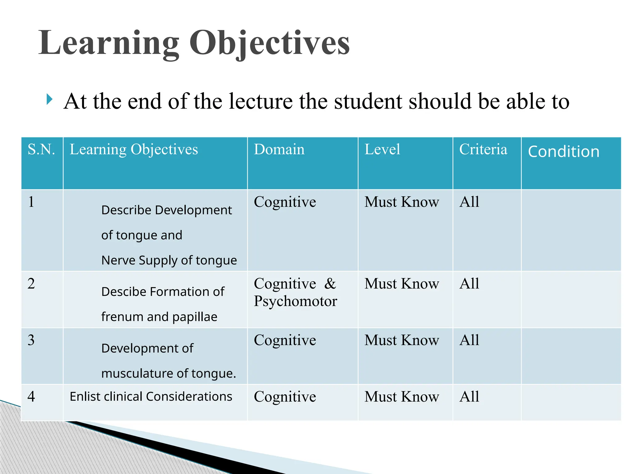
Atthe end of the lecture the student should be able to
S.N. Learning Objectives Domain Level Criteria Condition
1 Describe Development
of tongue and
Nerve Supply of tongue
Cognitive Must Know All
2 Descibe Formation of
frenum and papillae
Cognitive &
Psychomotor
Must Know All
3 Development of
musculature of tongue.
Cognitive Must Know All
4 Enlist clinical Considerations Cognitive Must Know All
4.
Pharyngeal ArchContributions
DEVELOPMENT OF TONGUE
Nerve Supply
CLINICAL CONSIDERATIONS
CONTENTS
5.
INTRODUCTION
Tongue is amass of striated muscle covered
with mucous membrane.
It is a highly muscular organ of deglutition,
taste and speech.
7.
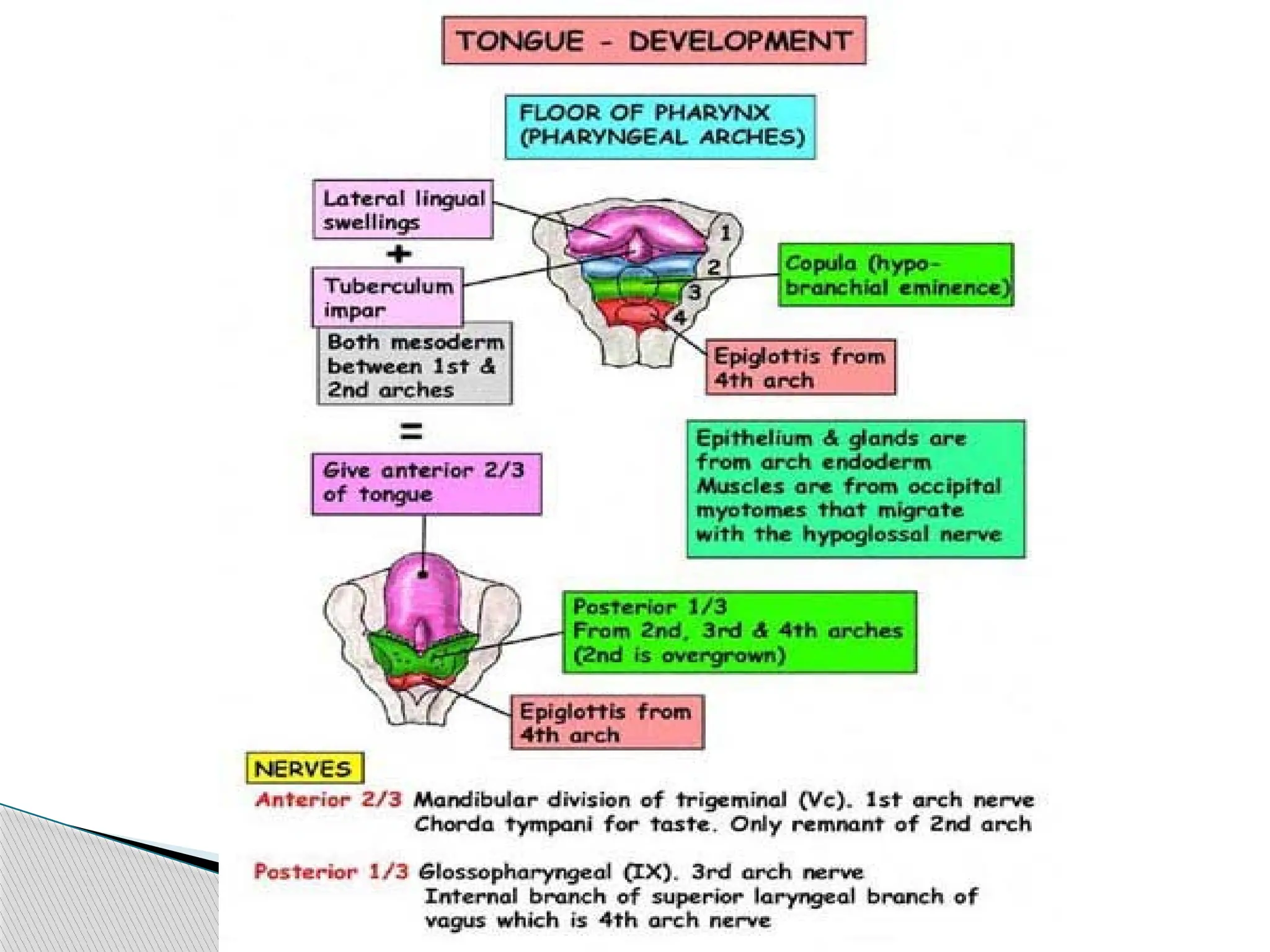
Overview Development ofthe Tongue
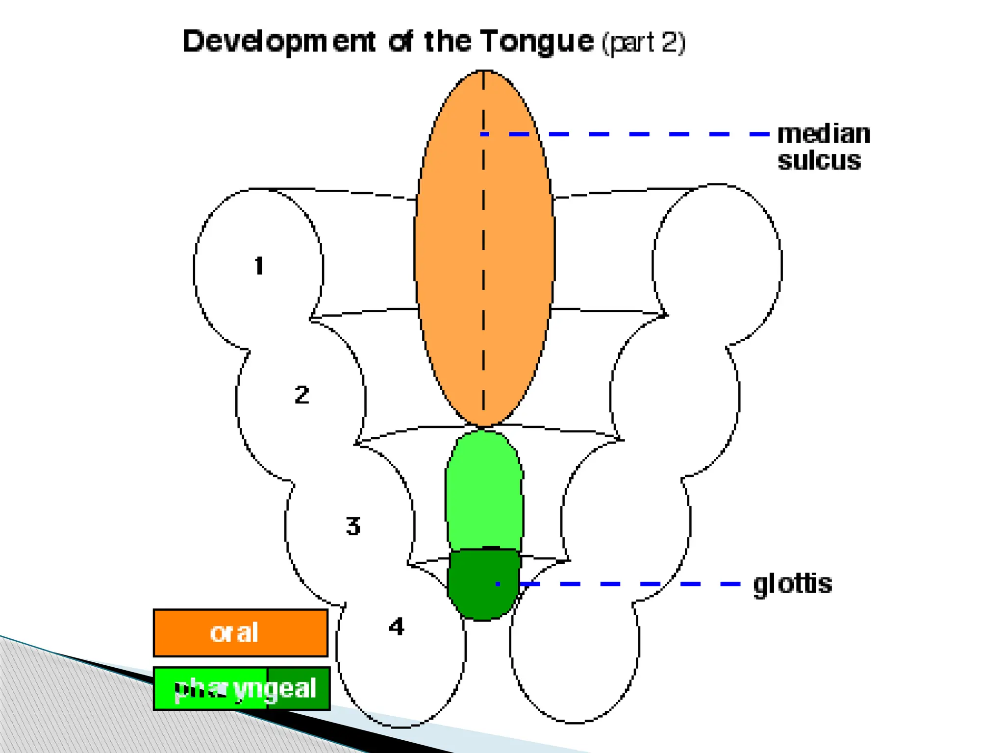
The tongue has contributions from all
pharyngeal arches which changes with time.
The tongue initially begins as swelling rostral
to foramen cecum, the median tongue bud.
8.
Pharyngeal Arch Contributions
Arch1 - oral part of tongue (anterior 2/3)
Arch 2 - initial contribution to surface is lost
Arch 3 - pharyngeal part of tongue
(posterior 1/3)
Arch 4 - epiglottis and adjacent regions
9.
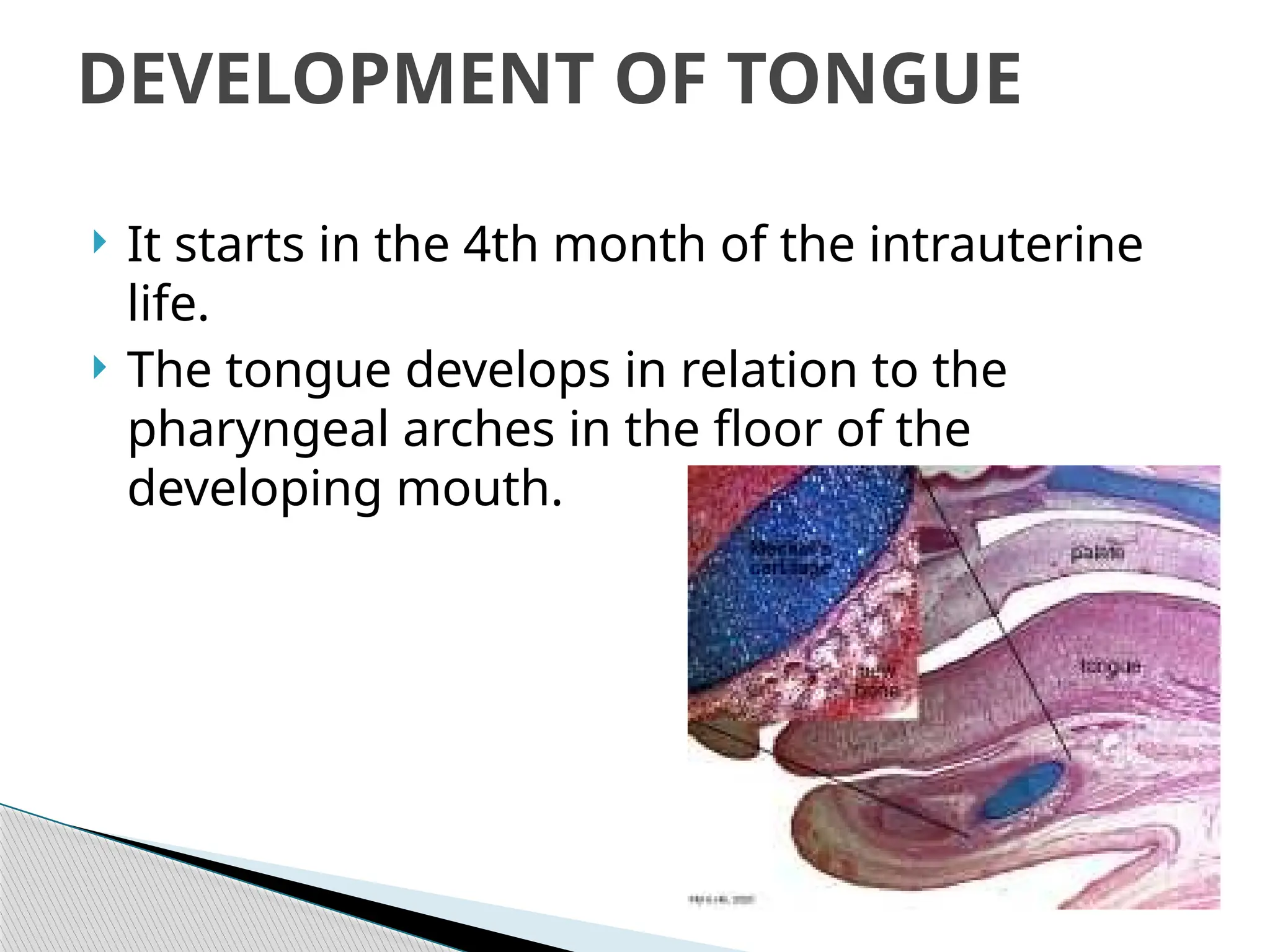
It startsin the 4th month of the intrauterine
life.
The tongue develops in relation to the
pharyngeal arches in the floor of the
developing mouth.
DEVELOPMENT OF TONGUE
11.
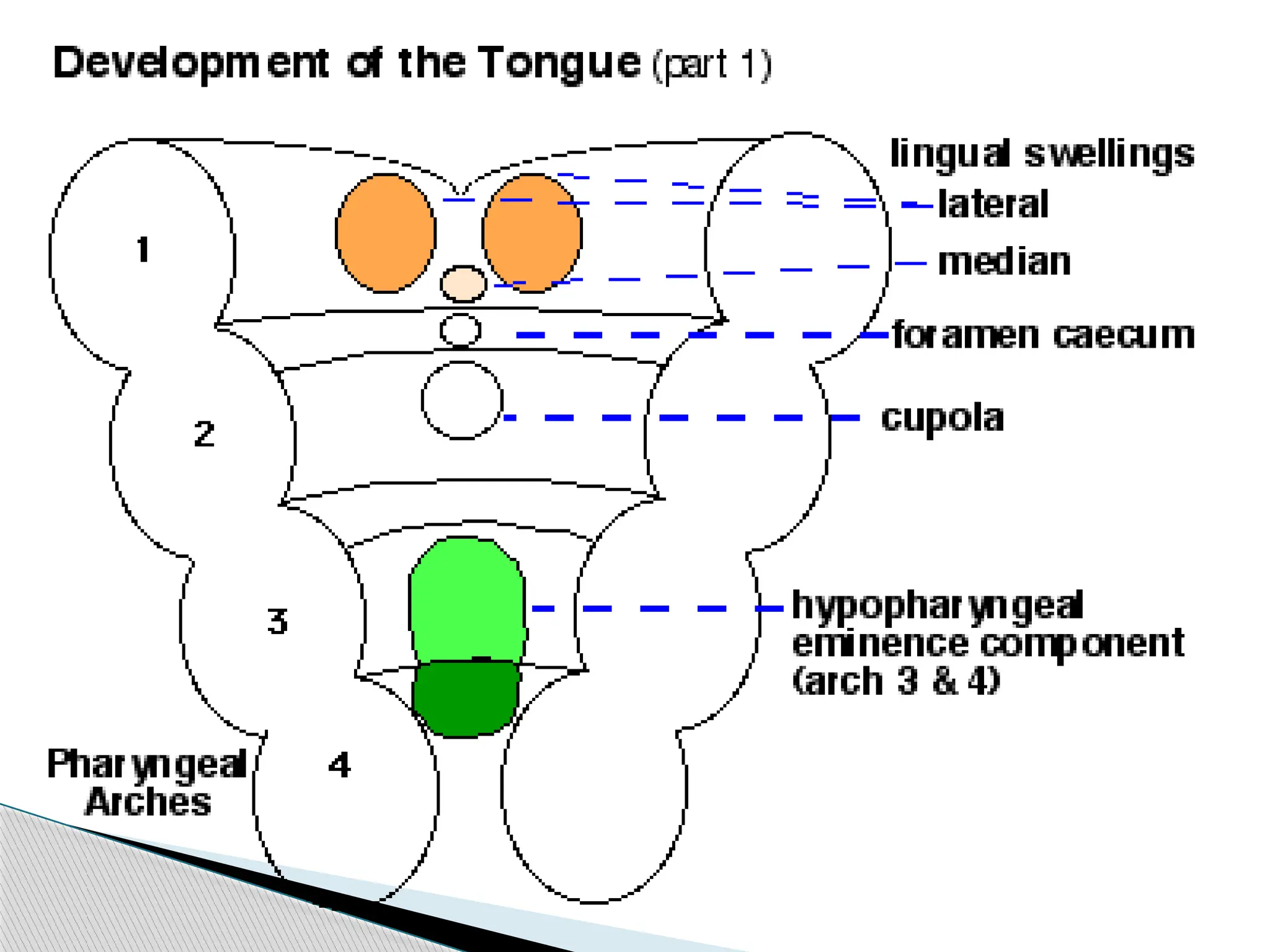
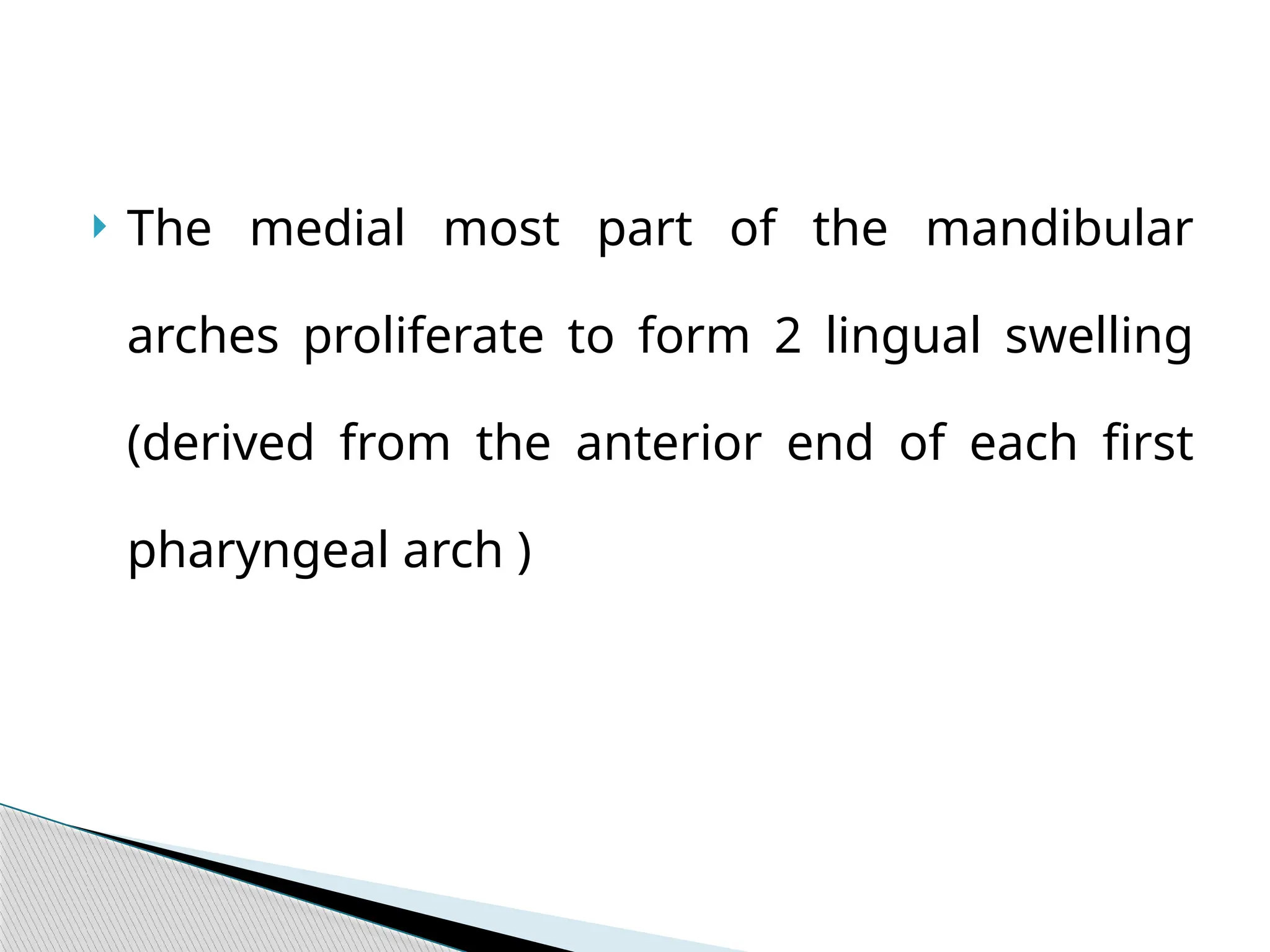
The medialmost part of the mandibular
arches proliferate to form 2 lingual swelling
(derived from the anterior end of each first
pharyngeal arch )
12.
The lingualswelling are partially separated
from each other by another swelling that
appears in midline , at about 4th
wk
The median swelling is called the tuberculum
impar .
13.
Immediately behindthe tuberculum impar , the
epithelium proliferates to form a down growth
( thyroglossal duct) from which thyroid gland
develops.
The site of this downgrowth is subsequently
marked by a depression called the foramen
caecum .
14.
The lateralswelling now enlarge,grow
medially and fuse with each other and the
tuberculum impar .
The lingual swelling thus form the anterior
2/3 or body of the tongue.
15.
Another midlineswelling is seen in relation to
the medial ends of the 2nd
, 3rd
, and 4th
arches.
This swelling is called the hypobrachial
eminence.
16.
The eminencesoon shows a subdivision into
a cranial part related to the 2nd
and 3rd
arches
(called the copula) and a caudal part related
to the 4th
arch.
The caudal part forms the epiglottis.
17.
The anterior2/3 of the tongue is thus
derived from the mandibular arch .
According to some the tuberculum impar
does not make any significant contribution
to the tongue.
18.
The posterior1/3 of the tongue is thus derived
from the cranial part of the hypobrachial
eminence (copula).
In this situation , the 2nd
arch mesoderm gets
buried below the surface .
19.
The 3rd
archmesoderm grows over it to fuse
with the mesoderm of the 1st
arch.
The posterior 1/3 of the tongue is thus
formed by 3rd
arch mesoderm .
The posterior most part of the tongue is
derived from the 4th
arch.
21.
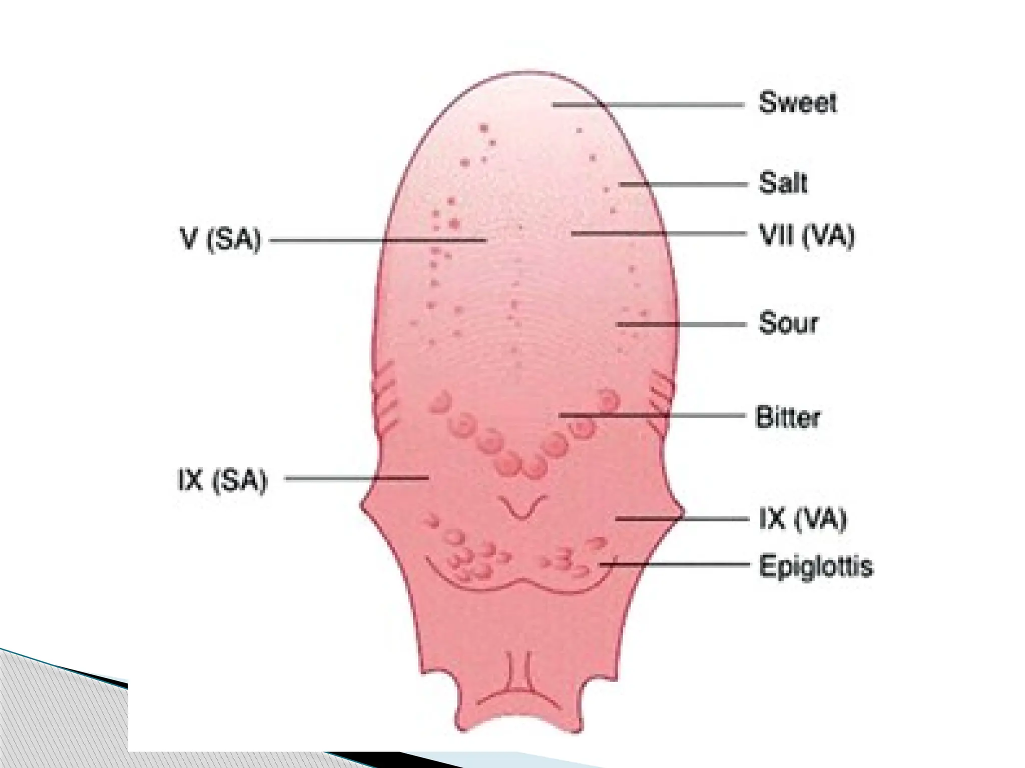
Anterior 2/3of the tongue -lingual branch of
the mandibular nerve, which is the post
trematic nerve of the 1st
arch and by the chorda
tympani which is the pretrematic nerve .
Posterior 1/3 of the tongue -superior laryngeal
nerve, which is the nerve of the 4th
arch.
Nerve Supply
23.
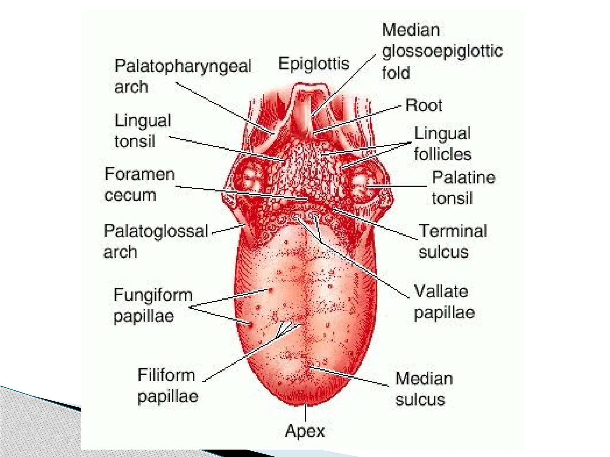
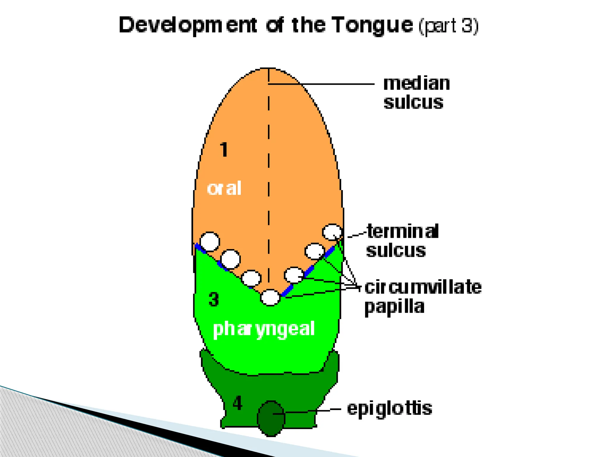
The sulcusterminalis represents the interval
between the lingual swellings of the 1st
pharyngeal arches & the anterior ends of the
3rd
pharyngeal arches.
Around the edge of the anterior 2/3 rd of the
tongue , the ectodermal cell proliferate & grow
inferiorly into the underlying mesenchyme.
Formation of frenum and papillae
24.
Later, thesecells degenerate so that this part
of the tongue becomes free. Some of the
entodermal cells remain in the midline and
help form the frenum of the tongue.
26.
It isderived from the occipital myotomes
and is thus supplied by the hypoglossal
nerve , which is the nerve of these
myotomes.
Development of musculature of tongue
27.
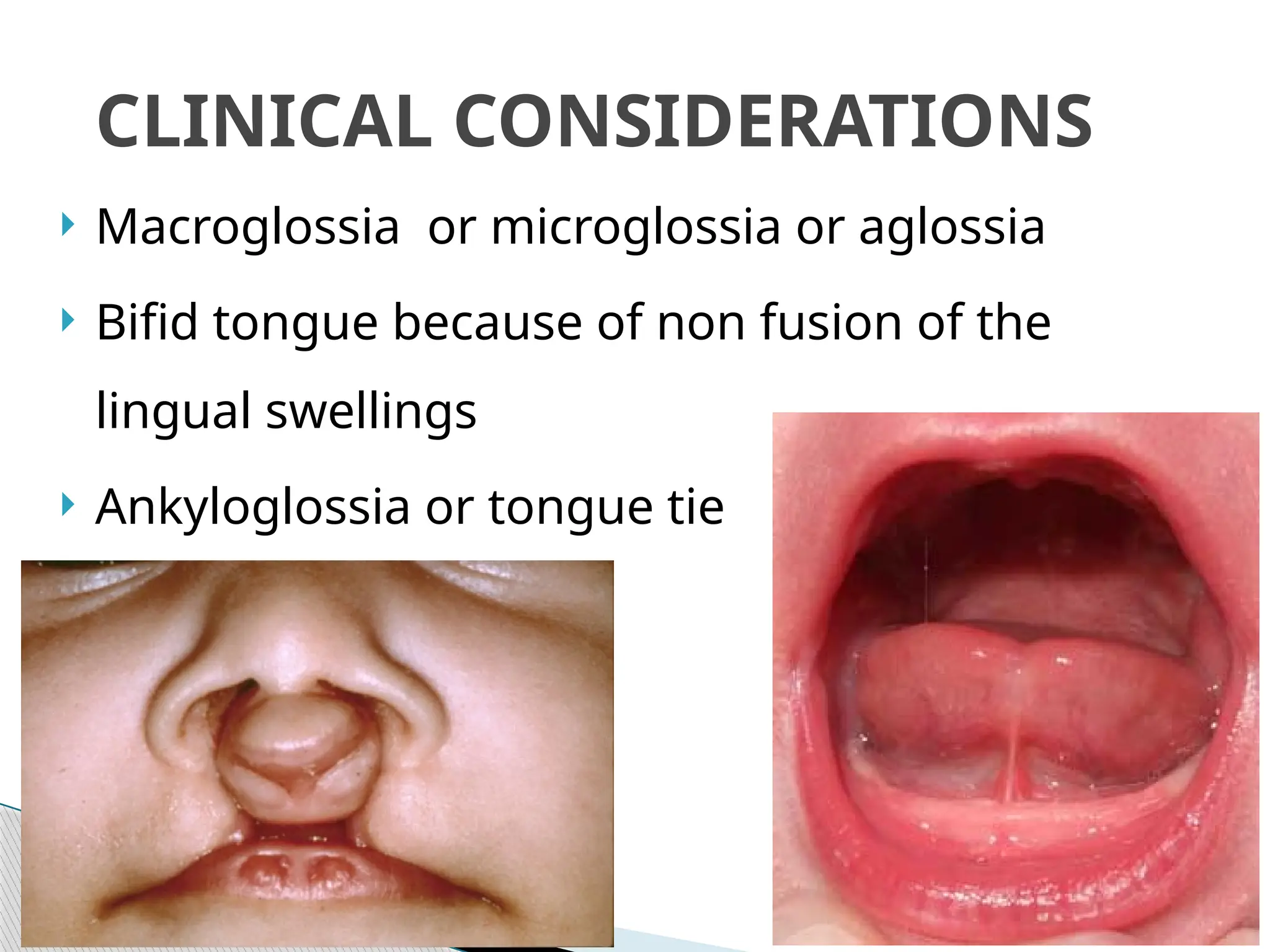
Macroglossia ormicroglossia or aglossia
Bifid tongue because of non fusion of the
lingual swellings
Ankyloglossia or tongue tie
CLINICAL CONSIDERATIONS
29.
Summary
Development oftongue
Nerve Supply of tongue
Formation of frenum and papillae
Development of musculature of tongue
Clinical Considerations
30.
BIBLIOGRAPHY
Color AtlasAnd Text Book Of Oral Anatomy,
Histology Berkovitz, B. 1ST
edition.
Oral Development and Histology Avery, j. K.1st
edition.
Oral Histology : Development, Structure and Function
Tencate, 4th
edition.
Dental Embryology, Histology & Anatomy. Marry
Bath- Balogh Inergaret. 2nd
edition.