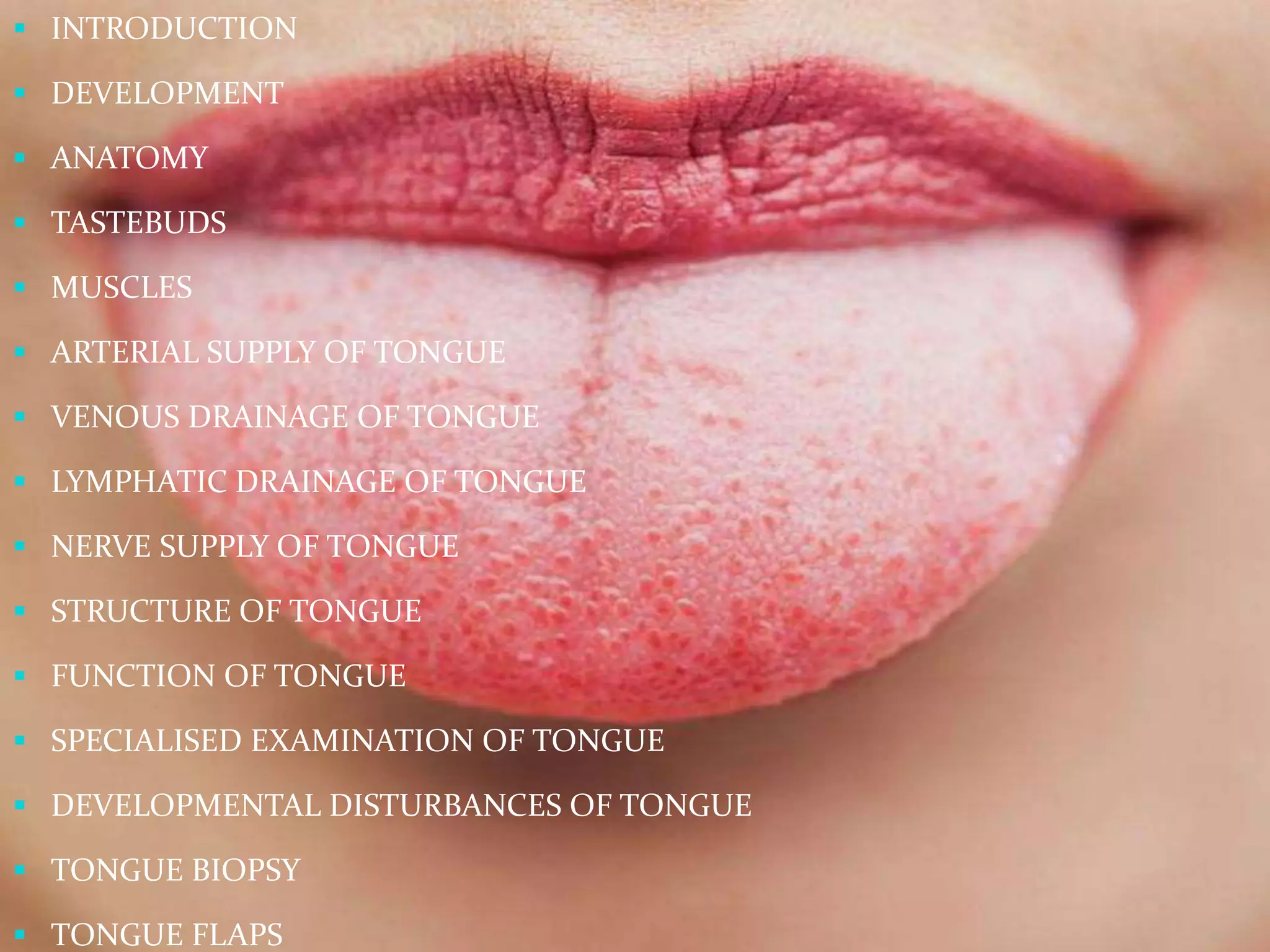
A
Dr. Amit T. Suryawanshi
Presentation
Created & Presented by
Dr. Amit T. Suryawanshi (MDS)
Facial Cosmetic Surgeon
Oral & Maxillofacial Surgeon
Dental Surgeon & Implantologist
Hair Transplant Surgeon (Germany)
Consulting Surgeon in Kolhapur, Sangli, Pune & Mumbai (India)
&
founder of
Face Art International Super speciality
at Kolhapur
Cell Phone no. +91 9405622455
Clinic Landline - +91 7758976097
Email– amitsuryawanshi999@gmail.com
 INTRODUCTION
 DEVELOPMENT
 ANATOMY
 TASTEBUDS
 MUSCLES
 ARTERIAL SUPPLY OF TONGUE
 VENOUS DRAINAGE OF TONGUE
 LYMPHATIC DRAINAGE OF TONGUE
 NERVE SUPPLY OF TONGUE
 STRUCTURE OF TONGUE
 FUNCTION OF TONGUE
 SPECIALISED EXAMINATION OF TONGUE
 DEVELOPMENTAL DISTURBANCES OF TONGUE
 TONGUE BIOPSY
 TONGUE FLAPS
DEVELOPMENT OF TONGUE
TONGUE DEVELOPS DURING 4TH – 8TH WEEK OF
PRENATAL DEVELOPMENT.
IT DEVELOPS FROM INDEPENDENT SWELLINGS
FORMED BY THE FIRST FOUR BRANCHIAL ARCHES.
BODY OF THE TONGUE : 1ST ARCH
BASE OF THE TONGUE : 3RD & 4TH ARCH
Click here
www.faceart-clinic.com
Click here
www.faceart-clinic.com
Click here
www.faceart-clinic.com
Click here
www.faceart-clinic.com
Click here
www.faceart-clinic.com
Some Recent Findings
 Yamane A. Embryonic and postnatal
development of masticatory and tongue
muscles.
 "Tongue myogenesis follows a similar
regulatory program to that for limb
myogenesis. Myogenesis and synaptogenesis in
the masticatory muscles are delayed in
comparison with other muscles and are not
complete even at birth,
whereas the development of tongue muscles
proceeds faster than those of other muscles
and ends at around birth.
 The regulatory programs for masticatory and
tongue myogenesis seem to depend on the
developmental origins of the muscles, i.e., the
origin being either a somite or somitomere,
whereas myogenesis and synaptogenesis seem
to progress to serve the functional requirements
of the masticatory and tongue muscles."
 The tongue has contributions from all
pharyngeal arches which changes with time.
The tongue initially begins as swelling rostral
to foramen cecum, the median tongue bud.
Click here
www.faceart-clinic.com
ANATOMY OF TONGUE
INFERIOR SURFACE OF TONGUE
DORSUM OF THE TONGUE
PAPILLAE OF TONGUE
MUSCLES-INTRINSIC
MUSCLES-EXTRINSIC
ARTERIAL SUPPLY OF TONGUE
VENOUS DRAINAGE
LYMPHATIC DRAINAGE
NERVE SUPPLY
STRUCTURE OF THE TONGUE
 MUCOUS MEMBRANE
 TASTEBUDS
Click here
www.faceart-clinic.com
FUNCTIONS OF THE TONGUE
 Speech
 Mastication
 Deglutition
 Digestion
 Taste
 Barrier function
 Jaw development
 Secretion
 Defence mechanism
 Maintainance of oral hygiene
 Sucking
 General sensitivity
SPECIALIZED EXAMINATIONS OF TONGUE
 CINERADIOGRAHY
 COMPUTER ASSISTED TOMOGRAPHY
 PULSED(DOPPLER) ULTRASOUND
 REAL TIME ULTRASOUND
 ISOTOPIC SCANNING TECHNIQUE
 ELECTROMYOGRAPHY
 SCANNING ELECTRON MICROSCOPE
 TRANSMISSION ELECTRON MICROSCOPY
MICROGLOSSIA
ANKYLOGLOSSIACLEFT-TONGUE
FISSURED TONGUE
GEOGRAPHIC TONGUE
MEDIAN RHOMBOID GLOSSITIS
LINGUAL THYROID NODULE
LINGUAL VARICOSITIES
HAIRY TONGUE
MACROGLOSSIA
 DEVELOPMENTAL DISORDERS OF TONGUE
MICROGLOSSIA
5/6/2016 27
MACROGLOSSIA
28
• The terms 'ankyloglossia', 'short fraenum', 'short fraenulum',
or
'tongue tie’, refer to a restricted lingual fraenum due to
consolidation of tissue. Leading to reduced mobility of the
tongue.
• Ankyloglossia occurs as a result of the fusion of the lingual
frenum
to the floor of the mouth.
• Partial ankyloglossia or "tongue-tie" is a much more common
condition, because complete fusion rarely occurs.
• This leads to a myriad of speech problems such as lisping
and
stuttering , periodontal and swallowing problems.
ANKYLOGLOSSIA
• This is the gross appearance
of the tongue when the patient
was asked to Stick out
tongue.
• Note the classic symptom
of a bifid or bilobed lingual
apex with a corresponding
midline "cleft" or septal
limitation.
Clinical features
 This photograph shows
the tongue's anatomy as
the patient is asked to
pull tongue back into
mouth as far as
possible.
Click here
www.faceart-clinic.com
• Manual elevation of
the lingual apex by
the examiner.
• The treatment is to surgically sever the connection between
the frenum and the floor of the mouth.
• In young children treatment is postponed until 4 – 5 years
since it is difficult to access severity of disorder in early life.
Frenectomy
Photographs are added
in the presentation with
patient’s permission.
34
CLEFT-TONGUE
• Cleft tongue is a condition where the tongue has a
cleft running right across it horizontally.
• Complete clefting (Diglossia) is extremely rare and
occurs as a result of lack of developmental forces to
push both halves of the tongue towards each other.
• Partial clefting presents as a deep groove in the
middle of the tongue and is a common feature in the
oro-facial-digital syndrome (thick fibrous bands in
lower anterior mucobuccal fold and clefting of
hypoplastic mandibular alveolar process).
35 5/6/2016
36
FISSURED TONGUE
 SCROTAL TONGUE
 FURROWED TONGUE
 LINGUA FISSURATA
 LINGUA PLICATA
 LINGUA SCROTALIS
 PLICATED TONGUE
 CEREBRIFORM TONGUE
 GROOVED TONGUE
5/6/2016
37 5/6/2016
38
Benign migratory glossitis or Erythema migrans
or Psoriasiform mucositis is a benign condition
that occurs in about 3% of the general
population.
GEOGRAPHIC TONGUE
5/6/2016
CLINICAL FEATURES
5/6/2016 39
40
TREATMENT
 No medical intervention is required because the lesion is
benign and most often asymptomatic.
 In severe cases topical corticosteroids in the form of
fluocinonide & beta methasone gel with zinc suppliments
can be prescribed.
5/6/2016
41
MEDIAN RHOMBOID GLOSSITIS
 Median rhomboid glossitis or central papillary atrophy or posterior
lingual papillary atrophy is a focal area of susceptibility to recurring
or chronic atrophic candidiasis.
 Prompting a recent movement toward the use of posterior midline
atrophic candidiasis as a more appropriate diagnostic term.
5/6/2016
CLINICAL FEATURES
5/6/2016 42
43
TREATMENT
 No treatment is necessary for median rhomboid glossitis, but
nodular cases are often removed for microscopic evaluation.
 Recurrence after removal is not expected, although those cases
with pseudoepitheliomatous hyperplasia should be followed
closely for at least a year after biopsy to be certain of the benign
diagnosis.
 Antifungal therapy (topical troches or systemic medication)
will reduce clinical erythema and inflammation due to candida
infection.
 Some lesions will disappear entirely with antifungal therapy.
5/6/2016
44
LINGUAL THYROID NODULE
Accessory accumulation of thyroid tissue that is usually
functional within the body of the posterior tongue.
5/6/2016
45
CLINICAL FEATURES
 The lingual thyroid is four times more common in females than in males.
 It presents as an asymptomatic nodular mass of the posterior lingual
midline, usually less than a centimeter in size but sometimes reaching more
than 4 cm in size .
 Larger lesions can interfere with swallowing and breathing, but most
patients are unaware of the mass at the time of diagnosis, which is usually in
the teenage or young adult years.
 Up to 70% of patients with lingual thyroid have hypothyroidism and 10%
suffer from cretinism.
 Other sites of ectopic thyroid deposition include the cervical lymph nodes,
submandibular glands and the trachea. Rarely, parathyroid glands are
associated with the ectopic thyroid tissue.
5/6/2016
47
TREATMENT
 Surgical excision or radioiodine therapy are effective
treatments for lingual thyroid, but no treatment should be
attempted until an 131iodine radioisotope scan has
determined that there is adequate thyroid tissue in the neck.
 Endocrine evaluation for hypothyroidism should, therefore,
be done in such cases. In this light, it is important to know
that three of every four patients with infantile
hypothyroidism have ectopic thyroid tissue
 Occasional patients with parathyroid tissue associated with
their lingual thyroid have developed tetany after their
inadvertent removal.
5/6/2016
48
 In those patients lacking thyroid tissue in the neck, the
lingual thyroid can be excised and autotransplanted to
the muscles of the neck.
 However, Most cases require no treatment and in cases
where biopsy is necessary it should be considered with
caution because of the potential for hemorrhage,
infection or release of large amounts of hormone into the
vascular system (thyroid storm).
 Rare examples of thyroid carcinoma arising in the mass
have been reported, almost always in males.
5/6/2016
49
LINGUAL VARICOSITIES
 Prominent lingual veins, usually observed on the ventral
and lateral surface of the tongue are called lingual
varicosities.
 These are hemmorhoid (dialated veins) caused by the
decrease in the amount of surrounding connective tissue.
 A normal variant in adults over 60 years of age believed to
be related to the aging process.
 It's occurance increases with age or increased blood
pressure
5/6/2016
50
• Enlarged veins usually purple or red or clusters on ventral and
lateral surface of the tongue.
5/6/2016
51
HAIRY TONGUE
 Commonly observed condition of defective desquamation
of the filiform papillae that results from a variety of
precipitating factors.
5/6/2016
CLINICAL FEATURES
5/6/2016 52
53
TREATMENT
 Treatment of hairy tongue is variable.
 In many cases, simply brushing the tongue with a toothbrush
or using a commercially available tongue scraper is sufficient
to remove elongated filiform papillae and retard the growth
of additional ones.
 Surgical removal of the papillae by using electrodesiccation,
carbon dioxide laser or even scissors is the treatment of last
resort when less complicated therapies prove ineffective.
5/6/2016
APPLIED ANATOMY OF TONGUE:
 INJURY TO HYPOGLOSSAL NERVE PRODUCES PARALYSIS OF
THE MUSCLES OF THE TONGUE.
 GLOSSITIS : USUALLY A PART OF GENERALIZED
ULCERATION. IN CERTAIN ANAEMIAS TONGUE BECOMES
BALD DUE TO ATROPY OF THE FILLIFORM PAPILLAE.
 DUE TO PRESENCE OF RICH NETWORK OF LYMPHATICS &
OF LOOSE AREOLAR TISSUE IN THE SUBSTANCE OF
TONGUE IS RESPONSIBLE FOR ENORMOUS SWELLING OF
THE TONGUE IN ACUTE GLOSSITIS.
 UNDERSURFACE OF TONGUE IS A GOOD SITE FOR
OBSERVATION OF JAUNDICE.
 IN UNCONSCIOUS PATIENT THE TONGUE MAY FALL
BACK & OBSTUCT THE AIR PASSAGES.
 IN PATIENT WITH GRAND MAL EPILEPSY THE
TONGUE IS COMMONLY BITTEN DURING ATTACK.
 CARCINOMA OF THE TONGUE IS QUITE COMMON . IT
IS BETTER TREATED WITH RADIOTHERAY THAN
SURGERY .BUT SINCE FACILITIES FOR IRRADIATION
ARE NOT ALWAYS AVAILABLE THE AFFECTED PART IS
REMOVED SURGICALLY. ALL THE DEEP CERVICAL
LYMPH NODES ARE ALSO REMOVED BECAUSE OF
RECURANCE OF MALIGNANT DISEASE OCCURS IN
LYMPH NODES.
 CARCINOMA OF POSTERIOR 1/3RD OF THE IS MORE
DANGEROUS DUE TO BILATERAL LYMPHATIC SPREAD.
Applied anatomy
•Carcinoma of the lat. part of ant 23rd of tongue spreads unilaterally.
Thus a hemiglossectomy with unilateral lymph node dissection can be
done.
•Gag reflex occurs on touching the post. 13rd of the tongue. IX n.provides
the afferent limb of the reflex.
•Injury to hypoglossal n. due to fracture mandible leads to unilateral
paralysis of tongue. Tongue deviates to the paralysed side on protrusion.
•Paralysis of genioglossus occurs when the patient is unconscious. The
tongue falls back & the patient may suffocate to death. Tongue is pulled
forward in an unconscios patient. Genioglossus is called a safety muscle
•Sublingual absorption of drugs – for fast absorption of drugs they are
placed sublingually because of the thin mucosa which allows quick
absorption
THYROGLOSSAL DUCT CYST
ABERRANT THYROID GLAND
TONGUE BIOPSY
INTRAORAL RECONSTRUCTION
WITH TONGUE FLAPS
 -FOR THE CLOSURE OF PERFORATIONS IN HARD
PALATE
 -PALATAL ALVEOLAR FISTULA AFTER NOMA
 DEFECT ON THE LOWER ASPECT OF TONGUE
AND FLOOR OF THE MOUTH
 CHEEK AREA RECONSTRUCTION
 MIDLINE GLOSSOTOMY
 RECONSTRUCTION OF PARTIAL GLOSSECTOMY
DEFECTS
 RECONSTRUCTION OF THE BASE OF TONGUE
AND TOTAL GLOSSECTOMY DEFECTS
Click here
www.faceart-clinic.com
Click here
www.faceart-clinic.com
Thank you
Feel
Free
to
Ask
Questions ??
as we always say
Follow us on SlideShare &
Click here
www.faceart-clinic.com

The Tongue. Everything about it.

  • 1.
    A Dr. Amit T.Suryawanshi Presentation
  • 2.
    Created & Presentedby Dr. Amit T. Suryawanshi (MDS) Facial Cosmetic Surgeon Oral & Maxillofacial Surgeon Dental Surgeon & Implantologist Hair Transplant Surgeon (Germany) Consulting Surgeon in Kolhapur, Sangli, Pune & Mumbai (India) & founder of Face Art International Super speciality at Kolhapur Cell Phone no. +91 9405622455 Clinic Landline - +91 7758976097 Email– amitsuryawanshi999@gmail.com
  • 3.
     INTRODUCTION  DEVELOPMENT ANATOMY  TASTEBUDS  MUSCLES  ARTERIAL SUPPLY OF TONGUE  VENOUS DRAINAGE OF TONGUE  LYMPHATIC DRAINAGE OF TONGUE  NERVE SUPPLY OF TONGUE  STRUCTURE OF TONGUE  FUNCTION OF TONGUE  SPECIALISED EXAMINATION OF TONGUE  DEVELOPMENTAL DISTURBANCES OF TONGUE  TONGUE BIOPSY  TONGUE FLAPS
  • 4.
    DEVELOPMENT OF TONGUE TONGUEDEVELOPS DURING 4TH – 8TH WEEK OF PRENATAL DEVELOPMENT. IT DEVELOPS FROM INDEPENDENT SWELLINGS FORMED BY THE FIRST FOUR BRANCHIAL ARCHES. BODY OF THE TONGUE : 1ST ARCH BASE OF THE TONGUE : 3RD & 4TH ARCH
  • 6.
  • 7.
  • 8.
  • 9.
  • 10.
  • 11.
    Some Recent Findings Yamane A. Embryonic and postnatal development of masticatory and tongue muscles.  "Tongue myogenesis follows a similar regulatory program to that for limb myogenesis. Myogenesis and synaptogenesis in the masticatory muscles are delayed in comparison with other muscles and are not complete even at birth, whereas the development of tongue muscles proceeds faster than those of other muscles and ends at around birth.
  • 12.
     The regulatoryprograms for masticatory and tongue myogenesis seem to depend on the developmental origins of the muscles, i.e., the origin being either a somite or somitomere, whereas myogenesis and synaptogenesis seem to progress to serve the functional requirements of the masticatory and tongue muscles."  The tongue has contributions from all pharyngeal arches which changes with time. The tongue initially begins as swelling rostral to foramen cecum, the median tongue bud. Click here www.faceart-clinic.com
  • 13.
  • 14.
  • 15.
  • 16.
  • 17.
  • 18.
  • 19.
  • 20.
  • 21.
  • 22.
  • 23.
    STRUCTURE OF THETONGUE  MUCOUS MEMBRANE  TASTEBUDS Click here www.faceart-clinic.com
  • 24.
    FUNCTIONS OF THETONGUE  Speech  Mastication  Deglutition  Digestion  Taste  Barrier function  Jaw development  Secretion  Defence mechanism  Maintainance of oral hygiene  Sucking  General sensitivity
  • 25.
    SPECIALIZED EXAMINATIONS OFTONGUE  CINERADIOGRAHY  COMPUTER ASSISTED TOMOGRAPHY  PULSED(DOPPLER) ULTRASOUND  REAL TIME ULTRASOUND  ISOTOPIC SCANNING TECHNIQUE  ELECTROMYOGRAPHY  SCANNING ELECTRON MICROSCOPE  TRANSMISSION ELECTRON MICROSCOPY
  • 26.
    MICROGLOSSIA ANKYLOGLOSSIACLEFT-TONGUE FISSURED TONGUE GEOGRAPHIC TONGUE MEDIANRHOMBOID GLOSSITIS LINGUAL THYROID NODULE LINGUAL VARICOSITIES HAIRY TONGUE MACROGLOSSIA  DEVELOPMENTAL DISORDERS OF TONGUE
  • 27.
  • 28.
  • 29.
    • The terms'ankyloglossia', 'short fraenum', 'short fraenulum', or 'tongue tie’, refer to a restricted lingual fraenum due to consolidation of tissue. Leading to reduced mobility of the tongue. • Ankyloglossia occurs as a result of the fusion of the lingual frenum to the floor of the mouth. • Partial ankyloglossia or "tongue-tie" is a much more common condition, because complete fusion rarely occurs. • This leads to a myriad of speech problems such as lisping and stuttering , periodontal and swallowing problems. ANKYLOGLOSSIA
  • 30.
    • This isthe gross appearance of the tongue when the patient was asked to Stick out tongue. • Note the classic symptom of a bifid or bilobed lingual apex with a corresponding midline "cleft" or septal limitation. Clinical features
  • 31.
     This photographshows the tongue's anatomy as the patient is asked to pull tongue back into mouth as far as possible. Click here www.faceart-clinic.com
  • 32.
    • Manual elevationof the lingual apex by the examiner. • The treatment is to surgically sever the connection between the frenum and the floor of the mouth. • In young children treatment is postponed until 4 – 5 years since it is difficult to access severity of disorder in early life.
  • 33.
    Frenectomy Photographs are added inthe presentation with patient’s permission.
  • 34.
    34 CLEFT-TONGUE • Cleft tongueis a condition where the tongue has a cleft running right across it horizontally. • Complete clefting (Diglossia) is extremely rare and occurs as a result of lack of developmental forces to push both halves of the tongue towards each other. • Partial clefting presents as a deep groove in the middle of the tongue and is a common feature in the oro-facial-digital syndrome (thick fibrous bands in lower anterior mucobuccal fold and clefting of hypoplastic mandibular alveolar process).
  • 35.
  • 36.
    36 FISSURED TONGUE  SCROTALTONGUE  FURROWED TONGUE  LINGUA FISSURATA  LINGUA PLICATA  LINGUA SCROTALIS  PLICATED TONGUE  CEREBRIFORM TONGUE  GROOVED TONGUE 5/6/2016
  • 37.
  • 38.
    38 Benign migratory glossitisor Erythema migrans or Psoriasiform mucositis is a benign condition that occurs in about 3% of the general population. GEOGRAPHIC TONGUE 5/6/2016
  • 39.
  • 40.
    40 TREATMENT  No medicalintervention is required because the lesion is benign and most often asymptomatic.  In severe cases topical corticosteroids in the form of fluocinonide & beta methasone gel with zinc suppliments can be prescribed. 5/6/2016
  • 41.
    41 MEDIAN RHOMBOID GLOSSITIS Median rhomboid glossitis or central papillary atrophy or posterior lingual papillary atrophy is a focal area of susceptibility to recurring or chronic atrophic candidiasis.  Prompting a recent movement toward the use of posterior midline atrophic candidiasis as a more appropriate diagnostic term. 5/6/2016
  • 42.
  • 43.
    43 TREATMENT  No treatmentis necessary for median rhomboid glossitis, but nodular cases are often removed for microscopic evaluation.  Recurrence after removal is not expected, although those cases with pseudoepitheliomatous hyperplasia should be followed closely for at least a year after biopsy to be certain of the benign diagnosis.  Antifungal therapy (topical troches or systemic medication) will reduce clinical erythema and inflammation due to candida infection.  Some lesions will disappear entirely with antifungal therapy. 5/6/2016
  • 44.
    44 LINGUAL THYROID NODULE Accessoryaccumulation of thyroid tissue that is usually functional within the body of the posterior tongue. 5/6/2016
  • 45.
    45 CLINICAL FEATURES  Thelingual thyroid is four times more common in females than in males.  It presents as an asymptomatic nodular mass of the posterior lingual midline, usually less than a centimeter in size but sometimes reaching more than 4 cm in size .  Larger lesions can interfere with swallowing and breathing, but most patients are unaware of the mass at the time of diagnosis, which is usually in the teenage or young adult years.  Up to 70% of patients with lingual thyroid have hypothyroidism and 10% suffer from cretinism.  Other sites of ectopic thyroid deposition include the cervical lymph nodes, submandibular glands and the trachea. Rarely, parathyroid glands are associated with the ectopic thyroid tissue. 5/6/2016
  • 47.
    47 TREATMENT  Surgical excisionor radioiodine therapy are effective treatments for lingual thyroid, but no treatment should be attempted until an 131iodine radioisotope scan has determined that there is adequate thyroid tissue in the neck.  Endocrine evaluation for hypothyroidism should, therefore, be done in such cases. In this light, it is important to know that three of every four patients with infantile hypothyroidism have ectopic thyroid tissue  Occasional patients with parathyroid tissue associated with their lingual thyroid have developed tetany after their inadvertent removal. 5/6/2016
  • 48.
    48  In thosepatients lacking thyroid tissue in the neck, the lingual thyroid can be excised and autotransplanted to the muscles of the neck.  However, Most cases require no treatment and in cases where biopsy is necessary it should be considered with caution because of the potential for hemorrhage, infection or release of large amounts of hormone into the vascular system (thyroid storm).  Rare examples of thyroid carcinoma arising in the mass have been reported, almost always in males. 5/6/2016
  • 49.
    49 LINGUAL VARICOSITIES  Prominentlingual veins, usually observed on the ventral and lateral surface of the tongue are called lingual varicosities.  These are hemmorhoid (dialated veins) caused by the decrease in the amount of surrounding connective tissue.  A normal variant in adults over 60 years of age believed to be related to the aging process.  It's occurance increases with age or increased blood pressure 5/6/2016
  • 50.
    50 • Enlarged veinsusually purple or red or clusters on ventral and lateral surface of the tongue. 5/6/2016
  • 51.
    51 HAIRY TONGUE  Commonlyobserved condition of defective desquamation of the filiform papillae that results from a variety of precipitating factors. 5/6/2016
  • 52.
  • 53.
    53 TREATMENT  Treatment ofhairy tongue is variable.  In many cases, simply brushing the tongue with a toothbrush or using a commercially available tongue scraper is sufficient to remove elongated filiform papillae and retard the growth of additional ones.  Surgical removal of the papillae by using electrodesiccation, carbon dioxide laser or even scissors is the treatment of last resort when less complicated therapies prove ineffective. 5/6/2016
  • 54.
    APPLIED ANATOMY OFTONGUE:  INJURY TO HYPOGLOSSAL NERVE PRODUCES PARALYSIS OF THE MUSCLES OF THE TONGUE.  GLOSSITIS : USUALLY A PART OF GENERALIZED ULCERATION. IN CERTAIN ANAEMIAS TONGUE BECOMES BALD DUE TO ATROPY OF THE FILLIFORM PAPILLAE.  DUE TO PRESENCE OF RICH NETWORK OF LYMPHATICS & OF LOOSE AREOLAR TISSUE IN THE SUBSTANCE OF TONGUE IS RESPONSIBLE FOR ENORMOUS SWELLING OF THE TONGUE IN ACUTE GLOSSITIS.  UNDERSURFACE OF TONGUE IS A GOOD SITE FOR OBSERVATION OF JAUNDICE.
  • 55.
     IN UNCONSCIOUSPATIENT THE TONGUE MAY FALL BACK & OBSTUCT THE AIR PASSAGES.  IN PATIENT WITH GRAND MAL EPILEPSY THE TONGUE IS COMMONLY BITTEN DURING ATTACK.  CARCINOMA OF THE TONGUE IS QUITE COMMON . IT IS BETTER TREATED WITH RADIOTHERAY THAN SURGERY .BUT SINCE FACILITIES FOR IRRADIATION ARE NOT ALWAYS AVAILABLE THE AFFECTED PART IS REMOVED SURGICALLY. ALL THE DEEP CERVICAL LYMPH NODES ARE ALSO REMOVED BECAUSE OF RECURANCE OF MALIGNANT DISEASE OCCURS IN LYMPH NODES.  CARCINOMA OF POSTERIOR 1/3RD OF THE IS MORE DANGEROUS DUE TO BILATERAL LYMPHATIC SPREAD.
  • 56.
    Applied anatomy •Carcinoma ofthe lat. part of ant 23rd of tongue spreads unilaterally. Thus a hemiglossectomy with unilateral lymph node dissection can be done. •Gag reflex occurs on touching the post. 13rd of the tongue. IX n.provides the afferent limb of the reflex. •Injury to hypoglossal n. due to fracture mandible leads to unilateral paralysis of tongue. Tongue deviates to the paralysed side on protrusion. •Paralysis of genioglossus occurs when the patient is unconscious. The tongue falls back & the patient may suffocate to death. Tongue is pulled forward in an unconscios patient. Genioglossus is called a safety muscle •Sublingual absorption of drugs – for fast absorption of drugs they are placed sublingually because of the thin mucosa which allows quick absorption
  • 58.
  • 59.
  • 60.
  • 61.
    INTRAORAL RECONSTRUCTION WITH TONGUEFLAPS  -FOR THE CLOSURE OF PERFORATIONS IN HARD PALATE
  • 62.
     -PALATAL ALVEOLARFISTULA AFTER NOMA
  • 63.
     DEFECT ONTHE LOWER ASPECT OF TONGUE AND FLOOR OF THE MOUTH
  • 64.
     CHEEK AREARECONSTRUCTION
  • 65.
     MIDLINE GLOSSOTOMY RECONSTRUCTION OF PARTIAL GLOSSECTOMY DEFECTS  RECONSTRUCTION OF THE BASE OF TONGUE AND TOTAL GLOSSECTOMY DEFECTS Click here www.faceart-clinic.com
  • 66.
  • 67.
  • 68.
  • 69.
    Follow us onSlideShare & Click here www.faceart-clinic.com